 |
 |
| [ Ana Sayfa | Editörler | Danışma Kurulu | Dergi Hakkında | İçindekiler | Arşiv | Yayın Arama | Yazarlara Bilgi | E-Posta ] | |
| Fırat Üniversitesi Sağlık Bilimleri Tıp Dergisi | |||||
| 2018, Cilt 32, Sayı 1, Sayfa(lar) 051-053 | |||||
| [ Özet ] [ PDF ] [ Benzer Makaleler ] [ Yazara E-Posta ] [ Editöre E-Posta ] | |||||
| Atipik Semptomlarla Aort Diseksiyonuna Bağlı Olarak Gelişen Serebrovasküler Olay | |||||
| Yahya AKALIN1, İbrahim ÇALTEKİN2, Emre GÖKÇEN2, Atakan SAVRUN3, Bülent DEMİR4, Şeyda Tuba SAVRUN3 | |||||
| 1Malatya Training and Research Hospital, Department of Neurology, Malatya, TURKEY 2University of Bozok, Training and Research Hospital, Department of Emergency Medicine, Yozgat, TURKEY 3University of Ordu, Faculty of Medicine, Department of Emergency Medicine, Ordu, TURKEY 4University of Celal Bayar, Faculty of Medicine, Department of Emergency Medicine, Manisa, TURKEY |
|||||
| Anahtar Kelimeler: Aort diseksiyonu, inme, yatak başı ultrason | |||||
| Özet | |||||
Aort diseksiyonu; aortun media tabakası ile intima tabakasının ayrılması sonucu oluşan yüksek mortalite ve morbidite ile seyreden kardiyovasküler acillerden biridir. Bazı vakalarda aort diseksiyonunun koma, stroke, mezenter iskemisi, renal yetmezlik, miyokard enfarktüsü gibi atipik bulgularla prezente olabileceği bilinmektedir. Bu olguda acil servise akut stroke semptomları ile başvuran ve etiyolojiye yönelik yapılan tetkikler sonucunda aort diseksiyonu tanısı saptanan 32 yaşında bir erkek hasta sunulmaktadır. |
|||||
| Giriş | |||||
Aortic dissection is a significant cardiovascular condition presenting high rate of mortality and morbidity and occurring due to the separation of aortic media from intima layer 1. The most common symptoms of aortic dissection can be listed as sudden chest and back pain; however, it is also seen in certain cases that the disease is accompanied with atypical findings such as coma, stroke, mesenteric ischemia, renal failure and myocardial infarction 2. Patients suffering from aortic dissection do not always go to the hospitals with typical symptoms such as sudden, severe and tearing chest, back and abdomen pains. Certain cases may involve atypical neurological findings such as syncope, hemiparesis-hemiplegia, paraparesis-paraplegia and acute stroke or atypical symptoms such as findings of myocardial infarction and gastrointestinal complaints 3. In our study, we have analyzed a case presenting to the emergency department with acute stroke symptoms and diagnosed with aortic dissection subsequent to the examinations made for etiology. |
|||||
| Olgu Sunusu | |||||
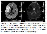
32-year-old male patient presented to the emergency department (ED) with the complaints of recently developed motor speech impairment and loss of strength on the left arm and the left leg. The patient did not complain of chest pain, abdomen pain or back pain. He did not have any other disease in the past medical history. The patient was conscious, cooperative and oriented. His vital signs were measured as follows: Blood Pressure: 134/85 mm/Hg, Pulse: 87 beat/minute, respiratory rate: 13 breath/minute, body temperature: 36.5 ºC. His Glasgow Coma Score was 15. No significant difference was found between two arms in terms of blood pressure; femoral pulses were taken from both legs. Pupillary light reflex was normal in neurological examination; however, slight deviation towards right side was observed in the eyes; motor speech impairment (dysarthria) was seen, and loss of strength was detected at the scale of 1/5 both in the upper extremities and lower extremities during the strength examination of the left extremities, and it was more demonstrable in upper extremities. Moreover, hypoesthesia and hypoalgesia were found in the left arm and leg of the patient. No murmur was heard during cardiac auscultation of the patient, and his ECG was in sinus rhythm. No pathological finding was detected in other results of the physical examination and the emergency laboratory tests. A non-contrasted cranial computerized tomography (CT) scan was performed shortly after the patient's arrival, due to his presenting signs and symptoms. This was done to rule out a hemorrhagic stroke or signs of cerebral infarction of a size or age that would preclude any indication for therapy with tissue plasminogen activator (t-PA). However, an image compatible with ischemic infarct involving the middle cerebral artery (MCA) site was detected in the diffusion-weighted cranial magnetic resonance imaging (MRI) (Figure 1A and B). A bedside trans-thoracic echocardiogram was performed as part of the routine work-up of potential causes of the patient's ischemic stroke. Via the parasternal long axis view of the heart, an aortic aneurysm with dissection could be appreciated. The aorta measured 6.0 cm in diameter at its widest area. In addition to the aneurysm, a dissection was evident, with a flap appearance spreading into the aortic lumen, seen in both the parasternal long axis and suprasternal examination views. CT angiography which was performed on the patient showed a Stanford A-type aortic dissection extending from ascending aorta to the distal of arcus aorta, and having dissection flab extending to proximal of brachiocephalic trunk (Figure 2A, B and C). The patient was transferred to another hospital at higher level in order to be operated, and it was observed that the patient was discharged from the hospital 25 days later subsequent to a successful operation without suffering from any neurological sequel.
|
|||||
| Tartışma | |||||
Sudden chest pain is observed in 85% of the patients suffering from aortic dissection, and the pain is generally described as sharp stabbing. Ischemic stroke resulting from aortic dissection is a rare condition (6-32%) in the cases of aortic dissection 4. Nevertheless, neurological incidents may develop at the rate of 18-30% in the cases of A-type aortic dissection occurring without any chest pain 5. It is quite difficult to diagnose the neurological conditions developing due to A-type aortic dissection without chest pain. The main reason for ischemic stroke is considered to be the reduction of the cerebral blood flow occurring as a result of the stenosis of main lumen by pseudo-lumen with thrombosis. Altered mental status, seizures or other neurological changes may be ultimately seen 6. Atypical formation of focal neurological findings in 10% of the cases may hinder the diagnosis and the rapid management of this rapid and fatal situation 7. Transthoracic echocardiography has been reported to have high sensitivity in the diagnosis of aortic dissection. For dilation of 40 mm or greater, its sensitivity is 77% and its specificity is 95%. For dilation of 45 mm or greater, its sensitivity is 64% and its specificity was 99% 8. CT angiography, on the other hand, is the most sensitive imaging method for the detection of aortic dissection, and it has a sensitivity rate of 98-100% 9. Conclusion, Aortic dissection should be taken into consideration for the patients presenting to the ED with certain indications unexpected to be seen in aortic dissection such as syncope, changes in consciousness, hypotension, atypical abdomen pain or loss of strength in the extremities. The patients should be examined for the detection of aortic dissection by means of bedside ultrasound in ED, which is a noninvasive method.
Acknowledgement |
|||||
| Kaynaklar | |||||
1) Mukherjee D, Eagle KA. Aortic dissection - an update. Curr Probl Cardiol 2005; 30: 287-325.
2) Scalia D, Rizzoli G, Scomparin MA. Aorto - right atrial fistula: A rare complication of aortic dissection type A. A report of two cases. J Cardiovasc Surg (Torino) 1997; 38: 619-622.
3) Rosenberg H, Al - Rajhi K. ED ultrasound diagnosis of a type B aortic dissection using the suprasternal view. Am J Emerg Med 2012; 30: 2084.
4) Gaul C, Dietrich W, Friedrich I, et al. Neurological symptoms in type Aaortic dissections. Stroke 2007; 38: 292-297.
5) Veyssier - Belot C, Cohen A, Rougemont D, et al. Cerebral infarction due to painless thoracic aortic and common carotid artery dissections. Stroke 1993; 24: 2111-2113.
6) Yamashiro S, Arakaki R, Kise Y, Kuniyoshi Y. Emergency operation for aortic dissection with ischemic stroke. Asian Cardiovasc Thorac Ann 2014; 22: 208-211.
7) Oon JE, Kee AC, Toh HC. A case report of Stanford type A aortic dissection presenting with status epilepticus. Am J Emerg Med 2011; 29: 243.
|
|||||
| [ Başa Dön ] [ Özet ] [ PDF ] [ Benzer Makaleler ] [ Yazara E-Posta ] [ Editöre E-Posta ] | |||||
 |
| [ Ana Sayfa | Editörler | Danışma Kurulu | Dergi Hakkında | İçindekiler | Arşiv | Yayın Arama | Yazarlara Bilgi | E-Posta ] |

