[ Ana Sayfa | Editörler | Danışma Kurulu | Dergi Hakkında | İçindekiler | Arşiv | Yayın Arama | Yazarlara Bilgi | E-Posta ]
Fırat Üniversitesi Sağlık Bilimleri Tıp Dergisi
2020, Cilt 34, Sayı 1, Sayfa(lar) 049-054
[ Özet ] [ PDF ] [ Benzer Makaleler ] [ Yazara E-Posta ] [ Editöre E-Posta ]
Ankilozan Spondilit Hastalık Aktivitesi Değerlendirmesinde Gözyaşı Ozmolaritesi
Hüseyin FINDIK1, Münevver SERDAROĞLU BEYAZAL2, Murat OKUTUCU1, Mehmet Gökhan ASLAN1, Hacı KOÇ3, Feyzahan UZUN1
1Recep Tayyip Erdoğan University, Training and Research Hospital, Eye Diseases Department, Rize, TURKEY
2Recep Tayyip Erdoğan University, Training and Research Hospital, Physical Medicine and Rehabilitation Department, Rize, TURKEY
3Private Ada Medical Hospital, Eye diseases, Sakarya, TURKEY
Anahtar Kelimeler: Ankilozan spondilit, tearlab, gözyaşı ozmolaritesi, kuru göz, schirmer testi, gözyaşı kırılma zamanı
Özet
Amaç: Ankilozan spondilitli (AS) hastalarda TearLab sistemi (TearLab Corp, San Diego, Calif.) ile gözyaşı ozmolaritesini (TO) ölçerek kuru göz sendromunu (DES) değerlendirmek ve göz kuruluğu ile AS arasındaki ilişkiyi saptamak.

Gereç ve Yöntemler: Çalışmaya 57 hastanın 57 gözü ve kontrol gurubu olarak 29 sağlıklı göz dahil edildi. Çalışmaya alınan hastalar, Bath Ankylosing Spondylitis Disease Activity Index (BASDAI) skorlarına göre; kontrol, hafif-orta (BASDAI ≤ 4), ve şiddetli (BASDAI>4) olarak; HLA B27’ye göre; kontrol, HLA B27 (+) ve HLA B27 (-) ve tedavi statüsüne göre kontrol, tedavi alan (+) ve tedavi almayan (-) olarak üç ana guruba ayrıldı. Bu üç ana gurubun her biri; TO, BUT ve Schirmer testleri bakımından karşılaştırıldı.

Bulgular: Bu üç ana grup’un her biri için yapılan karşılaştırmalarda; TO (p = 0.383), Schirmer testi (p = 0.108) ve BUT (p = 0.722) değerleri açısından anlamlı bir fark gözlenmedi. Ayrıca hastalık aktivite skor değerleri ile TO değerleri ( r=0,23, p=0,863), Schirmer skorları (r= –0.223, P= 0.095) ve BUT skorları (r = –0.044, P= 0.744) korele değildi.

Sonuç: Çalışmamızda; hastalık aktivitesi yüksek, HLA-B27 belirteci pozitif ve tedavi almayan hasta gruplarında elde edilen TO değerlerini daha yüksek olarak saptadık. Bu değerler her ne kadar istatistiksel anlamlılık düzeyine ulaşmamış olsa da, bulgularımıza gore yüksek inflamasyon düzeylerinde TO seviyesinin, AS hastalık aktivitesi için preliminer bir gösterge olarak kullanılabileceği kanaatindeyiz.

  • Başa Dön
  • Özet
  • Giriş
  • Materyal ve Metot
  • Bulgular
  • Tartışma
  • Kaynaklar
  • Giriş
    Ankylosing spondylitis (AS) is an immune-related rheumatic disease characterized by chronic inflammation and involving various organs 1,2. Instability occurs on the corneal surface in AS with the accumulation on the ocular surface of cytokines released by T cells and with the conversion of corneal Langerhans cells (LC), which play an important role in immune processes on the ocular surface, into an active participant in corneal immune response 3-5. This inflammation has been shown not only to cause disturbance on the ocular corneal surface, but also to cause dry eye syndrome (DES) by leading to dysfunction in the lachrymal gland under the effect of the cytokines 6,7.

    Secondary Sjögren’s syndrome (SS) has been shown to develop in 10% of patients with AS, and LC in primary and secondary SS have been shown to be associated with DES 8,9. DES thus occurs in inflammatory diseases as a result of involvement of the cornea together with increased tear osmolarity.

    DES is a multifactorial disease that causes injury to the ocular surface compromises tear film stability and can reduce ocular comfort 10. Various tests are used for the diagnosis of DES, including Schirmer’s test, tear break-up time (BUT) and vital staining methods (fluorescein and Rose Bengal). However, none of these alone is sufficient to diagnose DES and assess its severity 10,11. Studies have also evaluated LC morphology using confocal microscopy for the diagnosis of DES 12.

    TO has recently been used both to diagnose DES and as a quantitative and objective test for determining its severity 10,11,13. However, TO has not to date been used as an objective test for showing the presence and severity of DES in AS.

    The purpose of this study was therefore to investigate the presence and prevalence of DE in AS with low inflammatory activity and to reveal the association between the Bath Ankylosing Spondylitis Disease Activity Index (BASDAI) and severity of DES by measurement of TO.

  • Başa Dön
  • Özet
  • Giriş
  • Materyal ve Metot
  • Bulgular
  • Tartışma
  • Kaynaklar
  • Materyal ve Metot
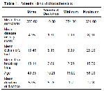
    This study was performed at Recep Tayyip Erdoğan University Medical Faculty Physical Medicine and Rehabilitation and Eye Diseases clinics, Turkey. The study protocol followed the guidelines of the Declaration of Helsinki. Recep Tayyip Erdogan University Clinical Researches Ethics Committee approval (07.10.2016 and 2016-65) and signed informed consent forms from patients were obtained. Fifty-seven eyes of 57 patients with AS and 29 healthy person(control) were included in the study. During diagnosis of DES all patients with AS exhibited a low level of inflammatory activity (CRP <0.5 mg/dL). Patients’ clinical characteristics are shown in Table 1. AS was diagnosed using modified New York criteria in the Physiotherapy and Rehabilitation Clinic 14. Seventeen patients were receiving anti-TNF alpha therapy. BASDAI was calculated during ocular examinations. BASDAI contains six questions showing disease activity and is the most widely used method of evaluating disease activity independently of inflammation status 15. Patients with SS, other immune diseases, endocrine diseases such as thyroid disorder and diabetes, ocular and systemic infections, or any ocular disease other than DES, or using ocular or systemic drugs were excluded from the study.


    Büyütmek İçin Tıklayın
    Table 1: Patients’ clinical characteristics

    All tests were performed by the same ophthalmologist. All patients underwent a 5-min Schirmer’s test without anesthetic, a BUT test and a routine ocular examination. The BUT test was administered using a cobalt blue light source slit lamp following fluorescein installation. The mean value from two consecutive measurements was used for analysis. Tests were performed 30 min apart. No examination was performed that might cause reflex tear secretion before measurements. The tests for the study were performed on the right eye in all cases. TO was measured for each selected eye 30 min after these tests using the TearLab Osmolarity system (TearLab Corp, San Diego, Calif.). Values exceeding 305 mOsm/L were regarded as DES 11.

    Erythrocyte sedimentation rate (ESR), C-reactive protein (CRP) concentration, blood chemistry, and complete blood count were requested from each patient. Serum levels of HLA-B27 were recorded as a qualitative (positive/negative) variable.

    AS disease activity was evaluated using BASDAI. The score range for this test is 0-8. We divided patıents with negatıve CRP into three groups based on their BASDAI scores as follows subgroups: control, mild-moderate (BASDAI ≤ 4), and severe (BASDAI>4), also into three groups based on HLA B27 status as follows subgroups: control, HLA B27 (+) and (-), also into three groups based on treatment status as follows subgroups: control, treatment (+) and (-).

    Statistical analyses were performed using version 22.0 of the SPSS software package for Windows (SPSS Inc., Chicago, Il, USA). The results were expressed as mean ±SD, median, minimum and maximum. Kolmogorov–Smirnov tests were used to determine whether variables were distributed normally. Sex was compared using the χ2 test. The one way ANOVA test and Kruskal–Wallis test were used to compare variables among subgroups. An overall p value less than 0.05 was considered as a statistically significant result. Spearsman’s correlation coefficients were calculated to evaluate relations between the Disease activity score values and TO, Schirmer’s, and BUT scores.

  • Başa Dön
  • Özet
  • Giriş
  • Materyal ve Metot
  • Bulgular
  • Tartışma
  • Kaynaklar
  • Bulgular
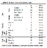
    The mean age of the 57 patients with AS and 29 heathy person who were included in this study was 40.35 (range 19–58) years. Twenty-seven (47.4%) of the patients were female, and thirty (52.6%) of the patients were male (Table 1, 2). DES was identified in 7 (12.3%) of the participating patients with AS on the basis of TO test results.


    Büyütmek İçin Tıklayın
    Table 2: Groups’ clinical characteristics

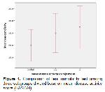
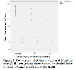
    The mean BASDAI value of the patients with AS was 4.91. The mild-moderate group consisted of 29 patients , the severe group of 28 and the control group of 29. No significant differences were determined between the three subgroups in terms of TO, Schirmer’s, and BUT test results (P>0.05 for all) (Table 3, Figure 1, 2).


    Büyütmek İçin Tıklayın
    Table 3: Comparison of dry eye tests among the groups


    Büyütmek İçin Tıklayın
    Figure 1: Comparison of tear osmolarity test among three subgroups divided base on mean disease activity score (BASDAI)


    Büyütmek İçin Tıklayın
    Figure 2: Comparison of Schirmer’s test and Break up time (BUT) test among three subgroups divided base on mean disease activity score (BASDAI)

    The patients were divided into three subgroups as the control, HLA B27(+) and (–), no significant differences were observed in terms of TO, Schirmer’s and BUT test results (all p> 0.05), (Table 3).

    The patients were divided into another three subgroups as the control, anti-TNF alpha treatment (+) and (–), no significant differences were observed in terms of TO and Schirmer’s and BUT test results (all P>0.05 ) (Table 3).

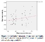
    Spearsman’s correlation coefficients demonstrated that disease activity score values were not correlated with TO values (r=0,23, P=0,863),Schirmer’s scores (r= –0.223, P= 0.095) and BUT scores (r = –0.044, P= 0.744), (Figure 3).


    Büyütmek İçin Tıklayın
    Figure 3: Correlation between mean tear osmolarity and mean disease activity (BASDAI) score in patients with ankylosing spondylitis

  • Başa Dön
  • Özet
  • Giriş
  • Materyal ve Metot
  • Bulgular
  • Tartışma
  • Kaynaklar
  • Tartışma
    In our study, there were no significant difference in terms of TO, BUT and Schirmer’s test results between the groups established on the basis of BASDAI, HLA B27 and treatment status with AS.

    A decrease in tear production and tear dysfunction on the ocular surface in DES, which develops as a result of various inflammatory diseases, lead to tear hyperosmolarity 16. Tear film hyperosmolarity in turn leads to osmotic stress in the surface epithelium that activates the inflammatory cascade on the ocular surface, and inflammatory mediators are released in the tear 17-19. In 2007, the International Dry Eye Workshop (DEWS) broadened the definition of DES to a multifactorial disease causing increased tear film osmolarity, ocular surface damage accompanied by inflammation, tear film instability and ocular irritability 19. TO measurement is therefore approved by many researchers as a valuable method in the diagnosis of DES, and it has for long been recommended as a potential gold standard in clinical practice 10,20,21.

    The prevalence of DES in the normal population ranges between 5.5% and 33.7% 22. Various subjective DES tests have been used in systemic inflammatory diseases in previous studies, such as Schirmer’s test, BUT, corneal and conjunctival staining, and the ocular surface disease index 23. The sensitivity and specificity of these tests are therefore lower than those of the test measuring TO using the TearLab system. In addition, these tests do not by themselves indicate severity and diagnosis. Definite diagnosis of DES in AS with secondary SS is therefore problematic. To the best of our knowledge, this is the first study to selectively evaluate TO in patients with AS. Additionally, the most important advantage of TO is its ability to reflect the diagnosis and severity of DES more objectively than other measurements 11,20.

    One study demonstrated correlations between TO findings and levels of inflammatory mediators in tears 24. The severity of DES may increase in line with the degree of increase in inflammatory activation. We therefore used TO in order to reveal the presence of DES and determine itself severity as a final objective. The TO cut-off value is important for the diagnosis of DES. Cut of values of 305 or 308 mOsm/L have generally been adopted in studies investigating the value of TO in the diagnosis of DES 10,25. In this study we adopted a cut-off value of 305 mOsm/L. This value is sensitive in identifying patients with DES. We identified DES at a level of 12.5% according to TO measurements at low levels of inflammation. The mean age of 40.3 years and low level of inflammatory disease may account for the low level of DES observed.

    Markovisky et al. 12 investigated the association between BASDAI levels as a disease activity score for AS and severity of DES using a confocal microscope and various subjective DES tests (OSDI, LIPCOF, Schirmer’s, and BUT). They observed a significant difference between low and high disease activity levels. All the patients in our study had low inflammation levels (CRP < 0,5 mg/dl), and we used the Schirmer’s, BUT and TO tests as tests for DES. There was no difference among mild-moderate (BASDAI ≤4), severe (BASDAI >4) and the control groups disease activity levels in the DES tests used. However, we think that if a significant difference can be determined in terms of TO between BASDAI groups in high levels of inflammation, TO can be used as an objective test for disease activity in AS.

    Markovisky et al. 12,26 investigated the probable effects of treatment on tear production and LC in patients with AS and rheumatoid arthritis (RA). Although the patients in the group received anti-TNF alpha therapy, that study determined pathology at subjective DES tests and confocal microscopy due to high CRP levels showing systemic inflammatory status. Theoretically, the presence of DES can be explained by increased cytokines in tears in AS with SS with high systemic inflammation impairing tear production.

    HLA-B27 is the gene predisposing to primary AS. In a previous study, whereas more than 90% of Caucasian AS patients was found HLA- B27(+), only about 50% of black AS patients was found positive for HLA- B27 27. Furthermore, in another study, The prevalence of HLA-B27 in the white population was stated around 6% to 8%, with HLA- B27-05 being the predominant subtype 28. The function of these molecules is to present antigenic peptides to T cells. They play a significant role in immunological reactions at the cellular level. Laval et al. reported higher relapse, severity and complications of uveitis when they compared HLA-B27-positive patients with HLA-B27-negative 29. In other studies, however, no difference has been determined between HLA-B27-positive and HLA-B27-negative patients either in tests regarding DES or at corneal analyses using confocal microscopy, and HLA B27 has been shown to have no effect on the corneal immune mechanism 4,26. Of the 57 patients with AS in our study, 64.9% were HLA B27-positive. No pathology was determined in either the HLA B27-positive or negative groups in terms of subjective tests as Schirmer’s and BUT or in more objective dry eye test as TO test.

    The main limitation of this study is that TO measurements were performed unilaterally. We performed TO measurements on the right eyes only in all patients due to the limited availability of TO kits.

    Conclusions; TO, BUT and Schirmer’s test results in the groups established on the basis of BASDAI, HLA B27 and treatment status were at normal levels in patients with a low level of systemic inflammation (CRP <0.5 mg/dl). Since AS is an inflammatory disease that progresses with remissions and activations, we think that objective screening of severity of DE using TO can represent a preliminary marker of AS disease activity in association with systemic inflammation level. However, since systemic inflammation levels were low in this study (CRP <0.5 mg/dL), we were unable to prove a relation between TO and disease activity (BASDAI). Further studies may now be planned to establish whether or not TO screening can be used for the definite diagnosis of DES and as a marker of AS disease activity in patients with high levels of systemic inflammation (CRP >0.5 mg/dL).

    Acknowledgments
    Financial support: No financial support was received for this submission.

    Conflict of interest: None of the authors has conflict of interest with this submission

  • Başa Dön
  • Özet
  • Giriş
  • Materyal ve Metot
  • Bulgular
  • Tartışma
  • Kaynaklar
  • Kaynaklar

    1) Braun J, Sieper J. Ankylosing spondylitis. Lancet 2007; 369: 1379-1390.

    2) Linder R, Hoffmann A, Brunner R. Prevalence of the spondyloarthritides in patients with uveitis. J Rheumatol 2004; 31: 2226-2229.

    3) Boehm N, Riechardt AI, Wiegand M, et al. Proinflammatory cytokine profiling of tears from dry eye patients by means of antibody microarrays. Investig Opthalmology Vis Sci 2011; 52: 7725.

    4) Goh L, Samanta A. Update on biologic therapies in ankylosing spondylitis: A literature review. Int J Rheum Dis 2012; 15: 445-454.

    5) Schaumburg CS, Siemasko KF, De Paiva CS, et al. Ocular surface APCs are necessary for autoreactive T cell-mediated experimental autoimmune lacrimal keratoconjunctivitis. J Immunol 2011; 187: 3653-3662.

    6) Szalai E, Berta A, Szekanecz Z, et al. Evaluation of tear osmolarity in non-sjögren and sjögren syndrome dry eye patients with the tearlab system. Cornea 2012; 31: 867-871.

    7) Bunya VY, Langelier N, Chen S, et al. Tear osmolarity in Sjögren syndrome. Cornea 2013; 32: 922-927.

    8) Kobak S, Celebi Kobak A, Kabasakal Y, Doganavsargil E. Sjögren’s syndrome in patients with ankylosing spondylitis. Clin Rheumatol 2006; 26:173-175.

    9) Lin H, Li W, Dong N, et al. Changes in corneal epithelial layer inflammatory cells in aqueous tear–deficient dry eye. Investig Opthalmology Vis Sci 2010; 51:122.

    10) Lemp MA, Bron AJ, Baudouin C, et al. Tear osmolarity in the diagnosis and management of dry eye disease. Am J Ophthalmol 2011; 151: 792-798.

    11) Sullivan BD, Whitmer D, Nichols KK, et al. An objective approach to dry eye disease severity. Investig Opthalmology Vis Sci 2010; 51: 6125.

    12) Marsovszky L, Németh J, Resch MD, et al. Corneal Langerhans cell and dry eye examinations in ankylosing spondylitis. Innate Immun. 2014; 20: 471-477.

    13) Liu H, Begley C, Chen M, et al. A Link between Tear instability and hyperosmolarity in dry eye. Investig Opthalmology Vis Sci 2009; 50: 3671.

    14) Linden S Van Der, Valkenburg HA, Cats A. Evaluation of Diagnostic Criteria for Ankylosing Spondylitis. Arthritis Rheum 1984; 27: 361-368.

    15) Garrett S, Jenkinson T, Kennedy LG, et al. A new approach to defining disease status in ankylosing spondylitis: The bath ankylosing spondylitis disease activity index. J Rheumatol 1994; 21: 2286-2291.

    16) Gilbard JP. Dry eye, blepharitis and chronic eye irritation: Divide and conquer. J Ophthalmic Nurs Technol 1999; 18: 109-115.

    17) Gilbard JP, Carter JB, Sang DN, et al. Morphologic effect of hyperosmolarity on rabbit corneal epithelium. Ophthalmology 1984; 91: 1205-1212.

    18) Li D-Q, Luo L, Chen Z, et al. JNK and ERK MAP kinases mediate induction of IL-1β, TNF-α and IL-8 following hyperosmolar stress in human limbal epithelial cells. Exp Eye Res 2006; 82: 588-596.

    19) Anonim. The definition and classification of dry eye disease: Report of the Definition and Classification Subcommittee of the International Dry Eye WorkShop. Ocul Surf 2007; 5: 75-92.

    20) Tomlinson A, Khanal S, Ramaesh K, et al. Tear film osmolarity: determination of a referent for dry eye diagnosis. Investig Opthalmology Vis Sci. 2006; 47: 4309.

    21) Chen W, Lin H, Dong N, et al. Cauterization of central cornea induces recruitment of major histocompatibility complex class II+ langerhans cells from limbal basal epithelium. Cornea 2010; 29:73-79.

    22) Villani E, Beretta S, Galimberti D, et al. In vivo confocal microscopy of conjunctival roundish bright objects: Young, older, and sjögren subjects. Investig Opthalmology Vis Sci 2011; 52: 4829.

    23) Versura P, Frigato M, Cellini M, et al. Diagnostic performance of tear function tests in Sjogren’s syndrome patients. Eye (Lond) 2007; 21: 229-237.

    24) VanDerMeid KR, Su SP, Ward KW, et al. Correlation of tear inflammatory cytokines and matrix metalloproteinases with four dry eye diagnostic tests. Investig Opthalmology Vis Sci 2012; 53: 1512.

    25) Frederick W, Kaleb M. Prevalence, risk, and risk factors for oral and ocular dryness with particular emphasis on rheumatoid arthritis. J Rheumatol 2008; 35: 1023-1030.

    26) Marsovszky L, Resch MD, Németh J, et al. In vivo confocal microscopic evaluation of corneal Langerhans cell density, and distribution and evaluation of dry eye in rheumatoid arthritis. Innate Immun 2013; 19: 348-354.

    27) Khan MA, Kushner I, Braun WE. Comparison of clinical features in HLA-B27 positive and negative patients with ankylosing spondylitis. Arthritis Rheum 1977; 20: 909-912.

    28) Haroon N. Does a positive HLA-B27 test increase your risk of mortality? J Rheumatol 2015; 42: 559-560.

    29) Laval SH, Timms A, Edwards S, et al. Whole-genome screening in ankylosing spondylitis: Evidence of non-MHC genetic-susceptibility loci. Am J Hum Genet 2001; 68: 918-926.

  • Başa Dön
  • Özet
  • Giriş
  • Materyal ve Metot
  • Bulgular
  • Tartışma
  • Kaynaklar
  • [ Başa Dön ] [ Özet ] [ PDF ] [ Benzer Makaleler ] [ Yazara E-Posta ] [ Editöre E-Posta ]
    [ Ana Sayfa | Editörler | Danışma Kurulu | Dergi Hakkında | İçindekiler | Arşiv | Yayın Arama | Yazarlara Bilgi | E-Posta ]