[ Ana Sayfa | Editörler | Danışma Kurulu | Dergi Hakkında | İçindekiler | Arşiv | Yayın Arama | Yazarlara Bilgi | E-Posta ]
Fırat Üniversitesi Sağlık Bilimleri Tıp Dergisi
2020, Cilt 34, Sayı 1, Sayfa(lar) 091-093
[ Özet ] [ PDF ] [ Benzer Makaleler ] [ Yazara E-Posta ] [ Editöre E-Posta ]
Maksiller Sinüs Orbita Tabanından Dentigeröz Kistle Birlikte Uzaklaştırılan Üçüncü Molar Diş: Bir Vaka Raporu
Serkan DÜNDAR1, Turgut KARLIDAĞ2, Erol KELEŞ2
1Firat University, Faculty of Dentistry, Department of Periodontology, Elazig TURKEY
2Fırat University, Faculty of Dentistry, Department of Otorhinolaryngology, Elazig, TURKEY
Anahtar Kelimeler: Dentigeröz kist, maksiller sinüs, gömülü diş
Özet
Dentigeröz kistler, daimi dişlerin kron kısmı ile ilişkili odontojenik kaynaklı, kötü huylu olmayan lezyonlardır. Gömülü dişleri çevreleyen dentigeröz kistler dişlerin ağızda olması gerektiğinden farklı yerlere sürmesine ve yer değiştirmesine neden olabilir. Üst çenede gömülü diş ile ilişkili dentigeröz kistlerde genellikle üst çene sinüs boşluğuna doğru bir yer değiştirme görülür. Bu lezyonlar Caldwell-Luc cerrahisi ve/veya endoskobik sinüs cerrahisi ile başarılı bir şekilde tedavi edilebilir. Bu vaka raporunda üst çene sinüs boşluğunda gömülü 3. büyük azı diş ile ilişkili dentigeröz kist rapor edilmiştir.
  • Başa Dön
  • Özet
  • Giriş
  • Olgu Sunusu
  • Tartışma
  • Kaynaklar
  • Giriş
    The development of the teeth is the result of the complex interactions between the oral epithelium and mesenchymal tissue. During development, abnormal tissue interactions may lead to ectopic tooth development and abnormal tooth eruption. Ectopic tooth eruption may occur due to developmental disorders, iatrogenic activity, or pathological processes such as tumors or cysts 1-3.

    Dentigerous cysts are odontogenic lesions. The development mechanism of these cysts is still not fully understood. It is thought that dentigerous cysts develop from the follicles of embedded teeth. As the dental follicles grow, fluid accumulates between the tooth crowns and epithelial components. Impaction pathogenesis is most common in the maxillary canines and mandibular third molars. Impacted teeth surrounded with dentigerous cysts often migrate and become ectopic. In the maxilla, these teeth are mostly displaced into the maxillary sinus 1-4.

    Dentigerous cysts have a slow progression character and may not be noticed for a few years. Symptoms usually occur when the lesion invades the maxillary sinus. These cysts can cause sequelae such as sinus obstruction and blindness. Standard treatment of a dentigerous cyst is the removal of the cyst and impacted tooth. These are usually easily removed from the maxillary sinus by a Caldwell-Luc procedure. Enucleation and tooth extraction are recommended following initial marsupialization to reduce the size of the bone defect in large cysts. In this paper, we report a case of a dentigerous cyst associated with an impacted maxillar third molar in the maxillary sinus 3-5

  • Başa Dön
  • Özet
  • Giriş
  • Olgu Sunusu
  • Tartışma
  • Kaynaklar
  • Olgu Sunusu
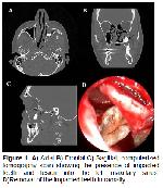
    A 43-year-old man with diabetes mellitus was diagnosed with a dentigerous cyst because of swelling and discharge in the posterior region of the left upper jaw after tooth extraction. Physical examination revealed a swelling in the sinus area of the left maxillary sinus. The swelling and malodor lasted for two to three months. It was determined that the patient had no upper maxillary third molar. Computerized tomography examination revealed a cystic lesion associated with the third molar adhered to the left maxillary sinus roof (Figure 1A, B, C, D). No abnormal findings were detected in the general examination. Caldwell-Luc and endoscopic sinus surgery were performed under general anesthesia after the regulation of the patient’s diabetes mellitus. The cyst was easily removed from the sinus floor of the left maxilla; the ectopic third molar was removed with a combination of endoscopic sinus and Caldwell-Luc surgeries.


    Büyütmek İçin Tıklayın
    Figure 1: A) Axial B) Frontal C) Sagittal, computerized tomography scan showing the presence of impacted teeth and lesion into the left maxillary sinus. D)Removal of the impacted teeth intraorally

    Endoscopic examination revealed that polypoid tissues were present in the left middle meatus and anterior ethmoid cells. These tissues were removed with cup forceps. The polypoid tissues originated from the maxillary sinus. A left uncinectomy, bullectomy, and anterior ethmoidectomy were done, and the maxillary sinus ostium was expanded. Then an incision of about 2 cm was made in the left gingivobuccal mucosa. After removing the bone tissues with a steel bur, approximately 3×3 cm of cystic structure within the maxillary sinus was reached. The cyst was infected, with teeth inside. The cyst was dissected from the surrounding tissues and removed en bloc. Then the ectopic third molar was removed. Bleeding control was provided and the incision sutured. Histopathologic examination revealed a dentigerous cyst. After the operation, antibiotics (penicillin and clavulanic acid, 1000 mg twice a day) and analgesics (dexopen, 25 mg twice a day) were prescribed for seven days. The patient’s postoperative period was uneventful.

  • Başa Dön
  • Özet
  • Giriş
  • Olgu Sunusu
  • Tartışma
  • Kaynaklar
  • Tartışma
    Ectopic eruption of teeth from the oral cavity to other regions—such as the nasal cavity, mandibular condyle, coronoid process, and palate—is rare. The maxillary sinus cavities are one of the areas where teeth show ectopic eruption. The etiology of the ectopic eruption of teeth has not yet been fully elucidated, but many theories have been suggested, including pathological conditions such as trauma, infection, developmental anomalies, and dentigerous cysts. It is thought that the dental buds are displaced by the expansion of dentigerous cysts, causing the teeth to become ectopic 3,6-8.

    Dentigerous cysts are the second most common cyst type to appear in jaws, after radical cysts. The majority (about 70%) of dentigerous cysts are found in the mandible, and a smaller proportion (about 30%) are found in the maxillary 3,4,7. Dentigerous cysts usually occur during the second or third decade of life and are rarely reported in childhood. However, according to some authors, most patients with dentigerous cysts are younger than 20. The age range of patients with dentigerous cysts in the literature varies from 4 to 57 years 3.

    Dentigerous cysts are usually painless, but they cause swelling in the face and may cause delay in tooth eruption. When dentigerous cysts appear in the maxillary sinus, symptoms often occur in the late period. In these cases, the cysts can be detected by routine radiographic examination 3,8-11. In other cases, the classic symptoms of sinus disease are seen in patients with cysts, including swelling, facial pain, headaches, and nasolacrimal obstruction. Cysts that are large enough to cover the maxillary sinus put pressure on the sinus walls and may cause nasal and ophthalmologic disorders 3. Altas et al. 12 reported epiphora due to nasolacrimal duct pressure originating from a dentigerous cyst associated with an unerupted ectopic canine tooth in the maxillary sinus. Avitia et al. 13 reported a case of orbital proptosis originating from a dentigerous cyst associated with an unerupted ectopic tooth in the maxillary sinus.

    There are also cases in the literature of dentigerous cysts that do not cause clinical symptoms. In radiological data, dentigerous cysts are seen as unilocular radiolucent cysts with well-defined sclerotic borders, and teeth are associated with the unerupted tooth crown. Waters radiography, orthopantomography, and plain skull radiography are useful and simple examination methods for clinical applications. Computed tomography can be used to determine the orbital or nasal invasion or involvement of a lesion. For this reason, computed tomography is not only necessary for a definitive diagnosis, but also an advantageous method for the evaluation of the related pathology, ectopic tooth localization, and determination of the appropriate treatment option 4,8,11.

    In this case, we observed the cyst and impacted tooth attached to the inner lateral wall of the maxillary sinus and roof of the maxillary sinus. Dentigerous cysts are associated with unerupted teeth. They usually contain third molar teeth and rarely contain deciduous, supernumerary teeth and odontomas. The patient’s third molar tooth was detected with the cyst, attached to the orbital base 3.

    In the differential diagnosis of dentigerous cysts, radical cysts, odontogenic keratocysts, odontogenic tumors, and other odontogenic cysts such as ameloblastoma, Pindborg tumors, odontoma, odontogenic fibroma, and cementomas should also be considered. However, mucoceles, retention cysts, and pseudocysts may also exist when an enlargement of the maxillary sinus is accompanied by cysts in the maxillary sinus. Odontogenic tumors such as ameloblastoma or epidermoid carcinoma that originate from a dentigerous cyst are sometimes seen. In the literature, metaplastic and dysplastic changes have not been reported from dentigerous cysts associated with ectopic teeth in the maxillary sinus 3.

    The standard treatment of a dentigerous cyst is the enucleation of the cyst and the removal of the cyst-related nonerupted tooth. Dentigerous cysts and embedded teeth in the maxillary sinus are usually easy to remove using a Caldwell-Luc operation. Removal of the entire cyst and the impacted tooth is important to prevent a recurrence of the cyst. The most significant disadvantage of marsupialization is the recurrence of the lesion. The traditional Caldwell-Luc procedure allows for visualization of the maxillary sinus, while causing more morbidity than a transnasal endoscope. Both techniques were used in the treatment of this case. Endoscopic surgical application was preferred with Caldwell-Luc surgery to remove the sinusoidal polypoid tissues in the left middle meatus and the anterior ethmoid cells, as well as to widen the maxillary sinus ostium. These polypoid tissues originated from the maxillary sinus 3,14-19.

    In conclusion, a differential diagnosis of a tooth impaction in the maxillary sinus should be considered for a patient with refractory sinusitis, maxillary pain, and swelling. Dentigerous cyst diagnosis is the most common pathology in these cases. Caldwell Luc and endoscopic sinus surgery are the preferred methods of treatment for such cases, and the techniques can also be used together.

    Conflict of Interest
    The authors declerate there is no conflict of interest.

  • Başa Dön
  • Özet
  • Giriş
  • Olgu Sunusu
  • Tartışma
  • Kaynaklar
  • Kaynaklar

    1) Bonardi JP, Gomes-Ferreira PH, de Freitas Silva L, et al. Large dentigerous cyst associated to maxillary canine. J Craniofac Surg 2017; 28: e96-e97.

    2) Nagori SA, Jose A, Bhutia O, Roychoudhury A. Large pediatric maxillary dentigerous cysts presenting with sinonasal and orbital symptoms: A case series. Ear Nose Throat J 2017; 96: E29-E34.

    3) Buyukkurt MC, Omezli MM, Miloglu O. Dentigerous cyst associated with an ectopic tooth in the maxillary sinus: A report of 3 cases and review of the literature. Oral Surg Oral Med Oral Pathol Oral Radiol Endod 2010; 109: 67-71.

    4) Tournas AS, Tewfik MA, Chauvin PJ, Manoukian JJ. Multiple unilateral maxillary dentigerous cysts in a nonsyndromic patient: A case report and review of the literature. Int J Pediatr Otorhinolaryngol Extra 2006; 1: 100-106.

    5) Ustuner E, Fitoz S, Atasoy C, Erden I, Akyar S. Bilateral maxillary dentigerous cysts: a case report. Oral Surg Oral Med Oral Pathol Oral Radiol Endod 2003; 95: 632-635.

    6) Goh YH. Ectopic eruption of maxillary molar tooth—an unusual cause of recurrent sinusitis. Singapore Med J 2001; 42: 80-81.

    7) Di Pasquale P, Shermetaro C. Endoscopic removal of a dentigerous cyst producing unilateral maxillary sinus opacification on computed tomography. Ear Nose Throat J 2006; 85: 747-748.

    8) Buyukkurt MC, Tozoglu S, Aras MH, Yolcu U. Ectopic eruption of a maxillary third molar tooth in the maxillary sinus: A case report. J Contemp Dent Pract 2005; 6: 104-110.

    9) Ko KS, Dover DG, Jordan RC. Bilateral dentigerous cysts—report of an unusual case and review of the literature. J Can Dent Assoc 1999; 65: 49-51.

    10) Murgod S, Girish HC, Savita JK, Varsha VK. Concurrent central odontogenic fibroma and dentigerous cyst in the maxilla: A rare case report. J Oral Maxillofac Pathol 2017; 21: 149-153.

    11) Dhupar A, Yadav S, Dhupar V, et al. Bi-maxillary dentigerous cyst in a non-syndromic child - review of literature with a case presentation. J Stomatol Oral Maxillofac Surg 2017; 118: 45-48.

    12) Altas E, Karasen RM, Yilmaz AB, et al. A case of a large dentigerous cyst containing a canine tooth in the maxillary antrum leading to epiphora. J Laryngol Otol 1997; 111: 641-643.

    13) Avitia S, Hamilton JS, Osborne RF. Dentigerous cyst presenting as orbital proptosis. Ear Nose Throat J 2007; 86: 23-24.

    14) Martínez-Pérez D, Varela-Morales M. Conservative treatment of dentigerous cysts in children: A report of 4 cases. J Oral Maxillofac Surg 2001; 59: 331-333.

    15) Vele DD, Sengupta SK, Dubey SP, Dokup MK. Cystic lesions of the nasal cavity and the paranasal sinuses: Report of two unusual cases. J Laryngol Otol 1996; 110: 1157-1160.

    16) Hasbini AS, Hadi U, Ghafari J. Endoscopic removal of an ectopic third molar obstructing the osteomeatal complex. Ear Nose Throat J 2001; 80: 667-670.

    17) Micozkadioglu SD, Erkan AN. Endoscopic removal of a maxillary dentigerous cyst. B-ENT 2007; 3: 213-216.

    18) Demirkol M, Keskinrüzgar A, Ege B, Mustafa R, Aras MH. Maksiller sinüs yerleşimli ektopik dişler: Üç olgu sunumu. Türkiye Klinikleri Dişhekimliği Bilimleri Dergisi 2016; 22: 147-151.

    19) Küçükkolbaşı H, Esen A. Maksiller sinüste ektopik 3. molar diş: Bir olgu sunumu. Selcuk Dental Journal 2014; 3: 134-137.

  • Başa Dön
  • Özet
  • Giriş
  • Olgu Sunusu
  • Tartışma
  • Kaynaklar
  • [ Başa Dön ] [ Özet ] [ PDF ] [ Benzer Makaleler ] [ Yazara E-Posta ] [ Editöre E-Posta ]
    [ Ana Sayfa | Editörler | Danışma Kurulu | Dergi Hakkında | İçindekiler | Arşiv | Yayın Arama | Yazarlara Bilgi | E-Posta ]