[ Ana Sayfa | Editörler | Danışma Kurulu | Dergi Hakkında | İçindekiler | Arşiv | Yayın Arama | Yazarlara Bilgi | E-Posta ]
Fırat Üniversitesi Sağlık Bilimleri Tıp Dergisi
2006, Cilt 20, Sayı 4, Sayfa(lar) 307-315
[ Özet ] [ PDF ] [ Benzer Makaleler ] [ Yazara E-Posta ] [ Editöre E-Posta ]
Paraoxonase and Arylesterase Levels in Behçet's Disease
Ahmet IŞIK1, Bilal ÜSTÜNDAĞ2
1Fırat Üniversitesi, Tıp Fakültesi, Romatoloji Anabilim Dalı, Elazığ -TÜRKİYE
2Fırat Üniversitesi, Tıp Fakültesi, Biyokimya Anabilim Dalı, Elazığ -TÜRKİYE
Keywords: Arylesterase, Behçet's Disease, Ceruloplasmin, Lipid Hydroperoxide, Paraoxonase
Summary
Lipid hydroperoxides (LOOHs) are by product of lipid peroxidation. Paraoxonase (PON), arylesterase (ARE), free sulphydryl (SH) groups and ceruloplasmin (CP) are enzymes or proteins with antioxidant characteristics. This study aims to determine the levels of LOOHs, the levels of SH, the activities of PON1, ARE and CP in active and inactive Behçet's disease (BD) patients.

The study included 21 BD cases (10 active and 11 inactive) and 23 healthy volunteers. The levels of LOOHs and SH and the activities of PON1, ARE and CP were determined.

The level of LOOHs was found to be significantly high, while level of SH and activities of PON1, ARE and CP were observed as significantly low in the BD group, when compared with the control group (p<0.001, for each). There was a significant negative correlation between the level of LOOHs and the activity of ARE in the BD group (r=-0.46, p<0.05). An increase in the level of LOOHs and a decrease in the activities of PON1, ARE and CP were observed in the active BD group when compared with the inactive BD group, but differences were not statistical significant (p>0.05, for each).

Results of the study indicate the impaired oxidant/antioxidant balance in BD. PON1 and ARE possess anti-atherosclerotic characteristics in addition to antioxidant ones. Accordingly, development of atherosclerosis in addition to tissue injury in BD seems to be inevitable due to the decrease in antioxidant levels resulting from increased oxidative stress.

  • Top
  • Summary
  • Introduction
  • Methods
  • Results
  • Disscussion
  • References
  • Introduction
    Behçet's Disease (BD) is a chronic, inflammatory and multisystemic autoimmune disease. It is characterized by recurrent oral aphthous ulcers, genital ulcer/scar, ocular inflammation, skin and joint signs 1. Although the exact etiology and pathogenesis of BD is not known yet, genetic, environmental and autoimmune factors are thought to be involved 2. In recent years increasingly more attention has been attracted for reactive oxygen species (ROS), which are formed as a result of normal aerobic metabolism and whose production is increased by active neutrophils during inflammation 3.

    Activated neutrophils lead to the production of ROS like hydrogen peroxide (H2O2), hydroxyl radical (OH), superoxide radical (O2•-) through Fenton reaction, and of nitrogen-involving radicals like peroxynitrite 3-7. Polymorphonuclear leukocytes increase the production of hypochlorous acid (HOCl) from H2O2 via myeloperoxidase enzyme 5-8. HOCl, which leads to sulphydryl oxidation and protein decarboxylation, takes part in tissue injury through oxidative modification of macromolecules in tissues 7,8. ROS which cause peroxidation of double-chain fatty acids found in the cell membrane, and thus cellular injury, also increase oxidative stress 3,4,7. Oxidants damage the antioxidant system, and decrease antioxidant levels. Antioxidant defense system can protect the cell against potentially detrimental effects of oxidant agents 5,7.

    Increase in ROS production and decrease the level of antioxidants due to oxidative stress in BD have been demonstrated in previous publications 3,6,9,10. It has been reported that the impaired oxidant/antioxidant balance has been responsible for tissue injury in BD 11,12. Moreover, the products of lipid peroxidation (LPO) take active parts in endothelial dysfunction and the pathogenesis of atherosclerosis 13,14. Chambers et al. 4 have pointed out to the presence of endothelial dysfunction in BD and have stated that endothelial dysfunction, a precursor of atherosclerosis, has been associated with oxidative stress and LPO products. Increase in ROS, LPO products and oxidative stress, together with the decrease in antioxidants may play roles in the development of atherosclerosis, beside be responsible for tissue injury 9-12. Moreover, it is well known that increased cytokines and oxidative stress trigger atherosclerosis by causing oxidative modification of LDL 15, and oxidative stress markers increase and antioxidants decrease in atherosclerosis and antioxidant treatments have protective effects on atherosclerosis 16,17.

    Paraoxonase (PON) and arylesterase (ARE) which are located on HDL are antioxidant and calcium dependent esterase enzymes 18. They can hydrolyze LPO products, as well as, organophosphates 18. It has been shown both experimentally and clinically that the levels of these enzymes decrease in many diseases associated with atherosclerosis 18-21. Glutathione, one of the main components of the antioxidant system, detoxifies oxidants using sulphydryl (SH) groups. Levels of SH have been shown to decrease in BD 7,22,23. Ceruloplasmin (CP), which is produced by hepatocytes and associated with copper metabolism has antioxidant characteristics, is an acute phase protein 24-26. It has been reported that the level and the activity of CP increase in inflammatory diseases like BD 11,23,27 and contrary this activity of CP is inhibited via ROS 28.

    The aim of this study is to investigate the changes in the level of lipid hydroperoxides (LOOHs), which are oxidative stress marker and the activities of PON1, ARE and CP and the level of SH, which have antioxidant characteristics in BD.

  • Top
  • Summary
  • Introduction
  • Methods
  • Results
  • Disscussion
  • References
  • Methods
    The study included 21 BD cases who applied to outpatient clinic of Rheumatology Department of Internal Medicine of Medical Center of Firat University and 23 healthy individuals who age and sex matched. The diagnosis of BD was made according to the International Study Group's criteria for BD 29. The patients were divided into two groups, as active and inactive, depending on the activity of their disease identified in the light of clinical and laboratory findings. The patients were interpreted as active if the patients with oral ulcer had at least two of below pathologies; genital ulcer, skin lesion, recent eye involvement, recent vascular involvement, recent neurological involvement, active arthritis, positive pathergy test and with high erythrocyte sedimentation rate (ESR) and/or C-reactive protein (CRP) 7,22. Histories of all individuals in the study were obtained, and systemic and rheumatological physical examinations were performed. Pathergy test was applied to all the BD groups, and 24-48 hours later, the patients were evaluated in terms of papulopustular lesions. All patients were assessed by an ophthalmologist for eye involvement.

    Approval of the local ethics committee and informed consent of the patients and healty individuals were obtained. Patients with dyslipidemia, cardiovascular diseases, diabetes mellitus, renal failure, chronic infection, alcohol abuse, and who used antilipidemic and antioxidant drugs were excluded from the study.

    Blood samples were obtained at 0800-0900 a.m. after 8-12 hours of fasting. The samples were centrifuged at 3000 rpm for 10 minutes to obtain serum samples. Ordinary biochemical assays were made in an Olympus AU 600 Autoanalyzer using Olympus kits (Olympus Corp., Tokyo-Japan). Erythrocyte sedimentation rate was determined by classical Westergren method immediately in whole blood with 1 mg/mL ethylene diamine tetraacetic acid (EDTA). The level of CRP was determined by immunoturbidimetric technique (Schiapparelli Biosystems, the Netherlands). The levels of LOOHs and SH, and the activities of PON1, ARE and CP, as the specific parameters, were measured using appropriate methods. Serums were stored at -20 ºC until the day of analysis.

    Measurement of sulphydryl groups: Free SH of sera were assayed according to the method of Elman 31. Briefly, 1 ml of buffer containing 0.1 M Tris, 10 mM EDTA, pH 8.2, and 50 μl serum was added to cuvettes, followed by 50 μl 10 mM DTNB in methanol. Blanks were run for each sample as a test, but there was no 55'-dithiobis-(2-nitrobenzoic acid) (DTNB) in the methanol. Following incubation for 15 min at room temperature, sample absorbance was read at 412 nm on a Cecil 3000 spectrophotometer. Sample and reagent blanks were subtracted. The concentration of SH was calculated using reduced glutathione as free SH standard.

    Measurement of the activities of PON1 and ARE: PON1 activity was determined using paraoxon as a substrate and measured by increases in the absorbance at 412 nm due to the formation of 4-nitrophenol, as already described 32. Briefly, the activity was measured, at 25 ºC, by adding 50 μl of serum to 1 ml Tris/HCl buffer (100 mM, pH 8.0) containing 2 mM CaCl2 and 5.5 mM of paraoxon. The rate of generation of 4-nitrophenol was determined at 412 nm. Enzymatic activity was calculated using the molar extinction coefficient 17 100 M−1 cm−1.

    ARE activity was measured spectrophotometrically. The assay contained 1 mM phenylacetate in 20 mM Tris/HCl, pH 8. The reaction was started by the addition of serum and the increase in absorbency was recorded at 270 nm, as already described 32. Blanks were included to correct for the spontaneous hydrolysis of phenylacetate. Enzyme activity was calculated using the molar extinction coefficient of 1310 M−1 cm−1.

    Measurement of ceruloplasmin activity: Serum ceruloplasmin activity was assessed by measuring its oxidase activity using p-phenylenediamine as substrate 33,34.

    Measurement of lipid hydroperoxides: Triiodide complex formed as a result of the reaction between lipid hydroperoxide and iodine was evaluated by spectrophotometry at 365 nm wavelength. The results were calculated using the extinction coefficient of triiodide (Є = 2,46 x 104.M-1.cm-1) 35.

    Statistics: All results were expressed as means±SD. Student's t-test and Spearman's correlation analysis were performed using SPSS for Windows Release 11.0 (SPSS Inc. Chicago, Illinois, USA). The P value less than 0.05 was considered to be significant.

  • Top
  • Summary
  • Introduction
  • Methods
  • Results
  • Disscussion
  • References
  • Results
    Twenty-one BD patients (8 female, 13 male; mean age, 34,4 ± 7,4 years; mean duration of disease, 3,7 ± 2,9 years) and 23 healthy controls (9 female, 14 male; mean age, 36,7 ± 4,2 years) were enrolled to the present study. Demographical characteristics and laboratory data of the BD and the control groups are presented in Table 1.


    Click Here to Zoom
    Table 1: Demographical characteristics and laboratory findings of the patient group and the control group

    There was no significant difference between the BD and control groups in terms of age, sex, hemoglobin, white blood cell count (WBC), fasting blood glucose, total cholesterol, LDL, HDL and triglyceride values (p>0.05, for each, Table 1). Although ESR was higher in the BD group, the difference from the control group was not significant (p>0.05); and the level of CRP was significantly higher in the former (p<0.05).

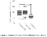
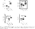
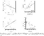
    The level of LOOHs was found significantly higher (p<0.001, Figure 1), while the level of SH and the activities of PON1, ARE and CP were significantly lower in the BD group vs. the control one (p<0.001, for each, Table 1, Figure 2). The activity of ARE negatively correlated with the level of LOOHs and positively correlated with the level of HDL (r=-0.46, p<0.05 and r=0.51, p<0.05, respectively) and the activity of CP positively correlated with the level of SH and negatively correlated with the level of CRP (r=0.46, p<0.05, r=-0.47, p<0.05, respectively and Figure 3) in the BD group.


    Click Here to Zoom
    Figure 1: The levels of LOOHs in the control and the BD groups.


    Click Here to Zoom
    Figure 2: Activities of PON1, ARE and CP and level of SH, which are all antioxidants, exhibit a statistically significant decrease in the active and inactive BD groups, relative to the control group. The same antioxidants show decreased in the active BD group, versus the in active one, without a statistically significant difference.


    Click Here to Zoom
    Figure 3: The result of correlation analysis in the BD group. The activity of ARE is negatively correlated with the level of LOOHs and positively correlated with the level of HDL, whereas the activity of CP is positively correlated with the level of SH and negatively correlated with the level of CRP.

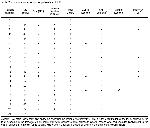
    Of the BD cases, 100% (n:21) had oral ulcer, 57,1% (n:12) genital ulcer or scar, 90,4% (n:19) skin lesion, 28,5% (n:6) ocular involvement, 9,5% (n:2) vascular involvement, 4,7% (n:1) neurological involvement and 42,8% (n:9) positive pathergy test (Table 2). Comparisons between the patients with and without positive pathergy test and patients with and without ocular lesions in the BD group did not show a significant difference in terms of the parameters concerned (p>0.05, for each).


    Click Here to Zoom
    Table 2: Clinical characteristic of patients with BD

    In seven cases in the BD group (33,3%) diagnosis were recently made in our department and they did not use any drugs. The rest 14 cases (66,7%) had been diagnosed previously and among these patients were using colchicine (6 cases), azothioprine, corticosteroid and colchicine combination (4 cases), infliximab and methotrexate (2 cases), salazopyrin and colchicine (1 case) and interferon (1 case). There was not any significant difference between recently diagnosed BD cases and BD cases previously diagnosed in terms of the parameters involved (p>0.05, for each).

    The comparison between active (n:10) and inactive (n:11) BD groups revealed significantly higher ESR and CRP values in the former (p<0.01 and p<0.05, respectively). An increase in the level of LOOHs and decrease in activities of PON1, ARE and CP were observed in the active BD group when compared with the inactive BD group, but differences were not statistical significant (p>0.05, for each, Table 3, Figures 1 and 2).


    Click Here to Zoom
    Table 3: Laboratory data of active BD and inactive BD groups

  • Top
  • Summary
  • Introduction
  • Methods
  • Results
  • Disscussion
  • References
  • Discussion
    It is known that activated inflammatory cells lead to ROS production in BD an autoimmune disease 3-7. Increased ROS production, in turn, enhances LPO products and causes tissue injury 3-7. It has been reported that serum levels of malondialdehyde (MDA) 5,10,36, and LOOHs 6, which are LPO products, increase in BD, and high serum MDA levels negatively correlated with antioxidants 10 and treatment with vitamin E, which is an antioxidant, significantly reduces MDA level 5. Similarly, it has been established that plasma and erythrocyte levels of thiobarbituric acid reactive substance (TBARS), an LPO product, are high in BD and there has been no significant difference between active and inactive BD groups in terms of TBARS levels 37. However, Orem et al. 3 have reported that LPO products in the active BD group are higher than those in the inactive BD group. In the present study, the level of LOOHs which are byproducts of LPO have been found higher in the BD group, too. Although when compared with inactive BD group, the level of LOOHs has been found slightly higher in the active BD group, this difference has been not statistically significant. This result we obtained is consistent with the study of Akar et al. 37. The inconsistency of the results about LPO products in active and inactive BD groups might be related with the differences in the methods of measurement used and/or in the methods used to determine the disease activity. It is known that currently there is no agreed activity criterias for BD.

    CP, which is synthesized by hepatocytes and which carries 90-95% of copper, is a protein with antioxidant characteristics 24. Oxidation of increased amounts of plasma homocysteine restores CP's redox state, leading to decreased copper transport into the cells 25. Previous studies have shown that CP is a powerful plasma antioxidant, when iron-stimulated reactions are involved and this has mainly been ascribed to its ferroxidase activity. The conversion of Fe2+ into Fe3+ can decrease oxidation by blocking the Fenton reaction through a decrease in the quantity of oxidant Fe2+ or sequestration of iron from apotransferrin 26. Human CP has recently been ascribed a thiol-linked peroxidase activity which can remove H2O2 and LOOHs 24. On the other hand, it has also been reported that ROS inhibit CP activity 28. It is noted that CP activity increases in BD and this is related with the acute phase response of CP 7,22,23. In the present study, however, it has been found that CP activity is reduced in BD and that CP negatively correlates with CRP, a marker of acute phase response, and positively correlates with SH which is known as antioxidant. CP has also antioxidant properties 24-26. The decrease in CP activity along with other antioxidants in BD supports this claim and it seems to be an expected result. However, it is also possible that increased oxidants may be responsible for the decrease in CP activity, as stated by Gutteridge et al. 28.

    Serum SH groups act as important cellular scavengers of peroxides and so help to protect cells from damage by these molecules. Decrease in SH level not only impairs cells' response to oxidants, but also changes the functions of inflammatory cells 38. It has been reported that in BD there is a decrease in SH level 7,22,23, which negatively correlates with MDA and CRP levels 22, and which is more marked in active BD, than in inactive BD 7. In our study, SH level has been found to be lower in the BD group, in comparison to the healthy controls, but there was no statistical difference between the SH levels of active and inactive BD groups. The positive correlation between the SH level and the activity of CP, another antioxidant, seems to support the claim that LPO products, together with other antioxidants, reduce SH levels. Lack of a significant difference between active and inactive BD groups with regard to SH levels may have resulted from the inadequacy of criteria used to determine the disease activity.

    It has been reported that PON1 activity decrease in BD 36 and RA 39,40 cases, when compared with the healthy individuals, and activity of ARE also decreased along with PON1 in RA cases with amyloidosis complication 41. Activities of PON1 and ARE enzymes have been found to be lower in the BD in the present study, too. It has been shown that PON1 hydrolyzes LPO products and H2O2 42. LPO products are not only formed via lipoprotein oxidation, but also lipids in the cell structure which undergo peroxidation in oxidative stress 18. It is known that LPO products increase in BD 3,5,6,10,36,37. In our study, a negative correlation has been found between ARE activity and LOOHs levels. We think that the decrease in the activities of PON1 and ARE enzymes may be important in terms of the progression of BD.

    PON1, which is a part of HDL cholesterol, is a strong antioxidant enzyme that is believed to have a protective role in the atherosclerotic process, by both contributing to HDL's protective effect against atherosclerosis and preventing lipoprotein peroxidation and oxidation of LDL cholesterol 18-20. Shih et al. 21 have demonstrated that rats with genetically PON1 deficiency are prone to atherosclerosis. It has been established that the activities of PON1 and ARE decline in atherosclerosis 18-20. Endothelial dysfunction and accelerated atherosclerosis have been emphasized in BD 4. In the light of this data, besides its contribution to disease progression, the decrease in PON1 and ARE enzyme activities suggest a possible increase in atherosclerosis incidence, in BD.

    It is known that PON1 structure includes three cysteine residues carrying sulphydryl groups. Of these, cysteinyl residues on the position 41st and 352nd are involved in the formation of intramolecular disulphide bonds, while cysteinyl residue on the position 283rd is free and responsible for activity 18-20,43. Aviram et al. 43 have demonstrated that LPO products are bound to cysteine residues found on the 283rd position of PON1, thereby inactivating PON1 and ARE activities. The negative correlation found between LOOHs level and ARE activity in our study supports the idea that LPO products are responsible for the decrease in the activities of PON and ARE in BD.

    Experimental studies have shown that cytokines like IL-1 and TNF-α reduce the production and the activity of PON1 44,45. It is known that the serum levels of many cytokines like IL-1, IL-6, IL-8 and TNF-α increase in BD 46-48. One probable reason of the decrease in activities of PON1 and ARE in BD might be the increase in these cytokines as pointed out by Feingold et al. 44 and Kumon et al. 45. Not finding significant correlation in PON1 and ARE activities with ESR, CRP and WBC values in our study seems to decrease the above probability. Activities of PON1 and ARE are known to be affected from HDL level 18-20. Although we have observed a positive correlation between HDL level and activity of ARE, not finding significant difference in HDL level between BD and healthy control reduces the probability of only HDL to be responsible from the decrease in activities of PON1 and ARE in BD.

    In conclusion, it has been established in the present study that the level of LOOHs, LPO product, have increased, and various antioxidants have decreased in the BD group, relative to the healthy control group. PON1 and ARE, which have antioxidant characteristics, are also known to possess protective effects against atherosclerosis. The decrease in the activities of these enzymes may have part in the development of atherosclerosis, as well as, tissue injury in BD. It is necessary to evaluate atherosclerosis in BD and to determine the role of decrease in antioxidants in atherosclerosis by controlled, prospective and multi-centered studies in large series.

  • Top
  • Summary
  • Introduction
  • Methods
  • Results
  • Discussion
  • References
  • References

    1) Sakane T, Takeno M, Suzuki N, Inaba G. Behcet's disease. N Engl J Med. 1999;341(17):1284-91.

    2) Onder M, Gurer MA. The multiple faces of Behcet's disease and its aetiological factors. J Eur Acad Dermatol Venereol. 2001;15:126-36.

    3) Orem A, Efe H, Deger O, Cimsit G, Uydu HA, Vanizor B. Relationship between lipid peroxidation and disease activity in patients with Behcet's disease. J Dermatol Sci. 1997;16:11-6.

    4) Chambers JC, Haskard DO, Kooner JS. Vascular endothelial function and oxidative stress mechanisms in patients with Behcet's syndrome. J Am Coll Cardiol. 2001;37:517-20.

    5) Kokcam I, Naziroglu M. Effects of vitamin E supplementation on blood antioxidants levels in patients with Behcet's disease. Clin Biochem. 2002;35:633-9.

    6) Orem A, Yandi YE, Vanizor B, Cimsit G, Uydu HA, Malkoc M. The evaluation of autoantibodies against oxidatively modified low-density lipoprotein (LDL), susceptibility of LDL to oxidation, serum lipids and lipid hydroperoxide levels, total antioxidant status, antioxidant enzyme activities, and endothelial dysfunction in patients with Behcet's disease. Clin Biochem. 2002;35:217-24.

    7) Yazici C, Kose K, Calis M, Demir M, Kirnap M, Ates F. Increased advanced oxidation protein products in Behcet's disease: a new activity marker? Br J Dermatol. 2004;151:105-11.

    8) Weiss SJ. Tissue destruction by neutrophils. N Engl J Med. 1989;320:365-76.

    9) Saglam K, Serce AF, Yilmaz MI, Bulucu F, Aydin A, Akay C, et al. Trace elements and antioxidant enzymes in Behcet's disease. Rheumatol Int. 2002;22:93-6.

    10) Kose K, Yazici C, Cambay N, Ascioglu O, Dogan P. Lipid peroxidation and erythrocyte antioxidant enzymes in patients with Behcet's disease. Tohoku J Exp Med. 2002;197:9-16.

    11) Dogan P, Tanrikulu G, Soyuer U, Kose K. Oxidative enzymes of polymorphonuclear leucocytes and plasma fibrinogen, ceruloplasmin, and copper levels in Behcet's disease. Clin Biochem. 1994;27:413-8.

    12) Niwa Y, Miyake S, Sakane T, Shingu M, Yokoyama M. Auto-oxidative damage in Behcet's disease--endothelial cell damage following the elevated oxygen radicals generated by stimulated neutrophils. Clin Exp Immunol. 1982;49:247-55.

    13) Witztum JL, Steinberg D. Role of oxidized low density lipoprotein in atherogenesis. J Clin Invest. 1991;88:1785-92.

    14) Yla-Herttuala S, Palinski W, Butler SW, Picard S, Steinberg D, Witztum JL. Rabbit and human atherosclerotic lesions contain IgG that recognizes epitopes of oxidized LDL. Arterioscler Thromb. 1994;14:32-40.

    15) Maziere C, Auclair M, Maziere JC. Tumor necrosis factor enhances low density lipoprotein oxidative modification by monocytes and endothelial cells. FEBS Lett. 1994;338:43-6.

    16) Madamanchi NR, Hakim ZS, Runge MS. Oxidative stress in atherogenesis and arterial thrombosis: the disconnect between cellular studies and clinical outcomes. J Thromb Haemost. 2005;3:254-67.

    17) Violi F, Cangemi R. Antioxidants and cardiovascular disease. Curr Opin Investig Drugs. 2005; 6:895-900.

    18) Mackness B, Durrington PN, Mackness MI. Human serum paraoxonase. Gen Pharmacol. 1998;31:329-36.

    19) Durrington PN, Mackness B, Mackness MI. Paraoxonase and atherosclerosis. Arterioscler Thromb Vasc Biol. 2001;21:473-80.

    20) Aviram M. Does paraoxonase play a role in susceptibility to cardiovascular disease? Mol Med Today. 1999;5:381-6.

    21) Shih DM, Gu L, Xia YR, Navab M, Li WF, Hama S, et al. Mice lacking serum paraoxonase are susceptible to organophosphate toxicity and atherosclerosis. Nature. 1998;394:284-7.

    22) Bekpinar S, Kilic N, Unlucerci Y, Akdag-Kose A, Azizlerli G, Ozbek-Kir Z. Evaluation of nitrosative and oxidative stress in Behcet disease. J Eur Acad Dermatol Venereol. 2005;19:167-71.

    23) Kose K, Dogan P, Ascioglu M, Erkilic K, Ascioglu O. Oxidative stress and antioxidant defenses in plasma of patients with Behcet's disease. Tohoku J Exp Med. 1995;176:239-48.

    24) Goldstein IM, Kaplan HB, Edelson HS, Weissmann G. Ceruloplasmin. A scavenger of superoxide anion radicals. J Biol Chem. 1979;254:4040-5.

    25) Rafter GW. Plasma thiols, copper and rheumatoid arthritis. Med Hypotheses. 1994;43:59-61.

    26) Gutteridge JM. Inhibition of the Fenton reaction by the protein caeruloplasmin and other copper complexes. Assessment of ferroxidase and radical scavenging activities. Chem Biol Interact. 1985;56:113-20.

    27) Taysi S, Kocer I, Memisogullari R, Kiziltunc A. Serum oxidant/antioxidant status in patients with Behcet's disease. Ann Clin Lab Sci. 2002;32:377-82.

    28) Gutteridge JM, Winyard PG, Blake DR, Lunec J, Brailsford S, Halliwell B. The behaviour of caeruloplasmin in stored human extracellular fluids in relation to ferroxidase II activity, lipid peroxidation and phenanthroline-detectable copper. Biochem J. 1985;230:517-23.

    29) The International Study Group for Behcet's disease. Evaluation of diagnostic ('classification') criteria in Behcet's disease--towards internationally agreed criteria. Br J Rheumatol. 1992;31:299-308.

    30) Ellman GL. Tissue sulfhydryl groups. Arch Biochem Biophys 1959;82:70-7.

    31) Hu ML, Louie S, Cross CE, Motchnik P, Halliwell B. Antioxidant protection against hypochlorous acid in human plasma. J Lab Clin Med. 1993;121:257-62.

    32) La Du BN, Eckerson HW. The polymorphic paraoxonase/arylesterase isozymes of human serum. Fed Proc. 1984;43:2338-41.

    33) Sunderman FW, Nomoto S. Measurement of human serum ceruloplasmin by its p-phenylenediamine oxidase activity. Clin Chem. 1970;16:903-10.

    34) Erel O. Automated measurement of serum ferroxidase activity. Clin Chem. 1998;44:2313-9.

    35) Gorog P, Kotak DC, Kovacs IB. Simple and specific test for measuring lipid peroxides in plasma. J Clin Pathol. 1991;44:765-7.

    36) Karakucuk S, Baskol G, Oner AO, Baskol M, Mirza E, Ustdal M. Serum paraoxonase activity is decreased in the active stage of Behcet's disease. Br J Ophthalmol. 2004;88:1256-8.

    37) Akar A, Arca E, Serdar MA, Akay C, Aydin A, Tastan HB, et al. Correlation between erythrocyte antioxidant activity, lipid peroxidation, and disease activity in patients with Behcet's disease. J Eur Acad Dermatol Venereol. 2003;17:482-3.

    38) McKeown MJ, Hall ND, Corvalan JR. Defective monocyte accessory function due to surface sulphydryl (SH) oxidation in rheumatoid arthritis. Clin Exp Immunol. 1984;56:607-13.

    39) Baskol G, Demir H, Baskol M, Kilic E, Ates F, Kocer D, et al. Assessment of paraoxonase 1 activity and malondialdehyde levels in patients with rheumatoid arthritis. Clin Biochem. 2005;38:951-5.

    40) Tanimoto N, Kumon Y, Suehiro T, Ohkubo S, Ikeda Y, Nishiya K, et al. Serum paraoxonase activity decreases in rheumatoid arthritis. Life Sci. 2003;72:2877-85.

    41) Maury CP, Junge W, Teppo AM. Serum esterase activity in reactive systemic amyloidosis and its relation to amyloid A degrading activity. J Lab Clin Med. 1984;104:761-6.

    42) Aviram M, Billecke S, Sorenson R, Bisgaier C, Newton R, Rosenblat M, et al. Paraoxonase active site required for protection against LDL oxidation involves its free sulfhydryl group and is different from that required for its arylesterase/paraoxonase activities: selective action of human paraoxonase allozymes Q and R. Arterioscler Thromb Vasc Biol. 1998;18:1617-24.

    43) Aviram M, Rosenblat M, Billecke S, Erogul J, Sorenson R, Bisgaier CL, et al. Human serum paraoxonase (PON 1) is inactivated by oxidized low density lipoprotein and preserved by antioxidants. Free Radic Biol Med. 1999;26:892-904.

    44) Feingold KR, Menon RA, Moser AH, Grunfeld C. Paraoxonase activity in the serum and hepatic mRNA levels decrease during the acute phase response. Atherosclerosis. 1998;139:307-15.

    45) Kumon Y, Nakauchi Y, Suehiro T, Shiinoki T, Tanimoto N, Inoue M, et al. Proinflammatory cytokines but not acute phase serum amyloid A or C-reactive protein, downregulate paraoxonase 1 (PON1) expression by HepG2 cells. Amyloid. 2002;9:160-4.

    46) Oztas MO, Onder M, Gurer MA, Bukan N, Sancak B. Serum interleukin 18 and tumour necrosis factor-alpha levels are increased in Behcet's disease. Clin Exp Dermatol. 2005;30:61-3.

    47) Katayama T, Tachinami K, Ishiguro M, Kubota Y. The relation between Behcet's disease and interleukin-1 beta production. Nippon Ganka Gakkai Zasshi. 1994;98(2):197-201.

    48) Mege JL, Dilsen N, Sanguedolce V, Gul A, Bongrand P, Roux H, et al. Overproduction of monocyte derived tumor necrosis factor alpha, interleukin (IL) 6, IL-8 and increased neutrophil superoxide generation in Behcet's disease. A comparative study with familial Mediterranean fever and healthy subjects. J Rheumatol. 1993;20:1544-9.

  • Top
  • Summary
  • Introduction
  • Methods
  • Results
  • Discussion
  • References
  • [ Başa Dön ] [ Özet ] [ PDF ] [ Benzer Makaleler ] [ Yazara E-Posta ] [ Editöre E-Posta ]
    [ Ana Sayfa | Editörler | Danışma Kurulu | Dergi Hakkında | İçindekiler | Arşiv | Yayın Arama | Yazarlara Bilgi | E-Posta ]