[ Ana Sayfa | Editörler | Danışma Kurulu | Dergi Hakkında | İçindekiler | Arşiv | Yayın Arama | Yazarlara Bilgi | E-Posta ]
Fırat Üniversitesi Sağlık Bilimleri Veteriner Dergisi
2009, Cilt 23, Sayı 3, Sayfa(lar) 141-146
[ Özet ] [ PDF ] [ Benzer Makaleler ] [ Yazara E-Posta ] [ Editöre E-Posta ]
Farklı Beslenme Uygulanmış İvesi Irkı Kuzu Derilerinin Histokimyasal ve Histometrik İncelenmesi
Berna GÜNEY SARUHAN1, Hakan SAĞSÖZ1, M. Aydın KETANİ1, Nihat TEKEL2, Deniz ŞİRELİ2
1Dicle Üniversitesi Veteriner Fakültesi, Histoloji ve Embriyoloji Anabilim Dalı, Diyarbakır, TÜRKİYE
2Dicle Üniversitesi Ziraat Fakültesi, Zootekni Bölümü, Diyarbakır, TÜRKİYE
Anahtar Kelimeler: Beslenme, deri, histokimya, kıl folikülü, kuzu
Özet
Koyun yetiştiriciliğinde et, süt ve yapağı verimi ön planda tutulmakta ve bu verimlerin artırılması yönünde çalışmalar sürdürülmektedir. Bu çalışmadaki amacımız, farklı beslenme uygulanmış İvesi ırkı kuzularda deri ve kıl folliküllerindeki histolojik ve histometrik değişimleri ortaya koymaktır. Çalışmada materyal olarak Güneydoğu Anadolu Tarımsal Araştırma Enstitüsünde yetiştirilen 24 adet 2.5 aylık tekiz erkek İvesi kuzular kullanıldı. Hayvanlar her grupta 8 adet olacak şekilde üç gruba ayrıldı ve 75 gün süre ile bakım ve beslenme uygulandı. I. grup sadece anne sütü, II. grup anne sağıldıktan sonra memede kalan sütle, III. Grup ise anne sütü ile beraber kaba yem ile beslenmiştir. Deri ve kıl foliküllerindeki histolojik ve histometrik farklılıklar belirlendi. Sonuç olarak, gruplar arasında bağdoku iplikleri dağılımı ve yoğunluğu bakımından herhangi bir farklılığın olmaması beslenmenin bu yönde etkili olmadığını, ancak anne sütü ile beslenen grupda en kalın dermis katmanının ve daha az sayıda kıl folliküllerinin varlığı, dericilik sanayisinde istenilen bir kalite kriteri olduğunu söyleyebiliriz.
  • Başa Dön
  • Özet
  • Giriş
  • Materyal ve Metot
  • Bulgular
  • Tartışma
  • Kaynaklar
  • Giriş
    Sheep breeding has an important place in Turkish economy. Domestic races hold a significant ratio in Turkey, which has a large sheep population. In sheep breeding, meat, milk and wool yields are sought after and therefore studies are conducted to increase these yields. Their leather is valuable as a product obtained after slaughtering1. Skin that covers the body makes up 7-12% of the live weight. Besides its biological and immunological importance, it has an economic value as a raw material in industry2. It consists of two layers with different development and features: Epidermis and dermis. Epidermis that covers the outer surface of the skin is covered with a stratified squamous keratinized epithelium and its thickness varies depending upon the body region3,4,5.

    Dermis is a connective tissue that supports epidermis and links to hypodermis. Histologically, it consists of two sub-layers which are papillary and reticular layers6. Reticular fibers, collagen fibers and elastic fibers are seen in papillary layer. Collagen fibers that belong to animals that completed growth period are more strongly structured1. This layer is rich in blood vessels and nerve endings1,2,6,7. Reticular layer is defined as the portion that remains under sweat glands and roots of hair follicles, extending to hypodermis. Connective tissue fibers that are mostly present in the nature of collagen also contain small amounts of connective tissue cells7.

    Sebaceous and sweat glands in skin are located close to hair follicles in papillary layer. Hair follicles are divided into two groups as primary and secondary, in terms of their development in the embryonic period. Secondary follicles develop later and at the necks of primary follicles. They are larger in number, and each primary follicle has a sweat and sebaceous gland, and arrector pili muscle that are rooted in it. Sebaceous glands can be seen in secondary hair follicles, but there are no sweat glands2,6.

    Many studies have been focused mostly on skins and structural, histological, histochemical and histometric characteristics of their hair follicles in various sheep races1,2,4,6-8. However no work until now or few work9 showed the effect of feeding on skin and their hair follicles in sheep races. Thus we aimed to investigate the effect of different feeding types in hair follicles and skin of Ivesi race lambs using histochemical methods.

  • Başa Dön
  • Özet
  • Giriş
  • Materyal ve Metot
  • Bulgular
  • Tartışma
  • Kaynaklar
  • Materyal ve Metot
    In the study, 24 male, 2.5 months old, Awassi Lamb, breeding in the Southern Anatolia Agricultural Research Institute, were used as material. Awassi lambs were suckled and fed for 75 days. The lambs were divided into 3 groups at the milking period and subjected to the following treatments; the first group fed only mothers milk (the lambs were separated from mothers only when mothers were fed during the suckling period and consequently lambs were fed only with his mother's milk during the suckling period), seconds group fed as creep feeding (lambs were suckle his mothers mammary gland remaining milk after the morning and evening suckling during the suckling period. Moreover, the 2 weeks old lams were feed gradually increased concentrate and they were reach 600 g concentrate consumption level at weaning. Lambs feed alfa alfa hay as ad libitum at the suckling period) and third group fed as mother's hand milked then lambs allowed to suckle for 0.5 hour in the morning and afternoon (lambs were suckle his mother as ad libitum during the suckling period. The 2 weeks old lambs were feed concentrate gradually increased amount during the suckling period and they were reach 600 g concentrate consumption level at weaning. Moreover Lambs were feed alfa alfa hay as ad libitum at the suckling period) and were supplemented with concentrate and alfalfa hay fed.

    After the treatments, animals were slaughtered at a local slaughterhouse. The skin samples were taken from the area between paralumbal fossa and midportion of end rib. Samples were fixed in 10% formaldehyde for 24 hours and blocked vertically to paraffin surface after the routine histological process. Crossman's triple, methyl blue-van Gieson's technique, James technique and aldehyde fuchsin technique10 were applied to the series of 5 µm thick sections were taken from paraffin blocks.

    Examination and photographing of preparations were made with Nikon Eclipse-400 research microscope. Follicle count was determined in 10x objective with 100 square ocular micrometers, epidermis thickness in 40x objective and thickness of sub-layer of dermis in 4x objective with ocular micrometer.

    Statistical analyses were performed with one way ANOVA, and Mann-Whitney U test between groups11.

  • Başa Dön
  • Özet
  • Giriş
  • Materyal ve Metot
  • Bulgular
  • Tartışma
  • Kaynaklar
  • Bulgular
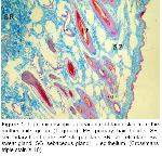
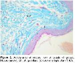
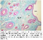
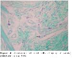
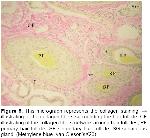
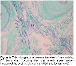
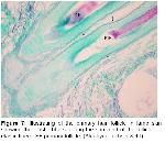
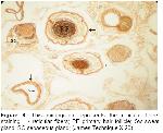
    When epidermis and dermis layers in skin sections of Awassi lambs were examined, histological and histometric differences among the groups were determined. In all groups, it was seen that epidermis was stratified squamous and keratinized; and stratum (str) lusidum was absent (Fig 1). Str. corneum was loose in the 1st group compared to the others; mitotic figures in str. bazale was more in the 1st and 3rd groups than those in the 2nd group (Fig 2). The connective tissue in the papillary layer was loose in 1st group (Fig 1) and tight in other groups (Fig 3). The number of the mast cells increased more 2nd group than in the others (fig 4). In all samples examined, it was established that end of hair follicles with bulbus pili in dermis, and medulla could be distinguished from cortex. In vertical sections to the surface, primary follicles as a cluster of 2 and 3 in papillary layer, secondary follicles as a cluster of 6 and 8, a sweat glands and a pair of sebaseous glands located immediately on the side of those were seen (Fig 1, 3). Collagen fibers in papillary layer become gradually thicker reticular layer and form larger fibrils (Fig 5). Elastic fibers were extended parallel to each other and to the surface between collagen fibers (Fig 6) and they were concentrated around hair follicles (Fig 7). It was seen that in all groups reticular fibers are located around sweat glands, sebaseous glands and hair follicles (Fig 8) and epidermis was lay on a rich basal membrane which was made by the reticular fibers.


    Büyütmek İçin Tıklayın
    Figure 1: Light microscopic appearance of lamb skin from the mother milk group (1 group). PF, primary hair follicle; SF, secondary hair follicle; SP, str. papillare; SR, str. reticulare; Sw, sweat gland; SG, sebaceous gland; → epithelium. (Crossmans triple stain X 10).


    Büyütmek İçin Tıklayın
    Figure 2: Appearance of mitosis from str.bazale (1 group). Mitosis (arrow); SP, str. papillare. (Crossmans triple stain X 40).


    Büyütmek İçin Tıklayın
    Figure 3: Light microscopic appearance of lamb skin from the mother milk with raw feed group (3 group).→ epithelium; SP str. papillare; * connective tissue cells and blood vessels; PF primary hair follicle; SF secondary hair follicle (Crossmans triple stain X 10).


    Büyütmek İçin Tıklayın
    Figure 4: Appearance of mast cells at group 2 (arrow) (Aldehyde fuchsin X40).


    Büyütmek İçin Tıklayın
    Figure 5: This micrograph represents the collagen staining. → illustrating of the collagen fibers surrounding the hair follicle; CF illustrating of the collagen fibers network around hair follicles; PF primary hair follicle; SF secondary hair follicle; SG sebaceous gland. (Methylene blue- van Gieson’sX20).


    Büyütmek İçin Tıklayın
    Figure 6: This micrograph represents the elastic fibers staining. EF lamb skin illusrating the long elastic fibers present throughout the depth of the dermis; (Aldehyde fuchsin X 40).


    Büyütmek İçin Tıklayın
    Figure 7: Illustrating of the primary hair follicle in lamb skin showing the elastic fibers a long the surround of the follicle. → elastic fibers; PF primary follicle; (Aldehyde fuchsin X 40).


    Büyütmek İçin Tıklayın
    Figure 8: This micrograph represents the reticular fibers staining. → reticular fibers; PF primary hair follicle; Sw sweat gland; SG sebaceous gland; (James Technique X 20).

    Statistical Findings: The thickness of epidermis was varied between 30.5-33.5 µm in the 1st group, 38.5-43 µm in the 2nd group and 28.5-31.5 µm in the 3rd group. Although the difference between the 1st and 2nd groups was found significant (P<0.05), the difference between the 1st and 3rd groups was found insignificant (P>0.05). In addition, the difference between the 2nd group and other groups were seen significant (P<0.05). The difference between the 3rd and 2nd group was significant (P<0.05) (Table 1).


    Büyütmek İçin Tıklayın
    Table 1: The statistically comparison of the histometric measurement among the groups.

    In the sectioned examined, it was determined that the thickness of papillary layer was varied between 2090-2220 µm in the 1st group, 1840-1930 µm in the 2nd group and 1910-1970 µm in the 3rd group. While the differences between the 1st group and other groups were found significant (P<0.05), the difference between the 1st and 2nd groups was found significant (P<0.05) and the difference between the 2nd and 3rd groups was found insignificant (P>0.05) (Table 1).

    Reticular layer varied between 1005-1060 µm in the 1st group, 835-900 µm in the 2nd group and 820-885 µm in the 3rd group. While the differences between the 1st group and other groups were found to be significant (P<0.05), the difference between the 2nd and 1st groups was significant (P<0.05) and the difference between the 2nd and 3rd groups was found insignificant (P>0.05) (Table 1).

    The range numbers of primary and secondary follicles in per square millimeter were found 2.83 and 6.45 in the 1st group, 3.73 and 8.95 in the 2nd group, 3.63 and 7.43 in the 3rd group, respectively. The differences in the number of the primary and secondary follicles between the 1st and other groups were found significant (P<0.05); while the number of primary follicles in the 2nd group was found significant (P<0.05) with the 1st group, and insignificant (P>0.05) with the 3rd group, the number of secondary follicles were found significant (P<0.05) in both groups. While the number of primary follicles in the 3rd group was found significant (P<0.05) with the 1st group and insignificant (P>0.05) with 2nd group, the difference in the number of secondary follicles was determined to be significant (P<0.05) with both groups (Table 1).

  • Başa Dön
  • Özet
  • Giriş
  • Materyal ve Metot
  • Bulgular
  • Tartışma
  • Kaynaklar
  • Tartışma
    General structure of skin is affected by factors such as climate, caring-feeding conditions, age and gender. Especially the race of the animal affects skin structure to a significant extent12. In all sections examined, it was determined that skin is generated in epidermis and dermis layers which are coherent with the skin structure and features of domestic mammals2,5,13,14. Kurtdede et al.6,15 observed that epidermis of Awassi sheep was thicker in summer compared to other seasons. The average epidermis thickness was determined as 42 µm in Merino rams13, 21.2 µm Akkaraman sheep7,16, 12.7 µm in Dağlıç sheep, and 21.32 µm by in Karacabey Merino sheep14. In the present study, the thickest epidermis was found 40 µm in the 2nd group. In addition, str. corneum was found thicker and, mitotic figure in str. basale was observed in less density compared to other two groups. This situation could be depend to inadequate feeding of lambs.

    Dermis, the essential layer of the leather used in industry, consists of two sub layers. The average thickness of papillary layer was reported as 1755 µm in Akkaraman sheep, 1149 µm in Dağlıç sheep7,16,17, 1614.58 µm in Kıvırcık sheep, 1490.97 µm in Karacabey Merino sheep14, 1790 µm in Awassi sheep, 2150 µm in Sakız sheep4. In this study, the papillary layer was determined as the thickest in the 1st group with 2151.25 µm and thinnest in 2nd group with 1906.25 µm. This condition could be depend to the thickness of papillary layer in the 1st group to loose structure in connective tissue and its thinner in the 2nd group to inadequate feeding.

    The thickness of reticular layer was determined as 968 µm in Akkaraman sheep, 808 µm in Dağlıç sheep7,16,17, 1247.92 µm in Kıvırcık sheep, 1415.97 µm in Karacabey Merino sheep14, 880 µm in Awassi sheep, 944 µm in Sakız sheep4. In their study of the dermis of skin samples, the reticular layer was determined as the thickest in the 1st group lambs with 1033.75 µm and thinnest in 3rd group lambs with 863.13 µm. This situation could be depend to inadequate feeding of lambs.

    It is important to obtain information about the collagen fiber orientation in biological fibrous tissues such as human and animal skins because the collagen fibers orientation in the skins may be closely related to the motional functions of the body. However, no reports are yet available on the distribution of collagen fiber orientation in a whole skin18. Özfiliz et al.2,14 observed papillary layer where thin collagen fibers and epidermal formations are present, whereas reticular layer where thicker collagen fibers form clusters and no epidermal formations exist. Dağlıoğlu and Bayramlar4 reported that the collagen fiber clusters were observed to have the thickest in surface portions of reticular layer in dermis. We can also say that, in our study, collagen fiber get thicker from papillary layer towards reticular layer and form larger clusters in all groups, and that this is consistent with the results reported by Özfiliz et al.2,14.

    Humans, mice, deer, cows, horses, pigs, and some kinds of dogs typically have this type of hair arrangement, with single, uniform hair follicles, each in communication with other hair follicles through long elastic fibers that extend out through the matrix19. Starcher et al.19 have earlier observed that elastic fibrils in mammal skins extend just under the epithelium parallel to each other and from one hair follicle to another; elastic fibers are attached from one follicle to another especially in deer skin and the elastic fiber array surrounding the hair follicles in sheep skin is very similar to that of the rabbit. Kurtdede et al. stated that in Lincoln, Akkaraman and Awassi sheeps, elastic fibers surround follicle weakly at the line of sebaceous glands, whereas elastic fibers surround primary and secondary follicles strongly at the bottom. Konya Merino, they surround follicles strongly at sebaceous glands level and at the bottom6,15. Özfiliz et al.2,14 observed that elastic fibers concentrate around sebaceous glands in Merino sheep. Dağlıoğlu and Bayramlar4 reported that elastic fibers that extend parallel to surface and each other are present in papillary layer of Awassi sheep. In our study, papillary layer elastic fibers are consistent with the results of Starcher et al.19, Dağlıoğlu and Bayramlar4.

    Özfiliz et al.2,14 reported that reticulum fibers of Awassi sheep are located around sweat glands and hair follicles, which is consistent with our results.

    Wool-bearing animals and animals with fine fur have two or more dissimilar types of hair follicles. Included in this group are the rabbit, sheep, fox, squirrel, raccoon, some kinds of dogs, and many other mammals19. The type and arrangement of follicles in the skin of all sheep regardless of breed are similar. The follicle population typically consists of a basic group of three primary follicles arranged in rows and a variable number of secondary follicles lying on one side of the trio of primaries. All primary follicles are associated with a sweat (sudoriferous) gland, an arrector pili muscle and a sebaceous gland, while secondary follicles possess a sebaceous gland only9. Corbett20 concluded that it was unlikely that maturation of primary follicles could be affected by malnutrition without also causing the death of the fetus and the dam. In examined sections, primary follicles in groups 2 and 3, secondary follicles in groups 6 and 8 and a sweat and a pair of sebaceous glands located just on the side were seen in str. papillare.

    The ratio of the total number of secondary follicles (S) to primary follicles (P), or S/P ratio is a measure of the potential number of wool fibers that can appear under favorable conditions9. Regardless of the experimental method used, these studies found that prenatal nutritional stress influences only the secondary follicle population. This is in agreement with Corbett20 who concluded that the primary follicle population is unaffected. There is however disagreement among the published authors as to whether the prenatal effects on the secondary follicle population are permanent9,14 or transitory9,21. This disagreement can be the result of three possible causes; namely breed differences, the severity of the nutritional stress and the timing of the nutritional stress. In our study, the number of primary and secondary follicles per square millimeter in the 2nd group was more than other groups.

    In conclusion, there was no difference in terms of the distribution of the connective tissue fibers and the density between the groups, for this reason, nutrition is not effective in this aspect. The groups to which were given mother milk had the thickest dermis layer and lesser hair follicle; this may be said to be important in leather industry.

  • Başa Dön
  • Özet
  • Giriş
  • Materyal ve Metot
  • Bulgular
  • Tartışma
  • Kaynaklar
  • Kaynaklar

    1) Papakçı S, Fırat A. Kıvırcık ve dağlıç ırkı kuzu derilerindeki kollagen miktarının tayini ve bunun mamul deri üzerine etkisi. İstanbul Univ Vet Fak Derg 2003; 29: 195-202.

    2) Özfiliz N, Balıkçıer M, Erdost H, Zık B. Histological and morphometric features of the skin of native and hybrid (f2) sheep. Turk J Vet Anim Sci 2002; 26, 429-438.

    3) Banks WJ. Applied Veterinary Histology. 2nd Edition, Baltimore: Williams and Wilkins, 1985.

    4) Dağlıoğlu S, Bayramlar S. Kıbrıs'ta yetiştirilen İvesi ve Sakız koyunlarının derileri üzerinde karşılaştırılmalı histolojik bir çalışma. İstanbul Univ Vet Fak Derg 1988; 29: 73-90.

    5) Dellman HD, Brown M. Integument. Textbook of Veterinary Histology. Philadelphia: Lea and Febiger, 1981.

    6) Kurtdede N. Investigation on the skin structure of lincoln longwoolx Konya merino cross-bred (f1 and b1) sheep. Turk J Vet Anim Sci 2002; 26: 709-714.

    7) Artan ME. Akkaraman ve Dağlıç koyun derilerinin histolojik yapısı üzerinde incelemeler: II. derinin histolojik yapı birimlerinin nitel olarak karsılaştırılması. İstanbul Univ Vet Fak Derg 1980; 29: 72-84.

    8) Aşti RN, Kurtdede N. Hampshire down koyun ırkı ile akkaraman, İvesi ve konya Merinosu arasındaki melezlemelerden elde edilen f1 ve g1 melezlerinin deri yapısı üzerinde araştırmalar. Turk J Vet Anim Sci 2002; 26: 701-708.

    9) Hatcher S, Johnson PR. Optimizing genetic potential for wool production and quality through maternal nutrition. AFBM Network Conference-Proceedings of Contributed Papers, 2004.

    10) Bancroft JD, Cook HC. Manual of Histological Techniques. First Published, Livingstone: Churchill 1984.

    11) Sümbüloğlu K, Sümbüloğlu V. Biyoistatistik. 1. Baskı, Ankara: Özdemir Yayıncılık, 1994.

    12) Akçapınar H. Koyun Yetiştiriciliği. 2. Baskı, Ankara, 2000.

    13) Kozlowski GP, Calhoun M. Microscobic anatomy of the integument of sheep. Am J Vet Res 1953; 14: 448-454.

    14) Özfiliz N, Özer A, Yakışık M, Erdost H. Kıvırcık ve Karacabey Merinos koyunlarının derilerinin histolojik ve morfometrik yönden karşılaştırmalı olarak incelenmesi. Turk J Vet Anim Sci 1997; 21: 125-133.

    15) Kurtdede N, Aştı RN Alman Siyah Baş, Hampshire Down, Lincoln Longwool, Akkaraman, İvesi ve Konya Merinosu deri yapısı üzerinde araştırmalar. Ankara Üniv Vet Fak Derg 1999; 46: 219-230.

    16) Artan ME. Akkaraman ve Dağlıç koyun derilerinin histolojik yapısı üzerinde incelemeler: I.histolojik yapı özellikleri. İstanbul Univ Vet Fak Derg 1980; 29: 73-84.

    17) Artan ME. Canlı koyun derilerinde (Akkaraman, Dağlıç, Merinos) alt katman kalınlıkları ile bağdoku iplik demetlerinin (kollogen ve elastik iplikler) sıklık ve kalınlıklarının işlenmiş deri kalitesi üzerine etkisi. Doğa Bilim Derg Vet Hay Tar Orm 1980; 6: 13-20.

    18) Osaki S. Distribution Map of Collagen Fiber Orientation in a Whole Calf Skin. Anat Rec 1999; 254: 147-152.

    19) Starcher B, Aycock RL, Hill CH. Multiple roles for elastic fibers in the skin. J Histochem & Cytochem 2005; 53: 431-443.

    20) Corbett J. Variation in Wool Growth with Physiological State, In, Corbett JL (Ed): Physiological and environmental limitations to wool growth. Armidale Australia: The University of New England Publishing Unit, 1979.

    21) Denney GD. Effect of pre-weaning farm environment on adult wool production of merino sheep. Aust J Exp Agr 1990; 30: 17-25.

  • Başa Dön
  • Özet
  • Giriş
  • Materyal ve Metot
  • Bulgular
  • Tartışma
  • Kaynaklar
  • [ Başa Dön ] [ Özet ] [ PDF ] [ Benzer Makaleler ] [ Yazara E-Posta ] [ Editöre E-Posta ]
    [ Ana Sayfa | Editörler | Danışma Kurulu | Dergi Hakkında | İçindekiler | Arşiv | Yayın Arama | Yazarlara Bilgi | E-Posta ]