 |
 |
| [ Ana Sayfa | Editörler | Danışma Kurulu | Dergi Hakkında | İçindekiler | Arşiv | Yayın Arama | Yazarlara Bilgi | E-Posta ] | |
| Fırat Üniversitesi Sağlık Bilimleri Tıp Dergisi | |||||||||||||
| 2011, Cilt 25, Sayı 3, Sayfa(lar) 137-140 | |||||||||||||
| [ Özet ] [ PDF ] [ Benzer Makaleler ] [ Yazara E-Posta ] [ Editöre E-Posta ] | |||||||||||||
| Renal Liposarkom: Olgu Sunumu | |||||||||||||
| Nusret AKPOLAT1, Enver ÖZDEMİR2, Bilge AYDIN TÜRK2, Ruhi ONUR2, Selami SERHATLIOĞLU3 | |||||||||||||
| 1Fırat Üniversitesi, Tıp Fakültesi, Patoloji Anabilim Dalı, Elazığ, TÜRKİYE 2Fırat Üniversitesi, Tıp Fakültesi, Üroloji Anabilim Dalı, Elazığ, TÜRKİYE 3Fırat Üniversitesi, Tıp Fakültesi, Radyoloji Anabilim Dalı, Elazığ, TÜRKİYE |
|||||||||||||
| Anahtar Kelimeler: liposarcoma, kidney, retroperitoneum | |||||||||||||
| Özet | |||||||||||||
Liposarkom, böbreğin nadir görülen malign mezanşimal tümörlerindendir. Malign böbrek tümörlerinin %1'in den azını oluşturur. Olgumuz 73 yaşında künt vasıfta göğüs ağrısıyla başvuran bir erkek hasta idi. Üst batın manyetik rezonans görüntüleme tekniği ile sol böbrek orta kutupta yerleşmiş yaklaşık 15 cm büyüklüğünde bir kitle tespit edildi ve hastaya radikal nefrektomi ameliyatı uygulandı. Makroskopik inceleme böbreğin alt kutbunda 18X12X11 cm büyüklüğünde etraf dokudan kısmen ayırt edilebilen bir tümör olduğunu gösteriyordu. Kesit yüzeyi kirli sarı renkte idi ve nekrotik bölgelerle birlikte olan nodüler görüntüsü vardı. Mikroskopik incelemede belirgin nükleer atipi içeren adipositler ve multivakuoler lipoblastlar nedeniyle, hastaya iyi differansiye renal liposarkom teşhisi kondu. Neoplastik olmayan bölgelerde ise xanthogranulomatous enflammasyon alanları vardı. Ameliyattan dört ay sonra hastada lokal nüks tespit edildi. Nadir rastlanan bir olgu olması nedeniyle bulgular literatür eşliğinde tartışıldı. |
|||||||||||||
| Giriş | |||||||||||||
Liposarcoma is a rare malignant mesenchymal tumor of the kidney1-3. It comprises 1% of malignant kidney tumors2. Though, retroperitoneal region is one of the frequent localization2-3.We discussed our case in the light of the available literature because isolated liposarcoma originated from the kidney is quite rare. |
|||||||||||||
| Olgu Sunusu | |||||||||||||
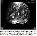
Our case was a 73 year-old male patient applied to outpatient department of urology with blunt chest pain. Inspection revealed abdominal asymmetry. A mass displacing intra-abdominal organs to the right side was found by physical examination. There was no abdominal tenderness. Color doppler ultrasound revealed a mass in the left kidney which was about 19 × 11 cm in size, heterogeneous, not well circumscribed and have cystic areas in the center. Magnetic resonance imaging of upper abdomen revealed a solid mass of around 15 cm in diameter with lipomatous areas originating from mid-lower pole of the left kidney (Figure 1). Serum urea and creatinine levels were higher and sedimentation was found to be 45 mm/hour.
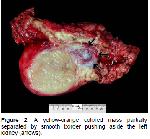
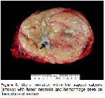
After completion of clinical and radiological evaluation, the patient underwent a left radical nephrectomy operation. Macroscopic examination of the specimen revealed a 18 × 12 × 11 cm in size and partly demarcated. The middle pole of the mass was not demarcated. Its cut surface was dirty yellow in color and nodular appearance with necrotic areas. The tumor was located within the kidney capsule (Figure 2, Figure 3).
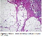
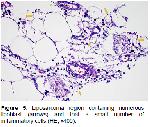
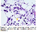
Microscopic examination showed lipocytes quite similar to normal but have significant differences in size. There were areas of xanthogranulomatous inflammation around the tumor.Adjacent to areas of inflammation, the more peripherally located, “adipocytes containing marked nuclear atypia and multivacuoler lipoblasts were seen. This highly cellular tumor which resembles normal lipomatous tissue was diagnosed as well differentiated renal liposarcoma” because of nuclear atypia, hyperchromasia and presence of lipoblasts (Figure 4 Figure 5, Figure 6).
Four months of follow-up MR imaging of the upper abdomen showed a solid mass with 62 × 29 × 42 mm in size, not discriminated with borders of psoas muscule, showing peripheral contrast enhancement and central necrosis. Histopathologic examination of the needle biopsy specimen of this lesion was in consistence with liposarcoma because it was containing fat necrosis and lipoblast like cells. Re-operation was offered the patient but the patient didn’t accept. The patient was alive without any complaint and follow-up examinations were performed by outpatient department of urology. |
|||||||||||||
| Tartışma | |||||||||||||
Primary renal sarcomas constitute 1-3% of kidney tumors1. Liposarcoma is a rare tumor of the kidney, but malignant mesenchymal tumors often localized in retroperitoneal space2-3. It is more common in 4th and 6th decades and in women2. Herewith, we present a 19-cm in great dimension liposarcoma mass with 3 mm distance from kidney capsule within the gerota. Renal liposarcomas can be asymptomatic for a long time and can stay asymptomatic before reaching a size large enough2,4. Patients, often present with symptoms such as pain, hematuria, renal mass, or weight loss3. Our patient’s chief complaint was blunt chest pain. It is difficult to determine the true radiologic prevalence of liposarcoma, because differentiation of retroperitoneal and primary renal liposarcoma is difficult and in the previous reports some liposarcomas are accidentally diagnosed as angiomyolipoma5. Definitive radiologically diagnosed cases have only been reported5. Renal liposarcoma is usually localized in the periphery and is placed between the kidney and the kidney capsule5. Tumor is typically large and it shows perirenal extension5. Tumor in our case was located within the kidney capsule and replaced most of the parenchymatous tissue, but no perirenal extention was observed. Most cases of liposarcomas in the kidney are well differentiated and are greater than 5 cm in diameter2,3. The most frequent localizations are capsule and perinephric regions, and less frequently, it is placed in the renal sinus2. In general, liposarcoma is a well-circumscribed lobulated mass7. Histological appearance is not different from the liposarcomas in the other organs2. According to histological type, liposarcomas are divided into 5 subtypes as well-differentiated, myxoid, round cell, dedifferentiated and pleomorphic6,7. These histological subtypes sometimes can be found together and is called as "mixed liposarcoma"7. 40-45% of the cases are well differentiated, 35% of myxoid and round cell and 5% of pleomorphic type7. Well differentiated liposarcoma frequently seen in 50-70 years of age, and in extremity (75%) and retroperitoneum; dedifferentiated liposarcoma seen in 50-70 years of age and in retroperitoneum (75%); myxoid / round cell liposarcoma seen in the 25-45 years of age and located in extremities (75%)6. There are 4 histologic subtypes of well differentiated liposarcomas. These are lipoma-like, sclerosing type, inflammatory type, and spindle cell type. In the lipoma-like type, unlike to lipoma there are atypical adipocytes with enlarged, irregular, hyperchromatic nuclei. Sclerosing type is characterized by fibrous septa and often multinucleated, hyperchromatic, pleomorphic cellswithin hyaline fibrous stroma. Spindle cell type is characterized by spindle cells in myxoid fibrous stroma. There are chronic inflammatory cells in the inflammatory type8. It is necessary to distinguish a primary renal liposarcoma from a retroperitoneal one. There are two important criteria for this. The first is to demonstrate the tumor location in the renal parenchyma and the second is to show all microscopic features of liposarcoma2. The other entity in the differential diagnosis is sarcomatoid carcinoma. Dedifferentiated and pleomorphic liposarcoma are usually confused with sarcomatoid carcinoma. For differential diagnosis, it is necessary to investigate the presence of carcinomatous component by taking a large number example from the tumor. However, positivity of cytokeratin for sarcomatoid carcinomas and positivity of S-100 in liposarcoma is important for the differential diagnosis2. Sarcomatoid renal cell carcinoma, anjiomyolipoma and other sarcomas should be considered in the differential diagnosis of primary renal liposarcama2,3. HMB-45 is available in differential diagnosis which is positive in anjiomyolipoma and negative in liposarcoma7. The most important prognostic factor for the liposarcoma is anatomical localization of the tumor7. With the excision of the tumor without leaving behind recurrence is greatly reduced7. In addition, the degree of tumor differentiation, size, histological type and stage are also important for the prognosis2,3. In the dedifferentiated liposarcomas local recurrence rate is quite high (40%) but in well-differentiated liposarcomas especially located in the extremities, mortality rate is very low3. The first option of treatment for primary renal liposarcomas is radical nephrectomy3. Radiotherapy or chemotherapy may be given for inoperable tumors2. Even without metastasis, local recurrence can be seen in 30% of cases9. In our case, the tumor was limited to the kidney and had microscopic features of liposarcoma and there were no other tumor focus in the radiodiagnostic studies. For these reasons, our case is diagnosed as primary renal liposarcoma. Because of very high local recurrence risk of liposarcoma and adipose tissue formation of second tumor mass, it is thought to be recurrence of the lesion. In conclusion, although the renal liposarcoma is rare, it should be in mind in the differential diagnosis of retroperitoneal tumors and renal sarcomas. |
|||||||||||||
| Kaynaklar | |||||||||||||
1) Fernández Aceñero MJ, Hernández Gómez MJ, Blanco González J, Suárez Aliaga B. Sarcomas of the kidney. Minerva Urol Nefrol 1997; 49: 145-149.
2) Kim MJ, Koo HY, Jun SY, Ro JY. Pleomorphic Liposarcoma of the kidney, A case report. The Korean Journal of Pathology 2003; 37: 210-213.
3) Bader DAL, Peres LAB, Bader SL. Renal liposarcoma. International Braz J Urol 2004; 30: 214-215.
4) Yaman Ö, Soygür T, Özer G, Arikan N, Yaman L.S. Renal liposarcoma of the sinus renalis. International Urology and Nephrology 1996; 28: 477-480.
5) Helenon O, Merran S, Paraf F, et al. Unusual Fat-containing tumours of the kidney: A diagnostic dilemma. RadioGraphics 1997; 1:129-144.
6) Weis SW, Goldblum JR. Liposarcoma. Enzinger and Weiss’s Soft Tissue Tumors. 4th Edition, 2001; 641.
7) Fletcher CDM, Unni KK, Mertens F. Adipositic tumours. Pathology and Genetics of Tumours of the Soft Tissue and Bone. Lyon: IARC Pres, 2002; 19-46.
|
|||||||||||||
| [ Başa Dön ] [ Özet ] [ PDF ] [ Benzer Makaleler ] [ Yazara E-Posta ] [ Editöre E-Posta ] | |||||||||||||
 |
| [ Ana Sayfa | Editörler | Danışma Kurulu | Dergi Hakkında | İçindekiler | Arşiv | Yayın Arama | Yazarlara Bilgi | E-Posta ] |
