
Kronik Böbrek Yetmezliği ile İlişkili Purtscher-like Retinopati: Olgu Sunumu
Cilt 29, Sayı 2
Ağustos 2015 · Sayfalar 095-096
Kronik Böbrek Yetmezliği ile İlişkili Purtscher-like Retinopati: Olgu Sunumu
Purtscher-like Retinopathy Associated with Chronic Renal Failure: Case Report
Sabiha GÜNGÖR KOBAT1, Burak TURGUT2
2Fırat Üniversitesi Tıp Fakültesi, Göz Hastalıkları Anabilim Dalı, Elazığ, TÜRKİYE
Purtscher-like retinopati etyolojide travmanın rol oynamadığı nadir bir hastalıktır. Karakteristik fundus bulguları multiple beyaz iskemik infarktlar veya atılmış pamuk benzeri eksudalar, retinal hemorajiler ve optik disk kabarıklığıdır.
Bu çalışmada kronik böbrek yetmezliği ile birlikte seröz retina dekolmanı olan nadir görülen bilateral Purtscher-like retinopatili bir olguyu sunmayı amaçladık.
Purtscher-like retinopathy is a rare condition with non-traumatic aetiology. Its fundus characteristics are multiple white ischemic infarcts or cottonwool spots (Purtscher fleckens), retinal hemorrhage, and optic disc swelling. We purposed to report an unusual case of bilateral Purtscher-like retinopathy with serous retinal detachment associated with chronic renal failure.
Giriş
Purtscher's retinopathy (PR) is a traumatic retinal angiopathy, most commonly caused by head trauma and compression of chest, malar bone fracture, mandibular fracture, and crush injury without any ocular trauma. The fundus characteristics of the disease are bilateral retinal signs include multiple white ischemic infarcts (cotton-wool spots of varying sizes or Purtscher-flecken) and hemorrhages (dot and blot, pre-retinal, or flame) and optic disc swelling. The patients with Purtscher-like retinopathy (PLR) have similar fundus findings to PR, however PLR is not associated with trauma[1-3]. PLR can be seen in patients with connective tissue disorder, HELLP syndrome, renal failure, long bone fracture, acute pancreatitis, pancreatic adenocarcinoma, the trombotic trombocytopenic purpura, preeclampsia-eclampsia and amniotic fluid embolia[4-7].We purposed to report an unusual case with Purtscher-like retinopathy with serous retinal detachment and chronic renal failure.
Olgu Sunumu
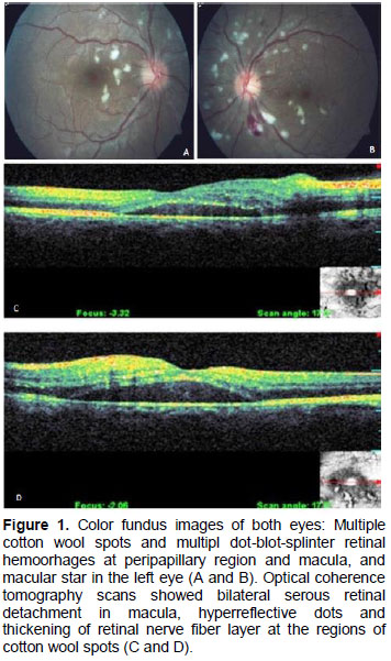
A 23-years old man presented to our outpatient clinic with the complaint of decreased vision in both eyes for a week. His best corrected visual acuities are 2/10 in right eye vision and 1/10 in left eye. Intraocular pressures are 14 mmHg in the right eye and 16 mmHg in the left eye. Slit-lamp biomicroscopy revealed no any pathological anterior segment sign. Fundus examination revealed multiple cotton wool spots and multipl dot-blot-splinter retinal hemoorhages at peripapillary region and macula (Figure 1A and B). Optical coherence tomography scans showed bilateral serous retinal detachment in macula, hyperreflective dots and thickening of retinal nerve fiber layer at the regions of cotton wool spots. (Figure 1C and D). His medical history revealed chronical renal failure and chronic controlled systemical hypertension but no acute hypertension. A diagnosis of Purtscher-like retinopathy was made. As an acute hypertension attack was not present, it was not considered the diagnosis of malign hypertension.The patient was directed to nephrology clinic for consultation. Blood tests revealed only that creatine level increased to 10.3 mg/dL. Our case was recommended to continue with the treatment nephrology and cardiology clinics have given and to attend further follow-up visits in our clinic. However, the follow-up visits were not possible because of patient inconsistency. Thus, we could not have possibility for more examination of the patient.
Tartışma
Purtscher's retinopathy which is firstly described by Otmar Purtscher is a traumatic retinal angiopathy which was associated with sudden blurring of vision and most commonly caused by head trauma and compression of chest, malar bone fracture, mandibular fracture and crush injury without any ocular trauma. Visual loss may be accompanied by field loss in the form of central, paracentral, or arcuate scotoma and peripheral visual function is often preserved[1-3).The fundus characteristics of disease are bilateral retinal signs include multiple white ischemic infarcts (cotton-wool spots in varying sizes or Purtscher-flecken) located around optic disk and at posterior pole and hemorrhages (dot and blot, pre-retinal, or flame) and sometimes optic disc swelling. The patients with PLR have similar fundus findings to PR, however PLR occurs in the patients with no relation to trauma. PLR is seen in patients with connective tissue disorder, HELLP syndrome, renal failure, long bone fracture, acute pancreatitis, pancreatic adenocarcinoma, the trombotic trombocytopenic purpura, preeclampsia eclampsia and amniotic fluid embolia In both PR and PLR, patients usually experience asymmetrical bilateral symptoms[1,2]. Although, the pathogenesis of these diseases is still uncertain, the pathogenesis involves microembolic occlusion of the precapillary arterioles caused by fat, air, leucocytes, fibrin, platelets by emboli, complement activation, elevated intracranial or intrathoracic pressure and extravasation of lymph, vasculitis due to free fatty acids[1].
The diagnosis is made on clinical findings and supported by fundus fluorescein angiography. OCT can provide details about optic disk and posterior pole involvement. FFA can reveal the blockade of choroidal flourescence, retinal arteriolar occlusion, capillary non-perfusion areas and perivenous staining. OCT can show macular edema or optic disk edema[1,2]. There are no specific the treatment of patients with Purtscher's retinopathy. The majority of patients with PR and PLR have some visual acuity without treatment. Application of hyperbaric oxygen and intravenous high-dose steroids may be useful for resisted and selected cases[1,8].
In conclusion, it should be considered that each case of Purtscher-like retinopathy could be a patient with renal failure.
References
- Agrawal A, McKibbin MA. Purtscher's and Purtscher-like retinopathies: A review. Surv Ophthalmol 2006; 51: 129-136.
- Lin YC, Yang CM, Lin CL. Hyperbaric oxygen treatment in Purtscher's retinopathy induced by chest injury. J Chin Med Assoc 2006; 69: 444-448.
- Cumurcu T, Yardım H, Güven M, et al. Purtscher's retinopathy: An unusual association with a complicated mandibular fracture. Turk J Med Sci 2008; 38: 373-376
- Stassen LF, Goel R, Moos KF. Purtscher's retinopathy: An unusual association with a complicated malar fracture. Br J Oral Maxillofac Surg 1989; 27: 296-300.
- Tabandeh H, Rosenfeld PJ, Alexandrakis G, Kronish JP, Chaudhry NA. Purtscher-like retinopathy associated with pancreatic adenocarcinoma. Am J Ophthalmol 1999; 128: 650-652.
- Stewart MW, Brazis PW, Guier CP, Thota SH, Wilson SD. Purtscher-like retinopathy in a patient with HELLP syndrome. Am J Ophthalmol 2007; 143: 886-887.
- Shukla D, Maheshwari R, Ramchandani B, Kanungo S. Purtscher-like retinopathy with serous retinal detachment in preeclampsia of pregnancy: Complications and management. Retin Cases Brief Rep 2010; 4: 332-335. 8. Wang AG, Yen MY, Liu JH. Pathogenesis and neuroprotective treatment in Purtscher's retinopathy. Jpn J Ophthalmol 1998; 42: 318-322.
- KRONİK BÖBREK YETMEZLİĞİNDE SERUM β- KAROTEN DÜZEYLERİ
- BÖBREK YETMEZLİĞİ GELİŞEN JOUBERT SENDROMLU HASTADA ANESTEZİ (Olgu Sunumu)
- Asetaminofen İntoksikasyonu Sonrası Karaciğer Yetmezliği Gelişmeden Renal Yetmezlik Gelişen Bir Olgu
- Ulcerative Pyoderma Gangrenosum and CIDP Associated with Chronic Renal Failure: a Case Report and Review of The Literature
- Yılan Sokması Sonucu Dissemine İntravaskuler Koagulasyon ve Akut Böbrek Yetmezliği Gelişimi