İntravitreal triamsinolon enjeksiyonu sonrası pseudohipopiyon gelişen iki olgunun sunulması.
Aynı operasyon gününde, 58 yaşında erkek hastanın sol gözüne ve 52 yaşında daha önce katarakt ameliyatı geçirmiş ve göz içi lensi yerleştirilmiş erkek hastanın sağ gözüne intravitreal triamsinolon enjeksiyonu (0.1 ml) yapıldı. Her iki hastada enjeksiyon sırasında pseudohipopiyon gelişitiği gözlendi.
Pseudohipopiyonun spesifik tedavi yapılmadan düzeldiği gözlendi.
Pseudohipopiyon, intravitreal triamsinolon enjeksiyonu sonrası gözlenen özel bir durumdur. Eğer gözde ilerleyici enflamasyon ve ağrı yok ise, hastanın yakın takibi yeterlidir.
To report two cases of pseudohypopyon that occurred during intravitreal triamcinolone injection.
In the same operating day, a 58-year-old man with left phakic eye and 52-year-old man with right pseudophakic eye underwent intravitreal triamcinolone injection (0.1 ml). In both patients, pseudohypopyon developed during intravitreal triamcinolone injection.
Pseudohypopyon resolved without specific treatment.
Pseudohypopyon seems to be a distinct clinical entity after intravitreal triamcinolone injection. If progressive intraocular inflammation and ocular discomfort do not occur, close observational management is indicated.
Giriş
Intravitreal triamcinolone has become a safe and effective treatment for diabetic maculopathy [1] and central retinal vein occlusion [2]. The most common side effects observed are glaucoma, cataract, retinal detachment and endophthalmitis. Pseudohypopyon has also been recently reported [3,4]. We present two cases of pseudohypopyon that occurred during intravitreal triamcinolone injection.
Olgu Sunumu
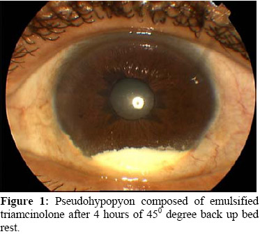
Case 1: A 58-year-old man with left phakic eye, who was suffering from unresolved macular edema due to central retinal vein occlusion, underwent intravitreal triamcinolone injection (0.1 ml) in the operating room, under topical anesthesia. The site of injection was in the temporal quadrant, 4 mm behind the limbus. Injection was done with a 30 gauge needle. Just after starting injection, triamcinolone was observed in the anterior chamber in an emulsified form. After 4 hours of 45º degree back up bed rest, slitlamp examination showed an anterior chamber deposition of triamcinolone without inflammatory activity (Figure 1). Posterior segment examination also showed suspended triamcinolone in the vitreous cavity.
Figure 1: Pseudohypopyon composed of emulsified triamcinolone after 4 hours of 45º degree back up bed rest.
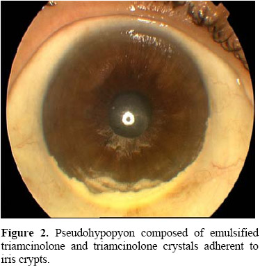
Case 2: In the same operating day, a 52-year-old man with right pseudophakic eye, who was suffering from unresolved diabetic macular edema, also underwent intravitreal triamcinolone injection (0.1 ml). Posterior capsule was intact. The site of injection was in the temporal quadrant, 4 mm behind the limbus. After seeing the tip of the 30 gauge needle in the vitreous cavity through the pupil, injection was started. Immediately after starting injection, we observed triamcinolone in the anterior chamber and stopped injection. After 4 hours of 45º degree back up bed rest, slitlamp examination also showed an anterior chamber deposition of triamcinolone without inflammatory activity (Figure 2). Posterior segment examination also showed suspended triamcinolone in the vitreous cavity.
Figure 2: Pseudohypopyon composed of emulsified triamcinolone and triamcinolone crystals adherent to iris crypts.
Postoperatively, there was neither ocular discomfort nor ciliary injection in both cases. So during close observation, only topical antiglaucomatous medication and topical antibiotic were given. In the tenth day of the follow up, pseudohypopyons were cleared spontaneously. At the end of the 2-months follow-up period, no other complications were seen. Intraocular pressures remained within normal limits at all times.
Tartışma
In the literature, pseudohypopyon was reported after intravitreal triamcinolone injection [3,4]. However only in one of these cases, pseudohypopyon developed immediately after the injection. In both of our cases, pseudohypopyon developed during the injection.
It is hard to differ pseudohypopyon from endophthalmitis unless it occurs such an early postoperative period. So, the clue is progressive intraocular inflammation and ocular discomfort. If progressive intraocular inflammation and ocular discomfort do not occur, close observational management is indicated.
Based on these findings, formation of pseudohypopyon is due to passage of emulsified triamcinolone to anterior chamber. The reason of this emulsification might be partial “jamming” [4] of crystalline triamcinolone in the barrel of the 30 gauge needle during injection. Using a smaller gauge needle or pushing the piston slowly and uninterruptedly may reduce this “jamming” [4] and emulsification in turn. This technique of injection also reduces fish-egg gas bubbling in intravitreal gas injections.
References
Martidis A, Duker JS, Greenberg PB, Rogers AH, Puliafito CA, Reichel E, Baumal C. Intravitreal triamcinolone for refractory diabetic macular edema. Ophthalmology 2002;109:920-927.
Greenberg PB, Martidis A, Rogers AH, Duker JS, Reichel E. Intravitreal triamcinolone acetonide for macular oedema due to central retinal vein occlusion. Br J Ophthalmology 2002; 86:247-248.
Campos VE, Maia A, Muccioli C, Belfort R Jr. Pseudohypopyon immediately after intravitreal triamcinolone acetonide-case report. Arq BrasOftalmology 2005;68:251-252.
Chen SD, Lochhead J, McDonald B, Patel CK. Pseudohypopyon after intravitreal triamcinolone injection for treatment of pseudophakic cystoid macular oedema. Br J Ophthalmology 2004; 88:843-844.