
Romatoid Artritli Hastalarda Total Antioksidan Cevap ve Oksidatif Stres
Cilt 21, Sayı 2
Aralık 2007 · Sayfalar 067-073
Romatoid Artritli Hastalarda Total Antioksidan Cevap ve Oksidatif Stres
Total Antioxidant Response and Oxidative Stress in Patients with Rheumatoid Arthritis
Ahmet IŞIK1, Şahabettin SELEK2
2Harran University Medicine Faculty Department of Biochemistry Şanlıurfa-TURKEY
Romatoid artrit (RA)'te çeşitli antioksidanların azaldığı bildirilmiştir. Total antioksidan kapasiteyi gösteren ve , total antioksidan cevap (TAR)'ın ölçüm metodu son zamanlarda belirlenmiş ve geliştirilmiştir olan total antioksidan cevap (TAR) tüm antioksidanların total antioksidan özelliklerini yansıtmaktadır. Bu çalışmanın amacı, RA'te total antioksidan kapasiteyi TAR ile ölçmek ve oksidatif stres ile ilişkilerini incelemektir.
Çalışmaya 47 aktif RA'li olgu ile kontrol grubu olarak yaş ve cinsiyetleri uyumlu 23 sağlıklı gönüllü alındı. Hasta ve kontrol gruplarında TAR, total peroksit (TP) uygun yöntemler ile çalışıldı ölçüldü ve oksidatif stres indeksi (OSI) hesaplandı.
TAR düzeyi hasta grubunda 1,02±0,09 mmol ve kontrol grubunda 1,16±0,08 mmol Trolox eq./L; TP düzeyi hasta grubunda 23,84±10,10 ve kontrol grubunda 7,72±1,86 µmol H2O2/L; OSI değeri ise hasta grubunda 2,34±1,19 ve kontrol grubunda 0,67±0,18 Arbitrary Unit olarak saptandı. Kontrol grubu ile karşılaştırıldığında, hasta grubundaki TAR düzeyi düşüklüğü, TP ve OSI düzeyleri yüksekliği anlamlı bulundu (herbirisi için, p<0.001). TAR düzeyi ile TP ve OSI düzeyleri arasında, hem hasta grubunda (sırasıyla; r=-0.534, p<0.01 ve r=-0.552, p<0.01), hem de kontrol grubunda (sırasıyla; r=-0.469, p<0.05 ve r=-0.382, p<0.05) anlamlı negatif korelasyon vardı.
Çalışma sonuçlarımız, RA'li olgularda oksidatif stres ve lipid peroksidasyon ürünlerinde artış ve antioksidan kapasitede azalma olduğunu göstermektedir.
It has been reported that various antioxidants decrease in rheumatoid arthritis (RA). Total antioxidant response (TAR), whose measurement method has been recently specified and developed, represents the total antioxidant properties of all antioxidants. This study aims to measure total antioxidant capacity in RA using TAR and evaluate its relations with oxidative stress.
The study included 47 active RA cases, and 23 age- and sex-matched healthy volunteers as the control group. TAR and total peroxide (TP) were studied using appropriate methods and oxidative stress (OSI) was calculated.
TAR level was 1,02±0,09 and 1,16±0,08 mmol Trolox eq./L in the patient group and the control group, respectively; TP level was 23,84±10,10 and 7,72±1,86 µmol H2O2/L in the patient group and the control group, respectively; and OSI value was 2,34±1,19 and 0,67±0,18 Arbitrary Units in the patient group and the control group, respectively. When compared with the control group, TAR level was significantly lower, and TP and OSI levels were significantly higher in the patient group (p<0.001 for each). TAR level was negatively correlated with TP and OSI levels both in the patient group (r=-0.534, p<0.01 and r=-0.552, p<0.01, respectively) and the control group (r=-0.469, p<0.05 and r=-0.382, p<0.05, respectively).
The results of our study indicate an increase in oxidative stress and lipid peroxidation products and a decrease in antioxidant capacity in RA cases.
Giriş
Rheumatoid arthritis (RA) is a progressive, relapsing and chronic inflammatory disease [1]. Although it affects about 1% of the adult population, its etiopathogenesis has not been fully revealed yet [1]. Nonetheless, reactive oxygen species (ROS) have been reported to play important roles in RA pathogenesis [2-4]. ROS, which can be produced as a result of normal aerobic metabolism and whose production is increased by active neutrophils during inflammation, have recently attracting increasing attention [5]. It is known that neutrophils in the joints affected by the disease are activated in RA cases. Activated neutrophils lead to the production of ROS like hydrogen peroxide (H2O2), hydroxyl radical (OH•) and superoxide radical (O2•-), as well as, nitrogencontaining radicals like peroxynitrite (ONOO-) [2-4].Polymorphonuclear leukocytes increase the production of hypochlorous acid (HOCl) from H2O2 via myeloperoxidase enzyme [2]. HOCl, in turn, causes tissue injury via sulphydryl oxidation and protein decarboxylation, leading to oxidative modification of macromolecules in the tissues [2].
The primary targets of ROS are lipids in the cell membrane. ROS increase lipid peroxidation (LPO), and thus, impair cell structure and functions [2-4]. Increase in LPO products has been reported in RA cases [6-8]. It has been demonstrated that increased LPO products and ROS inhibit antioxidant systems and impair oxidant/antioxidant balance in RA [9,10].
Blood contains many antioxidant molecules those prevent and/or inhibit harmful free radical reactions [11]. Exogenous antioxidants like vitamin C and vitamin E, and endogenous antioxidants like uric acid, bilirubin, albumin and superoxide dismutase and glutathione peroxidase as the scavenger enzymes can protect the cell against the potentially detrimental effects of oxidant agents [10,11]. Concentrations of these antioxidants in the plasma can be measured one by one, but this procedure is time-consuming, labour-intensive and costly and requires complicated techniques [12]. On the other hand, total antioxidant response (TAR), whose measurement method has been recently specified and developed, and which can indicate the total antioxidant state of the plasma [<12>,13]. In this method, TAR of plasma against especially potent free radical reactions which strongly lead to oxidative damage of biomolecules such as lipids, proteins and DNA, is measured.
In this study, we aimed to measure both the levels of some individual antioxidant components and the TAR values in plasma samples from active RA to evaluate their antioxidant status using a novel automated method [12,13]. As a reciprocal measure, the total peroxide (TP) levels of the same plasma samples were also measured [14]. The ratio of the plasma TP level to the TAR level was regarded as the oxidative stress index (OSI) [15].
Gereç ve Yöntem
The study included 47 active RA cases who applied to outpatient clinic of Rheumatology Department of Internal Medicine of Medical Center of Firat University and 23 healthy individuals who age and sex matched. RA was diagnosed according to the American Rheumatism Association (ARA) criteria [16]. Approval of the local ethics committee and informed consent of the patients and healthy individuals were obtained. Individuals who had pathologies those could lead to secondary lipid disorders, cardiovascular diseases (CVD), diabetes mellitus, renal failure, chronic infection, alcohol abuse, and those who used antilipidemic and antioxidant drugs were excluded from the study.Blood samples were obtained at 08.00-09.00 a.m. after a 8-12 hours of fasting. The samples were centrifuged at 3000 rpm for 10 minutes to obtain serums. Routine biochemical analyses were carried out in the serum samples by Olympus AU 600 Autoanalyzer using Olympus kits (Olympus Corp. Tokyo-Japan). Erythrocyte sedimentation rate (ESR) was determined by classical Westergren method immediately in whole blood with 1 mg/mL ethylene diamine tetraacetic acid. The level of Creactive protein (CRP) was determined by immunoturbidimetric technique (Schiapparelli Biosystems, the Netherlands). The level of rheumatoid factor (RF) was studied measured by nephelometric method (BNII, Dade Behring, Germany).
Levels of serum total protein, albumin, bilirubin and uric acid were measured by using commercial kits (Olympus AU Autoanalyzer). TAR and TP, the specific parameters, were measured in the plasma samples obtained by centrifugation of blood put in heparinized tubes, using appropriate methods. Plasma samples were stored at -80 20 ºC until the day of analysis.
Measurement of the total antioxidant status of plasma: The total antioxidant status of the plasma was measured using a novel automated colorimetric measurement method for the TAR developed by Erel [12,13]. In this method, the hydroxyl radical, the most potent biological radical, is produced by the Fenton reaction, and reacts with the colourless substrate Odianisidine to produce the dianisyl radical, which is bright yellowish-brown in colour. Upon the addition of a plasma sample, the oxidative reactions initiated by the hydroxyl radicals present in the reaction mix are suppressed by the antioxidant components of the plasma, preventing the colour change and thereby providing an effective measure of the total antioxidant capacity of the plasma. The assay results are expressed as mmol Trolox eq./L, and the precision of this assay is excellent, being lower than 3% [17].
Measurement of total plasma peroxide (TP) concentration: The total plasma peroxide concentrations were determined using the FOX2 method [14] with minor modifications [15]. The FOX2 test system is based on the oxidation of ferrous iron to ferric iron by the various types of peroxides contained in the plasma samples, in the presence of xylenol orange which produces a coloured ferric-xylenol orange complex whose absorbance can be measured. The FOX2 reagent was prepared by dissolving ammonium ferrous sulphate (9.8 mg) in 250 mM H2SO4 (10 ml) to give a final concentration of 250 µM ferrous iron in acid. This solution was then added to 90 ml HPLC-grade methanol containing 79.2 mg of butylated hydroxytoluene (BHT). Finally, 7.6 mg of xylenol orange was added, with stirring, to make the working reagent (250 µM ammoniumferrous sulphate, 100 µM xylenol orange, 25 mM H2SO4, and 4 nM BHT, in 90% (v/v) methanol in a final volume of 100 ml). The blank reagent contained all the components of the solution except ferrous sulphate.
Aliquots (200 µL) of plasma were mixed with 1.8 ml FOX2 reagent. After incubation at room temperature for 30 min, the vials were centrifuged at 12,000 g for 10 min. The absorbance of the supernatant was then determined at 560 nm. The TP content of the plasma samples was determined as a function of the difference in absorbance between the test and blank samples using a solution of H2O2 as standard. The coefficient of variation for individual plasma samples was less than 5%.
Oxidative stress index (OSI): The ratio of the TP to the total antioxidant potential gave the OSI, an indicator of the degree of oxidative stress [15].
Statistics: All results were expressed as means SD. Student's t-test and Spearman's correlation analysis were performed using SPSS for Windows Release 11.0 (SPSS Inc. Chicago, Illinois, USA). The P value less than 0.05 was considered to be significant.
Bulgular
The patient group included 40 females and 7 males, whose mean age was 50,4±12,5 years and mean duration of the disease was 4,4±6,2 (0-16) years. The patients were diagnosed as RA according to ARA criteria [16]. All RA patients were active according to the criteria for complete clinical remission of RA, as suggested by Pinals et al. [18]. The control group consisted of 17 females and 6 males healthy volunteers whose mean age was 49,7± 4,2 years. Of the patients in the RA group, 11 had been recently diagnosed and had not used corticosteroid and any disease-modifying antirheumatic drugs (DMARDs) in their history. Of the 36 cases who were diagnosed previously, 22 were receiving methotrexate, salazopyrin sulfasalazine, chlorochine and corticosteroid combination; 14 were using corticosteroid in addition to one of the DMARDs. RF was positive in 38 cases and negative in 9 cases. Nine cases in the patient group and 5 cases in the control group were diagnosed as having hypertension. Demographical characteristics and laboratory findings of the patient group and the control one are presented in Table 1.
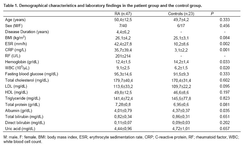
The comparison between the patient group and the control group revealed that white blood cell count (WBC), ESR and CRP values were significantly higher (p<0.05, p<0.01, p<0.001, respectively) and hemoglobin level was significantly lower (p<0.05) in the former. There was not any significant difference between the groups in terms of fasting blood glucose, total cholesterol, triglyceride, HDL and LDL levels (p>0.05, for each).
The level of albumin, an individual antioxidant, was significantly lower in the patient group (p<0.0501), but there was not any significant difference between uric acid and bilirubin levels of the groups (p>0.05, for each).
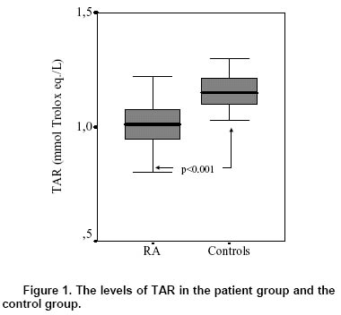
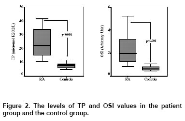
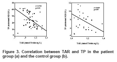
TAR level was 1,02±0,09 and 1,16±0,08 mmol Trolox eq./L in the patient group and the control group, respectively; TP level was 23,84±10,1 and 7,72±1,86 µmol H2O2/L in the patient group and the control group, respectively; and OSI value was 2,34±1,19 and 0,67±0,18 Arbitrary Units in the patient group and the control group, respectively. When compared with the control group, TAR level was significantly lower, and levels of TP and OSI were significantly higher in the patient group (p<0.001, for each, Table 2, Figure 1 and 2). TAR level was negatively correlated with TP (Figure 3) and OSI levels both in the patient group (r=-0.534, p<0.01 and r=-0.552, p<0.01, respectively) and the control one (r=-0.469, p<0.05 and r=-0.382, p<0.05, respectively).
There was no significant correlation among
biochemical findings, TAR, TP and OSI values of the
cases in the patient group in regard with gender, duration
of disease, RF positivity (p>0.05, for each).
Hydrogen peroxide and other derivatives of
peroxides, produced physiologically and which increase
in some conditions, diffuse into plasma. When TP is
measured, it means that the sum of many peroxides like
protein peroxide, lipid peroxide and H2O2 are measured
[19]. Although it is known that H2O2 and lipid peroxides
increase in RA [1-3,7,8], oxidative stress has not been
evaluated via measurement of TP. However, it has been
reported that TP level increases in passive smokers [21],
preeclampsia cases [22] and skin leishmaniasis [23]. It
was shown in our study that TP level also increased in
RA cases. Possible causes of this increase in TP might
be the inevitable increase in lipid peroxides and ROS
including H2O2 in RA.
Many antioxidant molecules found in blood prevent or
inhibit the harmful effects of free radicals [11]. Whenever
there is a decrease in antioxidants and/or an increase in
oxidants, oxidant/antioxidant balance is impaired in favor
of oxidants and this is known as oxidative stress [19,20].
It is known that oxidative stress is responsible for tissue
injury in many diseases and contributes to the
development of atherosclerosis [19,24,25]. Antioxidant
activity indicates the antioxidant characteristics of only
one antioxidant, whereas total antioxidant capacity (TAC)
represents the total antioxidant characteristics of all
antioxidants found in the plasma. TAR and total
antioxidant status (TAS) are used synonymously with
TAC [20]. It is doubtlessly more advantageous to
evaluate TAR, instead of individual antioxidant activities.
Many methods have been developed recently for this
aim. Total radical trapping antioxidant parameter (TRAP),
oxygen radical absorbance capacity (ORAC) and ferric
reducing antioxidant power (FRAP) are colorimetric
methods previously developed to assess TAC
[12,13,20]. It has been reported that TAR, a new
measurement method developed by Erel [12,13] has
correlated with the data obtained by other measurement
methods and has had some additional advantages
[12,13].
Blood has an important function in the
oxidant/antioxidant balance, as it carries and distributes
antioxidants through the body [20]. Plasma has various
antioxidant molecules. Albumin, uric acid, bilirubin and
ascorbic acid are the major antioxidant components of
plasma [12,13,20]. TAR represents practically all of them
[12,139. Albumin consists of about half of the TAC of the
plasma [12,13]. Albumin has several biological functions,
particular as a ligand binder [26]. Plasma thiol contents
originate from albumin. Thiol groups, on the surface of
albumin, bind oxidants [26]. Low level of albumin can
cause oxidative stress via leading to increase oxidants
like homocysteine [26]. In chronic inflammation, albumin
is reduced. We attribute the low albumin level in our
patient group to this protein's being a negative acute
phase reactant, but this decrease is also expected to
negatively influence antioxidant capacity. Bilirubin, a
powerful endogenous antioxidant, is one of the catabolits
of heme oxygenases [27]. However, Harma et al. [22]
have reported that bilirubin did not correlated with TAR,
in their clinical study. Uric acid is another well-known low
molecular weight water-soluble plasma antioxidant
[12,13]. Uric acid has a strong antioxidant activity and its
concentration in the plasma is about 10 fold than
antioxidants like vitamin C and vitamin E [20]. In the
present study, although a significant decrease was found
in the level of albumin, an individual antioxidant, in the
patient group, uric acid and bilirubin levels were not
significantly different from those in the control group. A
positive correlation was reported amongst uric acid,
bilirubin and total protein levels, and TAC level [20].
However, uric acid concentration changes depending on
gender, diet, heavy exercise, renal failure, and in some
metabolic diseases [20,28]. Methotrexate treatment
might alter uric acid level has been also reported in RA
cases [29]. We suggest that uric acid might not
appropriately reflect the TAC. However, it has been also
reported that uric acid was not a strong antioxidant and
might not protect against free radicals [30].
Orem et al. [31] have reported a decrease in TAR
level in Behcet's disease. It was established in our study
that TAR level decreased, while TP and OSI levels
increased and these increases negatively correlated with
TAR level in the patient group. We think that the
decrease in TAR level may have resulted from increased
oxidative stress. As uric acid and bilirubin levels have not
been significantly different in the patient and control
groups, it has seemed reasonable to assume that the
decrease in TAR level is a result of the decrease in other
antioxidants. It has been reported that PON1 activity
[32,33] and SH level [34], both of which are antioxidants, have decreased in RA. Ece et al. [35] have noted that
PON1 is positively correlated with TAR and negatively
correlated with TP and OSI in cases with nephrotic
syndrome. The decrease in TAR level in RA may be
associated with the decrease in PON1 and other
antioxidants.
Plasma TAR level has been reported to be lower in
those with CVD, compared with without CVD, in
smokers, compared with non-smokers, in diabetic cases,
compared with non-diabetic cases, and in hyperlipidemic
cases, compared with those who have a normal lipid
profile [36]. Additionally, children who are exposed to
passive cigarette smoking have found to have a
decrease in TAR level, and an increase in TP and OSI
levels [21]. As TAR is a fairly good representative of
antioxidant capacity, while TP and OSI are indicators of
oxidant capacity, decreased TAR and/or increased TP
levels indicate oxidative stress [12,13,28].
It has been reported in the literature that
atherosclerotic diseases increase in RA cases, but the
causes of this accelerated atherosclerosis cannot always
be explained by classical risk factors [5]. Oxidative stress
is blamed to be responsible in the pathogenesis of
atherosclerosis [24,25]. Atherosclerosis must also be
considered when evaluating RA cases, as it has been
shown that oxidative stress relates with atherosclerosis
and TAR levels decrease in atherosclerosis [36,37].
It is possible that the administration of DMARDs and
steroids affect the levels of oxidants and antioxidants
either negatively or positively. Previous experimental
studies had demonstrated that using steroid impaired
antioxidant systems and leading to overproduction of
ROS [38,39]. In RA patients, it has been reported that
MTX alone [40] or combined with sulfasalazine [41] lead
to increased plasma homocysteine level. In our study, we
did not observe any difference between the cases with
use or without use of these medications with regard to
oxidant and antioxidant levels. Not observing this
difference might be resulted from that the cases enrolled
to the study were in their active phases.
The limitation of our study might be not to evaluate
relationships amongst drugs used in our cases, level of
cytokines like IL-1, Il-6 and TNF-α, rheumatological data
reflecting the activity, severity and function of RA patients
(pain, DAS-28, disability, articular index, radiological
severity etc.).
In conclusion, oxidant/antioxidant balance is impaired
in RA and TAR, a new measurement method, effectively
shows this balance.
Tartışma
ROS and other oxidants have been shown to be
formed even in the normal physiological process [19,20].
It is known that activated inflammatory cells lead to ROS
production in RA a systemic autoimmune disease [1,2].
Increased ROS, in turn, increase LPO products and
cause tissue injury [1-5].Figures
Tables

References
- MICROSCOPIC FINDINGS IN RHEUMATOID SYNOVIAL TISSUE OF DOG KNEE JOINT
- Total Antioxidant Response and Oxidative Stress in Patients with Behçet's Disease
- The Effect of Passive Tobacco Smoke Exposure on Diet Quality, Oxidative Stress Index and Nutritional Status in Young Adults
- Efficacy of Abatacept Therapy in Patients with Seronegative and Seropositive Rheumatoid Arthritis