Screening of newborn diarrheal clinical samples for the ALPSpot assay validation: For the validation of the ALPSpot assay, fecal specimens were obtained from newborn calves presenting with diarrhea at the emergency unit of the animal hospital, Erciyes University, Turkey. To perform total viral RNA isolation from newborn diarrheal samples, fecal specimens were diluted (1:10 v:v) with PBS and centrifuge clarified at 10.000 × g for 15 min. Clarified supernatants were applied to the acid guanidinium thiocyanatephenolchloroform extraction method as previously described
18. Rotavirus genomes that consisted of 11 dsRNAs were visualized after running them in ethidium bromide-stained agarose gel to observe intact dsRNA integrity under a constant voltage of 100 V for less than 20 min. Molecular identification was directed to amplify the partial region of the VP6 gene of rotavirus with the following primer pairs: VP6-F sense (5′ GACGGVGCRACTACATGGT 3′) and anti-sense 157-R (5′ GTTTTCCAAGAGTDATHAHYTCAGC 3′) with modifications by Iturriza G. et al.
19. Primer pairs can amplify 405 bp of the rotavirus VP6 gene region. Reaction conditions were applied to the one-step amplification procedure as follows: 10 min incubation at 50°C; pre-denaturation at 95°C for 5 min; a thermal cycle loop run 35 times, including the denaturation of cDNAs at 95°C for 15 sec; an annealing step for primers at 55°C for 30 sec, and 30 sec at 72°C. Finally, the extension step was carried out at 72°C for a further 7 min. The reaction was cooled down to 4°C. RT-PCR amplified products were visualized using EtBr-treated medium-melt agarose gel (1%) electrophoresis with a constant voltage of 100 V for 15 min.
Cells and viruses for assay optimization: To optimize the ALPSpot assay, nine different BRV isolates were tested during assay development as positive controls concurrently with the TCID50 method. For the assay positive controls, virus stocks (n=3) obtained from the Erciyes University Faculty of Veterinary Medicine, Department of Virology cell and virus collection stocks and included to optimization studies. The virus titers were measured previously which are ranged from 104 to 107 TCID50/mL 3. Virus stocks were thawed on ice and then activated enzymatically with trypsin (10 μg/mL; SIGMA, St Louis, MO) in a 37°C water bath for 30 min. Virus suspensions were enzymatically activated and used for virus propagation on MA-104 (ATCC) cell cultures as described previously 20. Cells were maintained with 10% FBS (SIGMA, St Louis, MO) and a 1X antibiotic mix (SIGMA, St Louis, MO) containing growth media. Cells were infected when cell density reached sub-confluence in 6-well tissue culture plates (Costar, Cambridge). For the virus propagation experiments, MA-104 cells were incubated with an enzyme-conditioned media (1 μg/mL trypsin containing M199 media w/o FBS) for 3 h before the infection. Virus suspensions were diluted 10 times (1:10 v:v) in enzyme-conditioned media, and cells were infected by the virus adsorption cell-binding method at 37°C in a cell culture chamber (5% CO2 supplied and humidified) for 90 min. The virus infection was stopped by washing the cell surface with PBS, and flasks were kept with rotavirus growth media (0.5 μg/ml trypsin and 1% FBS containing M199 media). Plates were left at 37°C in a cell culture chamber until cells showed robust CPEs or became nearly detachable from the bottom of the plate, between 3 and 5 days post-inoculation (dpi). Cells with virus growth media were removed entirely from the plates by manually pipetting, and cellvirus content was stored at -80°C until use. Virus suspensions were repeatedly freezethawed three times. Cellular contaminants were separated from virus suspensions by centrifugation at 10.000 × g for 30 min. Virus-containing upper-clear phases were pooled and mixed, then equally dispensed in smaller volumes (100 μl/tube) to maintain an equal amount of virus titer for each assay and the repeats. Harvested BRV-containing supernatants were dispensed in several aliquots and kept at -80°C until further use. The working virus aliquots and virus-containing inoculums were discarded properly according to the waste management for biohazard safety guidelines of the Erciyes University Veterinary Medicine biosafety standard operation procedures.
Tissue Culture Infectious Dose-TCID50 assay: BRV working stock titer was calculated after performing a SpearmanKarber TCID50/mL assay in MA-104 cells 21. This method is based on a microscopic CPE observation and is well-established to determine infectious viruses in a given volume. MA-104 cells were transferred to a 96-well plate for 16 h before infection with 30X103 cells in ml-1 density. Cells were washed twice with M199 media before the virus inoculation and treated with an enzyme-conditioned medium for 3 h before the infection. To determine virus TCID50, working virus stocks were diluted ten times logarithmically in a dilution plate starting from 10-1 to 10-8 in quadruple rows. Cells were incubated in inoculums (0.05 mL) as described, and inoculums were then replaced with rotavirus growth media. Cells were left in the cell culture chamber and observed on the third dpi under an inverted microscope. Undiluted virus working stocks and PBS-mock diluted inoculums were used as assay controls.
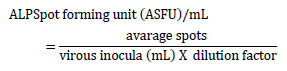
ALPSpot detection assay: MA-104 cells were transferred into 24-well tissue culture plates with 30X104 cells ml-1 density and incubated for 16 h in a cell culture chamber. Cells were washed twice with M199 media and treated with conditioned media for 3 h before the virus inoculation. Enzymatically activated (10 μg/mL of trypsin) and log10 serially diluted (10-1 to 10-6) working virus stocks were kept in wells in a triplicate manner with 0.2 mL volume. PBS-mock diluted and undiluted inoculums were included to plate for assay controls. The plate was kept at 37°C for 90 min and followed up by hand-shaking with 30 min intervals. Inoculums were replaced with overlay media (2% carboxymethyl cellulose growth media was equally mixed with 2X M199 and supplemented with the 1X antibiotic mix; 1% of FBS; 0.5 μg/ml trypsin). Agar overlay was maintained in the wells for the next three days in the cell culture chamber. The cells were formalin-fixed (10%) at RT for 30 min without removing overlay media. Then, wells were washed with PBS. Fixed cells were permeabilized with 1% of Nonidet-P 40 in PBS (perm-wash solution) for 15 min at RT. Cells were incubated with 5% skimmed milk containing Tris-buffered (100 mM) PBS (pH 8.0) at RT for 30 min for the blocking. The blocking solution was washed away with a perm-wash solution to clear the milk residues. To detect surface-expressed viral antigens, a polyclonal mouse serum (obtained from Erciyes University Faculty of Veterinary Medicine, Department of Virology), which was raised against the whole inactivated and alum adjuvant virus (1:25600 IgG titer), was used. Sera were diluted (1/2500; v:v) in perm-wash solution and incubated with cells at RT for 45 min by slow platform agitation. Wells were washed three times and incubated with ALP-conjugated anti-mouse IgG (SIGMA, St Louis, MO) antibody (1/2000; v:v) in perm-wash for 60 min at RT by slow platform agitation. Wells were carefully washed twice with a perm-wash solution and subsequently reacted with alkaline phosphatase buffer (100 mM Tris, 100 mM NaCl, and 5 mM MgCl2 containing alkali-pH 9.5 PBS). The chromogenic substrate NBT/BCIP (Thermo, USA) was added to wells and immediately kept in a dark place for color development. The incubation was stopped when the desired color development was observed under the inverted microscope, which varied between 15 min and 1 h. The following formula was used to enumerate virus titers:
Home-made sandwich ELISA for detection of BRV: Polyclonal rabbit anti-BRV antibody (obtained from Erciyes University Faculty of Veterinary Medicine, Department of Virology) was diluted (1 μg/ml) in a carbonate buffer (pH 9.6) and mobilized in detachable 96-well plates (Thermo, USA) overnight. Wells were washed twice with PBST-20 buffer, then subsequently blocked with skimmed milk (5%) for 1 h at 37°C. The plate was extensively washed with PBST-20 (0.01% Tween-20 in PBS) five times. Test samples were collected from infected cell supernatants at the fifth dpi and diluted in blocking buffer (1:10; v:v) before being added to wells consecutively. The plate was incubated at 37°C for 1 h. The plate washed as described previously, and HRP-conjugated monoclonal VP6 antibody (1D6 VP6 mAb; obtained from Erciyes University Faculty of Veterinary Medicine, Department of Virology) was used for the detector antibody (1:3000). The plate was incubated as before and subsequently washed with PBS-T buffer. The HRP substrate was prepared with the dissolving of a TMB tablet (1 mg; SIGMA, St Louis, MO) in DMSO followed by dilution in PBS up to 10 mL as described in the instructions. Peroxidase (30%, H2O2) was added (0.1%) to substrate buffer before use and the plate was incubated in a dark place for 10 min at RT for chromogenic color development. The reaction was stopped by adding 2 M H₂SO₄ to the wells. The plate was read with a spectrophotometer set to 450 nm (Allsheng, China) to obtain the optical densities. The cut-off value of the ELISA was calculated by three times the multiplication of negative control absorbance values.
Validation of ALPSpot assay: To validate the ALPSpot assay sensitivity, RT-PCR was confirmed, and cell culture-isolated virus stocks were tested in an ALPSpot titration assay along with TCID50 virus titration. ALPSpot titers were obtained and statistically compared to the TCID50 titer results to determine the ALPSpot assay sensitivity. For this purpose, fecal samples were briefly diluted in PBS (1:10 v:v) and clarified in a centrifuge at 10,000 × g for 30 min. The upper-clear phase was filter-sterilized (0.22 μm) and equally mixed with M199 cell media supplemented with 2X antibiotics. Virus cultivation was carried out as described above in MA-104 permissive cells with at least three blind passages (P0 to P3). Cell culture supernatants were harvested from each passage and tested using an ELISA to confirm virus propagation and isolation. The collected supernatants were molecularly confirmed with RT-PCR as described in this study. Cell culture-isolated BRV stocks were kept at -80°C for ALPSpot validation experiments, and virus titers were obtained both from TCID50 and ALPSpot assays.
Statistical analysis: Linear regression analysis was used to determine the correlation efficiency of the newly developed ALPSpot titer range of viral stocks by comparing it with the TCID50 titer for the accuracy of the assay. The correlation matrix of the linear regression analysis and graphical outputs were obtained by GraphPad software (GraphPad PRISM 7.0; La Jolla, CA). To visualize the ELISA results, OD values were obtained using a spectrophotometer set at 450 nm and assigned in grouped table format columns using GraphPad PRISM. To compare ALPSpot assay with TCID50 method, a non-parametric Wilcoxon two-sample paired rank test was performed for not normally distributed titer data (22) using GraphPad software (n=7). The significance level was considered as p<0.05 for all analyzes. To obtain TCID50 titers, a tabulated Excel TCID50 calculator based on the SpearmanKärber method was used as defined and validated previously 23.


)
)
)
)
)
)
)
