[ Ana Sayfa | Editörler | Danışma Kurulu | Dergi Hakkında | İçindekiler | Arşiv | Yayın Arama | Yazarlara Bilgi | E-Posta ]
Fırat Üniversitesi Sağlık Bilimleri Tıp Dergisi
2023, Cilt 37, Sayı 1, Sayfa(lar) 022-028
[ Özet ] [ PDF ] [ Benzer Makaleler ] [ Yazara E-Posta ] [ Editöre E-Posta ]
Non-Fermantatif Gram Negatif Bakterilerde Biyofilm Oluşumu ve Antibiyotik Duyarlılıklarının Belirlenmesi
Muhammed Fatih TURSUN1, Pınar ÖNER2, Zülal AŞÇI TORAMAN3
1Veterinary Control Institute, Biology, Elazığ, TÜRKİYE
2Elazığ Fethi Sekin City Hospital, Department of Microbiology, Elazığ, TÜRKİYE
3Firat University, Faculty of Medicine, Department of Microbiology, Elazığ, TÜRKİYE
Anahtar Kelimeler: Biyofilm, non-fermentatif Gram-negatif bakteri, kongo kırmızılı agar, standart tüp, mikropleyt
Özet
Amaç: Bu çalışmada çeşitli klinik örneklerden izole edilen Non-Fermantatif Gram Negatif (NFGN) bakterilerde biyofilm üretimi ve antibiyotik duyarlılıklarının belirlenmesi amaçlandı.

Gereç ve Yöntem: Çalışmaya, hastanemiz Mikrobiyoloji Laboratuvarı’na gönderilen 81 yara, 29 idrar, 12 steril vücut sıvısı, 12 kan, 16 solunum yolu örneğinden izole edilmiş, konvansiyonel yöntemler ve VİTEK 2 (Biomerieux, Fransa) otomatize sisteminde tanımlanmış toplam 150 adet NFGN bakteri (Pseudomonas aeruginosa (P. aeruginosa)) (42), Acinetobacter baumanii (A. baumanii) (104), Burkholderia cepacia (B. cepacia) (4)) dahil edildi. Biyofilm oluşumunun belirlenmesinde Kongo Kırmızılı Agar (KKA), Standart Tüp (ST) ve Mikropleyt (MP) yöntemleri kullanıldı. Antibiyotik duyarlılıkları CLSI önerileri doğrultusunda disk difüzyon yöntemi ile araştırıldı.

Bulgular: 150 adet NFGN bakteri izolatının KKA yöntemine göre 71(%47.33)’i biyofilm pozitif olarak tespit edildi. Kristal viyole boyası kullanılarak yapılan ST yöntemine göre izolatların 57(%38)’si, MP yöntemine göre 61(%40.7)’i biyofilm pozitif olarak tespit edildi. Safranin boyası kullanılarak yapılan ST yöntemine göre izolatların 65(%43.3)’i, MP yöntemine göre 83(%55.3)’ü biyofilm pozitif olarak tespit edildi. Biyofilm pozitif olan A. baumanii suşlarının amoksisilin klavulonik asite %88.89, trimetoprim sulfametaksazole %87.04 oranında direnç gösterdiği tespit edildi. Biyofilm pozitif olan P. aeruginosa suşlarının amoksisilin klavulonik asite %82.86, trimetoprim sulfametaksazole %85.72 oranında direnç gösterdiği tespit edildi. B. cepacia izolatlarının kolistin ve sefoperazon-sulbaktam hariç tüm antimikrobiyallere %100 direnç gösterdiği tespit edildi.

Sonuç: Biyofilm oluşumunun belirlenmesine yönelik yapılan test yöntemleri arasında gold standart test olarak gösterilen mikropleyt yöntemi bizim çalışmamızda da en yüksek biyofilm pozitiflik oranına sahip yöntem olarak belirlendi.

  • Başa Dön
  • Özet
  • Giriş
  • Materyal ve Metot
  • Bulgular
  • Tartışma
  • Kaynaklar
  • Giriş
    Non-fermentative gram-negative (NFGN) bacteria cause infections as opportunistic pathogens in immunocompromised patients and particularly in patients receiving drug threapy 1. These microorganisms are present in the respiratory tract flora and oral flora, especially the bacterial flora of the skin in humans. In addition to being found in soil, water, and moist environments in nature, they are commonly found in hospital environments. NFGN bacteria are a group of organisms that can grow under minimal growth conditions and vary in virulence, and they tend to carry acquired drug resistance. This group includes the genera Pseudomonas, Stenotrophomonas, Acinetobacter, Burkholderia, Flavobacterium, and Alcaligenes 2.

    Biofilm is a tightly networked, immobile, and communicating cell community formed by microorganisms, embedded in an extracellular organic matrix in the polysaccharide structure they produce, clinging to each other and a solid surface. The biofilm structure, which differs from planktonic forms in terms of both growth and gene expression and antibiotic sensitivity, is more resistant than the planktonic forms and has a high relapse rate after treatment, and is a source of chronic infection in the host 3. It is known that approximately 99% of bacteria survive by forming biofilms, and only 1% are free or planktonic, and biofilm formation is responsible for at least 65% of bacterial infections in humans 4. Biofilm formation is known to have an important role in many diseases, especially in natural valve endocarditis, osteomyelitis, catheter-related blood circulation and urinary system infections, periodontitis, middle ear infections, and particularly chronic lung diseases with cystic fibrosis 5.

    While the risk of infection can be reduced by destroying free bacteria in the body with antibiotics, the bacteria in the biofilm often get rid of the host response and often show resistance to antibiotic treatment 6. It has been reported that biofilm formation has been observed since the antibiotic treatment was stopped. In In-vitro tests, it was found that biofilm-forming bacteria were resistant to antibiotic concentrations several hundred or thousand times higher than the minimum inhibition concentration (MIC) determined for free bacteria 4. Various experimental models in which resistance genes are transferred among microorganisms contained in a biofilm have also been demonstrated 7. Therefore, biofilm-induced infections are one of the most leading causes of treatment failure today 18.

    This study aims to determine the biofilm formation and resistance to antibiotics in NFGN bacteria, which are known as opportunistic pathogens, causing hospital infections with high mortality and morbidity.

  • Başa Dön
  • Özet
  • Giriş
  • Materyal ve Metot
  • Bulgular
  • Tartışma
  • Kaynaklar
  • Materyal ve Metot
    Research and Publication Ethics: This study was carried out after obtaining the Clinical Research Ethics Committee of Firat University's approval, with the protocol number 2010/12-12 dated 02/11/2010.

    This study was carried out using conventional methods and isolated from 81 wounds, 29 urine, 12 sterile body fluids, 12 blood, 16 respiratory tract samples sent to the Microbiology Laboratory of Firat University Faculty of Medicine Hospital. A total of 150 NFGN bacteria, identified in the VITEK 2 (Biomerieux, France) automated system; Pseudomonas aeruginosa (P. aeruginosa) (42), Acinetobacter baumannii (A. baumanii) (104), Burkholderia cepacia (B. cepacia) (4) were included. CRA, ST, and MP methods were used to determine the phenotypic biofilm formation of the identified strains.

    Determination of biofilm presence by the MP method: In order to determine the biofilm formations quantitatively, P. aeruginosa, A. baumannii, S. maltophilia, and B. cepacia strains were taken from the stock culture and incubated overnight at 37°C in the BHIB containing 0.25% glucose to be activated. As a result of incubation, it was diluted at a rate of 1:20 with freshly prepared and pre-warmed with BHIB with 0.25% glucose. Then 200 μl was added to each well of a sterile 96-well flat-bottomed microplate and incubated at 37°C for 24 hours. The negative control was filled only with a liquid medium. After incubation, the wells were emptied, and the wells were washed 3 times with PBS. In the study conducted in two groups, 200 μl 0.1% crystal violet was placed in the first group of microplates that were dried by inversion. In the second group, 200 μl of the prepared 0.1% saffron solution was placed. Both microplates were incubated at room temperature for 15 minutes. After the staining period, both groups were washed again with PBS (PH 7.2) 3 times and left to dry by turning them upside down on the blotting paper. Then a 200 μl (80:20) ethanol-acetone mixture was transferred to each well of the first group of plates stained with crystal violet, and the microplates were read at 590 nm in the spectrophotometer device. 200 μl of 50% acetic acid solution was added to the wells of the second group of microplates painted with 0.1% saffron and read at 470 nm wavelengths in the spectrophotometer device. According to optical density values, the results were evaluated as 1 positive (+), 2 positive (++), 3 positive (+++) and negative (-) (Figure 1, 2) 9. Since the phenotypic expression in biofilm formation is highly susceptible to change under in vitro conditions, each test was run twice to minimize errors and ensure the reliability of the information.


    Büyütmek İçin Tıklayın
    Figure 1: Biofilm formation in the microorganisms isolated by the MP (Crystal violet) method


    Büyütmek İçin Tıklayın
    Figure 2: Biofilm formation in the microorganisms isolated by the MP (Safranine) method

    Determination of Biofilm Presence by the ST Method: The modified tube adherence method, also known as the Christensen method 10, was used for the qualitative determination of biofilm formation. NFGN bacteria were inoculated in Brain Heart Infusion Broth (BIBI) and incubated at 37 °C for 24-48 hours. After incubation, the tube contents were emptied and washed with phosphate buffer solution (PBS). In the study, which was carried out in two sets, an equal volume of 3 ml of safranine or crystal violet was placed in each tube and mixed slowly. After waiting for a while, the paint in the tube was poured and the emptied tubes were left to dry by turning them upside down on blotting paper. The presence of a colored layer on the inner wall of the tubes was accepted as ibofilm positive, and the biofilm-forming capacity of the strains was graded as very strong, strong, and weak according to the darkness and thickness of the color. The absence of color change was recorded as a negative result (Figure 3-6). In addition, the result was evaluated as negative despite the presence of dye residue in the part where the medium came into contact with air.


    Büyütmek İçin Tıklayın
    Figure 3: Biofilm formation in the microorganisms isolated according to the ST (Crystal Violet) Method. 1: Biofilm (+++) positive. 2: Biofilm (++) positive. 3, 4: Biofilm (+) positive. 5: Biofilm negative


    Büyütmek İçin Tıklayın
    Figure 4: Biofilm-positive samples in the microorganisms, isolated according to the ST (Crystal Violet) method


    Büyütmek İçin Tıklayın
    Figure 5: Biofilm formation in the microorganisms isolated according to the ST (Safranine) method. 1, 2, 3: Biofilm positive samples. 4: Biofilm negative sample


    Büyütmek İçin Tıklayın
    Figure 6: Biofilm-positive samples in the microorganisms isolated according to the ST (Safranine) method

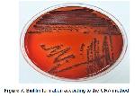
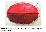
    Determination of Biofilm Presence by the CRA Method: NFGN bacterial strains were inoculated on CRA media using the single colony method. After sowing, the plates were incubated at 37ºC for 48 hours. The presence of bright black colonies on the plates after the incubation period was evaluated as biofilm (+) and the presence of pink colonies was evaluated as biofilm (-) (Figure 7, 8).


    Büyütmek İçin Tıklayın
    Figure 7: Biofilm formation according to the CRA method


    Büyütmek İçin Tıklayın
    Figure 8: According to the CRA method; 1: Biofilm negative, 2: Biofilm positive

    Antibiotic Susceptibility Tests: The routine antibiotic susceptibilities of bacteria isolated at the end of sufficient incubation periods and defined at the species level were checked in sheep blood and EMB media. Their susceptibility was investigated by the disc diffusion method in line with CLSI recommendations. Müller-Hinton agar was used for antibiotic susceptibility tests.

  • Başa Dön
  • Özet
  • Giriş
  • Materyal ve Metot
  • Bulgular
  • Tartışma
  • Kaynaklar
  • Bulgular
    A total of 150 NFGN strains (A. baumannii 104 (69.33%), P. aeruginosa 42 (28%), B. cepacia 4 (2.67%), isolated from 81 (54%) wounds, 29 (19.33) urine, 12 (8%) sterile bodily fluids12 (8%) blood, 16 (10.67%) respiratory samples were included in the study.

    It was observed that the isolates formed the highest rate of biofilm 83 (55.33%) with the MP (Safranine) method. Also, it was observed that the isolates with strong positive biofilm were formed with the MP (Safranine) method at the highest rate. The comparison of biofilm positivity according to methods is shown in Table 1.


    Büyütmek İçin Tıklayın
    Table 1: Biofilm positivity of isolates according to the methods used.

    Biofilm positivity was detected in 54 (51.9%) of A. baumannii and 28 (66.6%) of P. aeruginosa strains by the MP (Safranine) method. The biofilm distribution of the isolates included in the study according to the methods used in included is shown in Table 2.


    Büyütmek İçin Tıklayın
    Table 2: Biofilm distribution of isolates according to the methods used

    Biofilm positivity was detected in 54 (66.6%) of 81 wound samples by the ST (Safranine) method. Biofilm positivity was detected in 19 (65.5%) of 29 urine samples by the MP (Safranine) method. The distribution of biofilm results, according to the methods used and laboratory samples, is shown in Table 3.


    Büyütmek İçin Tıklayın
    Table 3: Distribution of biofilm results according to the methods used and laboratory samples.

    Acinetobacter baumannii isolates were found to be 35.18% sensitive to CT, 35.18% to SCF, and 88.89% resistant to AMC. It was determined that P. aeruginosa isolates were 57.14% sensitive to CT and 75% resistant to imipenem (IPM). The antimicrobial susceptibility distribution of biofilm-positive isolates according to the MP (Safranine) method is shown in Table 4.


    Büyütmek İçin Tıklayın
    Table 4: Antimicrobial susceptibility distribution of biofilm-positive isolates according to MP (Safranine) method.

  • Başa Dön
  • Özet
  • Giriş
  • Materyal ve Metot
  • Bulgular
  • Tartışma
  • Kaynaklar
  • Tartışma
    This study showed that the highest rate of biofilm formation with A. baumannii and P. aeruginosa isolates were detected by MP (Safranine) method.

    NFGN bacteria are among the leading agents of community-based infections, particularly hospital infections 11. Nosocomial infections, especially in patients hospitalized in intensive care units, are difficult to treat and have high mortality. In cases with inappropriate antibacterial therapy, mortality rates of serious infections such as ventilator-associated pneumonia (VIP) and bacteremia reach 60% 12. The most important virulence agents of NFGN bacterial species are biofilm formation 13. Biofilm is an important structure for bacterial adherence, antibiotic resistance, and phagosytosis. The fact that bacteria come together to form a community and adapt to the environment through gene modulation (Quorum Sensing) has further increased the importance of biofilm structure 14.

    There are different methods used to detect the biofilm formation. In our study comparing biofilm formation in NFGN bacteria with different methods (MP, ST, CRA) and investigating antibiotic resistance developing in the biofilm media, 150 NFGN bacterial strains isolated from various clinical samples were evaluated. The biofilm formation of 17 A. baumannii strains isolated from different hospitals in Turkey by Can F. et al. was examined by quantitative method and biofilm formation was observed in 9 of the isolates 15. In a study conducted by Wang et al., biofilm formation was investigated in 273 A. baumannii isolates using the MP method and 71 (26%) isolates were observed to be biofilm positive 16. Yassein et al. detected the presence of biofilms in 20 of the 50 P.aeruginosa strains isolated from clinical samples 17. Rodríguez-Baño et al. investigated the biofilm formation in 92 A. baumannii isolates by the microtitration plate method and found that 56 (63%) insolates formed biofilms 18. Delissalde et al. investigated the biofilm presence in P. aeruginosa isolates, isolated from 162 clinical samples using the MP method, and found 18% biofilm presence at the end of 24 hours of incubation 19. Abdi-Ali et al. detected biofilm formation at the rate of 80% with the ST method and 75% with the MP method in 75 A. baumannii isolates 20. Considering the distribution of biofilm positivity of clinical samples included in our study according to different methods, MP (Safranine) was determined to be 83 (55.33%), MP (Crystal violet) 61(40.6%) 6), CRA 71 (47.33%), ST (Safranine) 65 (43.33%), ST (Crystal violet) 57 (38%). Our study results are in line with other studies on the subject, and it is considered that the differing results are due to the fact that the biofilm formation process is a quite dynamic process, besides various environmental factors such as heat, temperature, pH, and nutrient concentration in the environment, biofilm formation is basically a similar process in general terms. However, it is considered that this may be due to the fact that it contains significant differences between the species and strains it contains in terms of formation steps.

    In a study, the biofilm-forming properties of 60 A. baumannii isolates isolated from sputum, wound, and catheter samples were evaluated by the MP (crystal violet) method and it was determined that 10 (16.6%) of the 60 isolates had weak biofilms, 19 (31.6%) had moderately strong biofilm and 31 (51.6%) of them formed strong biofilm 21. In the study conducted by Babapour et al., 10.26% of 156 A. baumanii isolates were detected as biofilm positive by the CRA method, while the percentage of bacteria forming biofilm positive with ST, MP, and modified MP methods was determined as 48.72%, 66.66% and 73.72%, respectively 22. In another study, biofilm formation was qualitatively observed in 34 of 55 A. baumannii isolates, with the ST method, and strong biofilm formation was detected in 34 isolates and weak biofilm formation in 14 isolates by the MP method quantitatively 23. Our study with the MP method and ST method and two different dyestuffs used in these two methods is in parallel with other similar studies. In our study, biofilm formation was also evaluated by the CRA method, and 71 (47.33%) of the isolates were found to be biofilm positive and 79 (52.67%) biofilm negative. The majority of clinical samples in our study consisted of wound samples from the plastic surgery clinic (54%). NFGN bacteria are considered to have a higher risk of transmission from wounds than the other ways of transmission. Biofilm formation was determined in 47 (58%) of 81 wound samples by the CRA method and 54 (66.67%) by the ST (Safranine) method, and no biofilm formation was observed in the sterile body fluid samples. In our study, the highest biofilm positivity in A. baumannii and P. aeruginosa strains was determined by MP (Safranine) method, being 54 and 28, respectively. In addition, strong positive (+++) biofilm-forming strains were obtained with the MP (Safranine) method at the highest rate. Biofilm formation was detected in 44 (42.3%) of A. baumannii strains and 25 (59.5%) of P. aeruginosa strains by the CRA method. In the study conducted by Harika et al., the highest biofilm positivity rate in P. aeruginosa strains was obtained with the MP method, and either weak positivity or negative results were obtained with the ST and CRA methods 24. There are studies in which ST 25 and CRA 26 methods are not recommended to identify biofilm-producing isolates.

    According to some studies conducted in our country, resistance to carbapenems, which are usually used as the first choice in treatment, has been detected at levels of 90-94% in Acinetobacter and 25-33% in Pseudomonas 27,28. In the study of Rao et al., biofilm positive A. baumannii isolates showed 100% resistance to IPM, 89% to cephotaxime, 80% to AK, and 73% to CIP 23. In our study, the imipenem resistance rates of biofilm-positive strains were 75% in P. aeruginosa and 100% in B. cepacia, while this rate was 85.19% in A. baumannii. According to these results, it is understood that carbapenem-resistant strains are more likely to produce biofilms compared to carbapenem-sensitive strains. While the imipenem resistance in our study was consistent with other studies for A. baumannii, it was concluded that the percentage of P. aeruginosa was higher than the ones in other studies. Resistance levels vary by geographic region. For instance, between 1997 and 2000, gentamycin resistance in P. aeruginosa was 15.8% in North America, compared to 28.3% in Europe and 38.2% in Latin America 29. These differences between the data obtained suggest that the studies were conducted at different times and in different locations, the clinics where the samples were isolated are different, and the bacteria are becoming more resistant by the day. Since resistance rates may vary depending on the location and usage times, it is a natural result that the resistance rate increases day by day. In our study, biofilm-positive A. baumannii strains were found to have high levels of resistance to all antibiotics, especially AMC (88.89%). In contrast, A. baumannii strains were shown to be sensitive to CT and SCF with a maximum of 35.18%. The study, conducted by Schaber et al., showed that biofilm-positive P. aeruginosa strains became approximately 10 times more resistant to imipenem, betalactam, gentamicin, and piperacillin-tazobactam antibiotics than the planktonic form 30. In our study, it was concluded that there is a positive correlation between biofilm formation and multidrug resistance in NFGN microorganisms. As colistin resistance increases in A. baumannii strains, an increase in biofilm production capacity is observed 31. In this study, colistin resistance was determined as 43.37% according to the antimicrobial susceptibility results determined by the disc diffusion method of biofilm-positive strains. This study indicated that biofilm-positive Pseudomonas strains showed the most sensitivity to CT (57.14%) and SCF (53.57%), while they showed the most resistance to AMC (92.86%). Dizbay et al. reported resistance rates as 49% for meropenem, 62% for ceftazidime, and 56% for SXT for B. cepacia strains, which they identified as nosocomial infection agents in their five-year study 32. In our study, it was determined that B. cepacia strains showed 100% resistance to all antibiotics except SCF (25%).

    In conclusions today, the intensive or unnecessary use of antibiotics cause the formation of resistant bacteria. One of the leading causes of hospital infections, the non-fermentative gram-negative bacteria are protected from both host defense and antimicrobial agents by forming a biofilm, which may be considered as an important virulence factor. In this study, we have determined that the best method to show the formation of biofilm in NFGN bacteria is the MP (Safranine) method.

    Conflict of Interest: The authors declare that there is no conflict of interest regarding this article.

  • Başa Dön
  • Özet
  • Giriş
  • Materyal ve Metot
  • Bulgular
  • Tartışma
  • Kaynaklar
  • Kaynaklar

    1) Buzilă ER, Năstase EV, Luncă C, et al. Antibiotic resistance of non-fermenting Gram-negative bacilli isolated at a large Infectious Diseases Hospital in North-Eastern Romania, during an 11-year period. Germs 2021; 11: 354-362.

    2) Ozenen GG, Bal ZS, Umit Z, et al. Nosocomial Non-fermentative gram negative bacteria bloodstream infections in children; Risk factors and clinical outcomes of carbapenem resistance. J Infect Chemother 2021; 27: 729-735.

    3) Gedefie A, Demsis W, Ashagrie M, et al. Acinetobacter baumannii Biofilm Formation and Its Role in Disease Pathogenesis: A Review. Infect. Drug Resist 2021;14: 3711.

    4) Pontes JTCd, Borges ABT, Roque-Borda CA, Pavan FR. Antimicrobial peptides as an alternative for the eradication of bacterial biofilms of multi-drug resistant bacteria. Pharmaceutics 2022;14: 642.

    5) Donlan RM, Costerton JW. Biofilms: Survival Mechanisms of Clinically Relevant Microorganisms. Clinical Microbiology Reviews 2002; 15: 167-193.

    6) Sindhu S. Clinical significance, antibiotic resistance and biofilm formation of Acinetobacter baumannii. Rev Clin Microbiol 2018; 7(4): 1-2.

    7) Azizi O, Shahcheraghi F, Salimizand H, et al. Molecular analysis and expression of bap gene in biofilm-forming multi-drug-resistant Acinetobacter baumannii. Rep Biochem Mol Biol 2016; 5: 62-72.

    8) İştar EH, Alışkan HE, Başustaoğlu A. Metisiline duyarlı ve dirençli Staphylococcus aureus izolatlarının biyofilm oluşturma özelliklerinin konvansiyonel ve moleküler yöntemlerle belirlenmesi. Mikrobiyol Bul 2020; 54: 223-234.

    9) Stepanović S, Vuković D, Hola V, et al. Quantification of biofilm in microtiter plates: overview of testing conditions and partical recommendations for assessment of biofilm production by staphylococci. APMIS 2007; 115: 891-899.

    10) Christensen GD, Simpson WA, Younger JJ, et al. Adherence of coagulase-negative staphylococci to plastic tissue culture plates: A quantitative model for the adherence of staphylococci to medical devices. J Clin Microbiol 1985; 22: 996-1006.

    11) Tucaliuc D, Alexa O, Tuchiluş CG, et al. Antibiotic resistance spectrum of non fermenting Gram negative bacilli isolated in the Orthopedic Traumatology Clinic of “Sf. Spiridon” Clinical Emergency Hospital Iaşi. Rev Med Chir Soc Med Nat Iasi 2015;119: 536-543.

    12) Bacakoğlu F, Ekren PK, Taşbakan MS, ve ark. Solunumsal yoğun bakım ünitesinde çoklu antibiyotik dirençli Acinetobacter Baumannii enfeksiyonu. Mikrobiyol Bul 2009; 43: 575-585.

    13) Kovalchuk VP, Nazarchuk OA, Burkot VM, et al. Biofilm forming activity of non-fermenting gram-negative bacteria. Wiad Lek 2021; 74: 252-256. 14. Erdal B, Yalınay M, Elmas Ç, Yazıcı GN. Investigation of Pseudomonas aeruginosa biofilm formation and quorum sensing genes in piperacillin/tazobactam and ciprofloxacin sub-minimal inhibitory concentrations. Mikrobiyol Bul 2020; 54: 547-558.

    15) Can F, Kurt-Azap Ö, Demirbilek M, et al. Biofilm Formation By Acinetobacter Baumannii Strains Isolated in Hemocultures. Turkish Journal of Infection 2006; 20: 159-163.

    16) Wang YC, Huang TW, Yang YS, et al. Biofilm formation is not associated with worse outcome in Acinetobacter baumannii bacteraemic pneumonia 2018; 8: 7289.

    17) Yassein M, Khardori N, Ahmedy A, Toama M. Modulation of Biofilms of Pseudomonas aeruginosa by Quinolones. Antimicrobial Agents and Chemotheraphy 1995; 39: 2262 -2268.

    18) Rodríguez-Baño J, Martí S, Soto S, et al. Biofilm formation in Acinetobacter baumannii: associated features and clinical implications. Clin Microbiol Infect 2008; 14: 276-278.

    19) Delissalde F, Amabile-Cuevas CF. Comparison of antibiotic susceptibility and plasmid content, between biofilm producing and non-producing clinical isolated of Pseudomonas aeruginosa. Int J Antimicrobic Agents 2004; 24: 405-408.

    20) Abdi-Ali A, Hendiani S, Mohammadi P, Gharavi S. Assessment of biofilm formation and resistance to imipenem and ciprofloxacin among clinical 100 isolates of Acinetobacter baumannii in Tehran. Jundishapur J Microbiol 2014; 7: 1-5.

    21) Solmaz S. Acinetobacter ile İlişkili Kateter Enfeksiyonlarında Antibiyotik Kilit Tedavi Modelinin Değerlendirilmesi ve Biyofilm İlişkili Genlerin Araştırılması. Uzmanlık Tezi, Ankara: Gazi Üniversitesi Tıp Fakültesi, 2015.

    22) Babapour E, Haddadi A, Mirnejad R, Angaji S-A, Amirmozafari N. Biofilm formation in clinical isolates of nosocomial Acinetobacter baumannii and its relationship with multidrug resistance. Asian Pac J Trop Biomed 2016; 6: 528-533.

    23) Rao RS, Karthika Ru, Singh SP, et al. Correlation between biofilm production and multiple drug resistance in imipenem resistant clinical isolates of Acinetobacter baumannii. Indian J Med Microbiol 2008; 26: 333-337.

    24) Harika K, Shenoy VP, Narasimhaswamy N, Chawla K. Detection of biofilm production and its impact on antibiotic resistance profile of bacterial isolates from chronic wound infections. J Glob Infect Dis 2020; 12: 129-134.

    25) Hassan A, Usman J, Kaleem F, et al. Evaluation of different detection methods of biofilm formation in clinical isolates. Braz J Infect Dis 2011; 15: 305-311.

    26) Knobloch JK, Horsetkotte MA, Rohde H, Mack D. Evaluation of different detection methods of biofilm formation in Staphylococcus aureus. Med Microbial Immunol 2002; 191: 101-106.

    27) Karapınar BA, Yeşiloğlu C, Durmuş MA, ve ark. Alt solunum yolu örneklerinden izole edilen pseudomonas aeruginosa direnç profilinin incelenmesi. Bozok Med J 2020; 10: 48-52.

    28) Cesur S, Irmak H, Yalçın NA, et al. Yoğun bakım ünitesinde yatan hastaların çeşitli kültür örneklerinden izole edilen Acinetobacter baumannii suşlarının antibiyotik duyarlılıkları. Ortadoğu Tıp Dergisi 2017; 9: 51-55.

    29) French GL. Antimicrobial resistance in hospital flora and nosocomial infections. In: Hospital Epidemiology and Infection Control, 3rd Edition, Mayhall CG (ed.), Lippincott Williams & Wilkins, Philadelphia, 2004:1613-1638.

    30) Schaber JA, Hammond A, Carty NL, Williams SC. Diversity of biofilms produced by quorum-sensing-deficient clinical isolates of Pseudomonas aeruginosa. J Med Microbiol 2007; 56: 738- 748.

    31) Zeighami H, Valadkhani F, Shapouri R, Samadi E, Haghi F. Virulence characteristics of multidrug resistant biofilm forming Acinetobacter baumannii isolated from intensive care unit patients. BMC Infect Dis 2019; 19: 1-9. 32. Dizbay M, Tunccan OG, Sezer BE, Aktas F, Arman D. Nosocomial Burkholderia cepacia infections in a Turkish university hospital: A five-year surveillance. J Infect Dev Ctries 2009; 3: 273-277.

  • Başa Dön
  • Özet
  • Giriş
  • Materyal ve Metot
  • Bulgular
  • Tartışma
  • Kaynaklar
  • [ Başa Dön ] [ Özet ] [ PDF ] [ Benzer Makaleler ] [ Yazara E-Posta ] [ Editöre E-Posta ]
    [ Ana Sayfa | Editörler | Danışma Kurulu | Dergi Hakkında | İçindekiler | Arşiv | Yayın Arama | Yazarlara Bilgi | E-Posta ]