[ Ana Sayfa | Editörler | Danışma Kurulu | Dergi Hakkında | İçindekiler | Arşiv | Yayın Arama | Yazarlara Bilgi | E-Posta ]
Fırat Üniversitesi Sağlık Bilimleri Tıp Dergisi
2024, Cilt 38, Sayı 1, Sayfa(lar) 048-053
[ Özet ] [ PDF ] [ Benzer Makaleler ] [ Yazara E-Posta ] [ Editöre E-Posta ]
Ön Çapraz Bağ Cerrahisi Geçiren Hastalarda Devam Eden Rotasyonel İnstabilite Nedeni Anterolateral Ligament
Hüseyin KÜRÜM1, Hacı Bayram TOSUN2, Orhan AYAS2, Adnan AKCAN1, Kübra KÜRÜM 3
1Ergani State Hospital, Department of Orthopaedics and Traumatology, Diyarbakır, TÜRKİYE
2Fethi Sekin Training and Research Hospital, Department of Orthopaedics and Traumatology, Elazığ, TÜRKİYE
3İnönü University, Turgut Özal Medical Centre Physiotherapy and Rehabilitation Department, Malatya, TÜRKİYE
Anahtar Kelimeler: Anterior cruciate ligament, anterolateral ligament, magnetic resonance imaging
Özet
Amaç: Ön çapraz bağ (ÖÇB) rekonstrüksiyonu, diz eklemine stabiliteyi geri kazandırmayı, işlevini iyileştirmeyi ve nihayetinde bireyin spor aktivitelerine katılmaya devam etmesini sağlamayı amaçlayan cerrahi bir prosedürdür. Bu çalışmanın amacı, tek başına ÖÇB ameliyatı yapılan hastalarda rerüptür ve rotatuar instabilite sorunlarını belirlemek ve bu sorunların Anterolateral ligament (ALL) ile ilişkisini ve fonksiyonel sonuçlar üzerindeki etkisini incelemektir.

Gereç ve Yöntem: 2021 ve 2023 yılları arasında 48 hastadan oluşan bir grup üzerinde retrospektif analiz yapılmıştır.

Bulgular: ÖÇB ve ALL yaralanması olan 9 (%69.2) hastada lateral menisküs hasarı görülürken, sadece ÖÇB yaralanması olan 3 (%8.6) hastada lateral menisküs yırtığı görüldü (p<0.001). Lysholm diz skorlama ölçeği sadece ÖÇB yaralanması olan grupta 85.60±5.57 iken hem ÖÇB hem de ALL yaralanması olan grupta 63.54±5.04 idi (p<0.001). Tegner aktivite düzeyi sadece ÖÇB yaralanması olan grupta 8.14±1.03 iken hem ÖÇB hem de ALL yaralanması olan grupta 5.62±1.19 idi (p<0.001).

Sonuç: Ameliyat öncesi muayenenin yetersizliği ve tüm hastalarda MRG'de ALL tanısının konulamaması, ameliyat sonrası Tegner aktivite düzeyleri ve Lysholm diz skoru gibi ölçekler kullanılarak değerlendirilen fonksiyonel sonuçlar üzerinde ciddi bir etkiye sahip olan bu yırtığın gözden kaçmasına neden olmuştur. Ayrıca, mevcut bilgilerimize dayanarak, bu çalışma ALL lezyonları ile birlikte yüksek oranda lateral menisküs yırtığı olduğunu gösteren ilk çalışmadır.

  • Başa Dön
  • Özet
  • Giriş
  • Materyal ve Metot
  • Bulgular
  • Tartışma
  • Kaynaklar
  • Giriş
    Injuries to the anterior cruciate ligament (ACL), such as strains and ruptures, are widely reported in physically active individuals. ACL reconstruction (ACLR) is a surgical procedure that aims to restore stability to the knee joint, improve its function, and ultimately enable the individual to resume participating in sports activities. Regarding developments in surgical procedures and a more comprehensive knowledge of ACL anatomy and biomechanics, one out of each four patients 1-3.

    The anterolateral ligament (ALL) is significant due to its recently discovery as a ligament and its potential contribution to the anterolateral rotational stability of the knee 4,5.

    The failure rates of ACL ranges in 0% to 14%, however the rates of rerupture fall around the range of 2% to 26%. The rates of failure gradually expand to an excessive extent; the frequency of these rates is significantly elevated in young adults, athletes, or adults with ligament laxity 6,7. Recent research has demonstrated that certain failures can be related to rotational instability resulting from the rupture of the ALL.

    The anterolateral ligament is a fibrous structure that extends in the anteroinferior orientation between the upper part of the tibia and the lower part of the femur. It includes of branches made up of tightly packed collagen fibers that link to the lateral meniscus at the joint level 8. The ALL's meniscal insertion is placed between the anterior horn and the body of the lateral meniscus (Most exactly, it extends from 36% to 41.9% of the lateral meniscal circumference). On average, its attachment length measures 5.6 mm 8,9.

    Standardized protocols may provide more effective identification of ALL lesions. The ideal sequence for identifying ALL on magnetic resonance imaging (MRI) is the coronal plane with proton density weighted. The ALL is seen as a slender linear structure wrapped by adipose tissue, while the meniscus part is observed as an isolated point on coronal MRI scans and it arises near the inferior genicular artery. To establish a better understanding of the anatomy of ALL, it is useful to analyze axial and sagittal pictures by recognizing the reference to ALL in the coronal plane 10,11.

    The aim of this study was to identify the problems of rerupture and rotatory instability in patients who performed alone ACL surgery, and to examine the correlation between these problems and the ALL, as well as their impact on functional outcomes.

  • Başa Dön
  • Özet
  • Giriş
  • Materyal ve Metot
  • Bulgular
  • Tartışma
  • Kaynaklar
  • Materyal ve Metot
    Research and Publication Ethics: Approval for the study was given by the local Ethics Committee (2023/12-06). Data collection and evaluation was done in accordance with the Declaration of Helsinki.

    In this study retrospective analysis was conducted on a group of 48 patients, ranging in age from 17 to 41 years, who performed surgery for anterior cruciate ligament rupture between the years 2021 and 2023. All patients were informed about the treatment and their written consent was obtained. The imaging and medical information were obtained from the electronic patient records using the Picture Archiving and Communication System (PACS) software at our institution.

    Inclusion And Exclusion Criteria: All patient became a standard radiological evaluation using MRI during admission. The inclusion criteria consists of patients who exhibited rupture of the ACL and/or ALL on MRI images, patients who reported experiencing a feeling of rotation and unloading in the knee, and patients who demonstrated at least one positive result on the anterior drawer, pivot shift, or lachman test during physical examination.

    The study excluded patients who had undergone anterior cruciate ligament surgery in the past, had a follow-up time of less than 1 year, encountered unsuccessful tunnel deployment, had postoperative infection, or had reduced knee range of motion before the operation.

    Rehabilitation: Each patient was scheduled for follow-up examinations on the 15th day, 1st month, 3rd month, 6th month, 1st year, and subsequently annually. During these appointments, all patients performed the same exercise program. Passive activities to flex and extend the knee, straight leg raises, heel slides against the wall, and quadriceps stretching exercises were initiated on the first day after the surgery. During the first 20-day period, the patient was walked using crutches with partial weight-bearing. The period of this time was 30 days in patients who underwent meniscal repair. Leg press workouts were initiated at the conclusion of the second month, with a steady increment in the weight. Patients were advised to avoid from high-impact activities, such as running, for the initial 3 months and from playing football until the 7th month, in order to reduce strain on the knee joint.

    The Lysholm knee scoring system, which ranges from 100 to 90 points for an excellent result, 89 to 80 points for a good result, 79 to 70 points for a fair result, and less than 70 points for a poor result, was utilized for clinical evaluation. Additionally, the Tegner activity scale, which ranges from 0 to 10, was also employed. The study recorded the presence of a meniscal tear along with the treatment method used for the tear, rates of retear, MRI findings including rupture of the ACL increased signal in the anterior root of the lateral meniscus, Segond fracture, and any additional meniscal tears. Persistent instability complaints and postoperative functional outcomes were also recorded.

    Surgical Technique
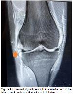
    ACL Repair: The surgical procedure was performed on all patients while they were under spinal anesthesia in a supine position. Tendons from the musculus semitendinosus and musculus gracilis were taken from the same knee. An arthroscope was introduced into the knee and the medial portal was used as the procedure portal. Removed fragments of damaged cruciate ligaments. Anatomic single bundle anterior cruciate ligament reconstruction surgery was carried out. The anatomical ACL footprint for the femoral tunnel was utilized. The positioning of the femoral tunnel was assessed following the insertion of the endobutton using radiographic imaging (Figure 1). The tibial tunnel was again created using the anatomical ACL footprint. The Biorci screw and U nail were utilized for fixing to the tibial tunnel. Following the graft insertion, the arthroscope demonstrated that the graft did not experience compression when the patient's knee was in either a flexed or extended position. Ultimately, the strain of the graft was examined using a probe.


    Büyütmek İçin Tıklayın
    Figure 1: Increased signal intensity in the anterior horn of the lateral meniscus in a patient with an ALL lesion

    Meniscus Repair: Concurrently with the rupture of the ACL, a partial meniscectomy was performed using an arthroscopic punch and shaver to address deteriorated tears in the white area that had a beak-like morphology and were not receptive to repair. In cases of non-displaced root lacerations, the injured ends were regenerated with a shaver and let to heal by conservative treatments. The Bucket handle, horizontal, displaced root tears, and tears in the red area were repaired with outside inside all inside, outside to inside, inside outside all inside techniques as well as with the assist of a suture passer.

    Statistical Analysis: Histogram and Q-Q plots were examined, Shapiro-Wilk’s test was applied to assess the data normality. Levene test was used to test variance homogeneity. To compare the differences between groups, either a two-sided independent samples t test were applied for continuous variables, Pearson’s χ2 test was applied for categorical variables. Analysis were conducted using R 4.3.2 softwares. Significance level was accepted as p<0.05.

  • Başa Dön
  • Özet
  • Giriş
  • Materyal ve Metot
  • Bulgular
  • Tartışma
  • Kaynaklar
  • Bulgular
    The study comprised a total of 48 individuals, consisting of 35 patients with isolated ACL injury and 13 patients with both ACL and ALL injuries. Out of the individuals, 39 (81%) were male and 9 (19%) were female. The average age of patients with only ACL damage was 24.89±7.36 years while patients with ACL and ALL injuries had an average age of 29.62±5.35 years (p<0.021). More demographic information is provided in Table 1.


    Büyütmek İçin Tıklayın
    Table 1: Baseline characteristics and demographic data

    MRI revealed oedema of the lateral tibial plateau in 13 (37.1%) individuals with just ACL damage and in 6 (46.2%) patients with both ACL and ALL injuries. This observation was statistically significant (p<0.001). 3 (23.1%) patients with ACL and ALL injuries were determined to have segond fractures (p<0.017). 9 (69.2%) patients with ACL and ALL injuries demonstrated lateral meniscal damage, while only 3 (8.6%) patients with ACL injury alone showed such damage (p<0.001). Additional MRI imaging results are provided in Table 2.


    Büyütmek İçin Tıklayın
    Table 2: MRI findings and concomitant meniscal lesion

    None of the patients with only ACL injury experienced a recurrent rupture. However, 3 (23.1%) patients with both ACL and ALL injury experienced a re-injury of the ACL (p<0.017) (table 2).

    The lysholm knee scoring scale was used to evaluate patients with ACL injury. Out of the total number of patients, 11 (31.4%) achieved outstanding outcomes, 18 (51.4%) achieved good results, and 6 (17.1%) achieved bad results. 2 (15.4%) with ACL and ALL injuries exhibited low result, while 11 (84.6%) patients demonstrated poor result (p<0.001) (Table 3). The lysholm knee scoring scale was 85.60±5.57 in the group consisting solely of ACL injuries and 63.54±5.04 in the group with both ACL and ALL injuries (p<0.001). The Tegner activity level was 8.14±1.03 in the group of individuals with just ACL injuries and 5.62±1.19 in the group of individuals with both ACL and ALL injuries (p<0.001) (Table 1).


    Büyütmek İçin Tıklayın
    Table 3: Lysholm Knee Scoring Scale

  • Başa Dön
  • Özet
  • Giriş
  • Materyal ve Metot
  • Bulgular
  • Tartışma
  • Kaynaklar
  • Tartışma
    The study associated chronic instability issues following ACL surgery to the ALL. The inadequacy of preoperative examination and lack of diagnosis of the ALL on MRI in all patients resulted in the oversight of this tear, which had a severe impact on the functional outcomes assessed using scales such as Tegner activity levels and Lysholm knee score post-surgery. Additionally, based on our present knowledge, this study is the first to show a high incidence of lateral meniscal tears in association with ALL lesions.

    Tavlo M. et al. 12 reported that anterolateral ligament injuries occurring with anterior cruciate ligament injuries can lead to ongoing rotational instability if only treated with anterior cruciate ligament reconstruction. ALL ruptures were seen in 13 out of 48 patients who underwent solitary anterior cruciate ligament restoration in the study. Every one of these patients experienced persistent otational instability issues. These patients were offered repeat surgery for postoperative ALL rupture. Before to ACL surgery, it is significant to appropriately detect and address any concurrent rupture of the ALL.

    MRI is the most effective imaging technique for evaluating ALL and other diseases among several diagnostic modalities. The MRI detection rate of ALL has been established to range from approximately 80% to 100% 10,13. MRI sections showed the development of ALL rupture in some patients during the research (Figure 2). Additionally, a total of 6 (46.2%) patients with ALL lesions and 13 (37.1%) patients with isloe ACL rupture exhibited enhanced signal at the lateral tibial plateau. This disparity was shown to be statistically significant. The limited ability to detect ALL rupture on MRI in all patients in the research was due to the use of 4 mm imaging slices. For individuals with ALL lesions, it is necessary to use MRI imaging with narrower slices, such as 2 mm.


    Büyütmek İçin Tıklayın
    Figure 2: Coronal MRI image of the anterolateral ligament

    Helito CB. et al. 8 observed that ALL spreads in an anteroinferior direction, extending between the proximal tibia and distal femur. They found that this region has branches composed of tightly wrapped collagen fibers that are attached to the lateral meniscus at the level of the joint. In the study, it was shown that 11 (84.6%) patients with ALL experienced an increased signal in the anterior horn of the lateral meniscus when examined using MRI (Figure 3). Lateral meniscal injury was more commonly detected in cases of ACL rupture accompanied by ALL lesion compared to cases of singular ACL rupture. In the study, the co-incidence rate of ACL rupture, ALL rupture and lateral meniscus tear triad was observed to be high.


    Büyütmek İçin Tıklayın
    Figure 3: Intraoperative AP-Lateral arthroscopic image of the knee

    The failure rates of ACL surgery range from 0% to 14%, whereas the rates of rerupture range from 2% to 26%. The frequency of these rates is elevated in young adults, athletes, or individuals with ligament laxity 6,7. Recent studies have established that ALL reconstruction significantly decreases the risk of ACL recurrence 14,15. In the study ACL rerupture reported in 3 (23.1%) participants with ALL lesions. ALL rupture leads to rotational instability and raises the probability of recurrent ruptures.

    Segond fracture referred to as bone avulsion, can be associated to ALL lesions originating from the proximal lateral tibia 16. Segond fracture was observed in 3 (23.1%) patients with ALL lesion in the study. The absence of segond fracture in all ALL lesions can be attributed to the fact that the rupture of the ALL was observed in the trunk region rather than at the tibial attachment site.

    Sonnery-Cottet et al. 17 shown that a portion of the ACL injury did not result in a substantial raise in tibial internal rotation at a flexion angle of 20°, but did lead to an increase in tibial internal rotation at 90°. Similarly, the study found that patients with ALL lesions exhibited higher levels of internal rotation and pain compared to the intact side during the internal rotation challenge test. This test was conducted with the knee flexed at a 90° angle while the person was lying down.

    The study utilized the Tegner activity level scale and Lysholm scoring scale to assess subjective complaints, measure satisfaction levels, and evaluate activity levels. Our study found that patients with only ACL rupture had significantly higher Lysholm score points and Tegner activity level scale scores compared to patients with both ACL and ALL rupture during the postoperative period. The Lysholm scoring scale and Tegner activity level scale scores following arthroscopic ACL restoration align with the findings reported in the literature 14,18,19.

    The study is limited by its retrospective design and the unequal and limited number of study groups.

    In conclusion, while there has been a notable rise in research on ALL in recent times, it is still possible for the diagnosis of ALL to be overlooked. Additionally, patients who undergo ACL surgery may continue to experience problems related to rotational movements, even after the surgery. This study has demonstrated that an undiagnosed rupture of the ALL is one of the contributing factors to the failure of ACL reconstruction. The study examined the impact of ALL on the functional results following ACL reconstruction, as well as its association with meniscal lesions and MRI findings. Successful anterior cruciate ligament surgery necessitates additional biomechanical investigations and long-term clinical trials of the anterolateral ligament.

  • Başa Dön
  • Özet
  • Giriş
  • Materyal ve Metot
  • Bulgular
  • Tartışma
  • Kaynaklar
  • Kaynaklar

    1) Chambat P, Guier C, Sonnery-Cottet B, Fayard JM, Thaunat M. The evolution of ACL reconstruction over the last fifty years. Int Orthop 2013; 37: 181-186. <2> Ristanis S, Stergiou N, Patras K, et al. Excessive tibial rotation during high-demand activities is not restored by anterior cruciate ligament reconstruction. Arthroscopy 2005; 21: 1323-1329.

    3) Tashman S, Collon D, Anderson K, Kolowich P, Anderst W. Abnormal rotational knee motion during running after anterior cruciate ligament reconstruction. Am J Sports Med 2004; 32: 975-983.

    4) Vundelinckx B, Herman B, Getgood A, Litchfield R. Surgical indications and technique for anterior cruciate ligament reconstruction combined with lateral extra-articular tenodesis or anterolateral ligament reconstruction. Clin Sports Med 2017; 36: 135-153.

    5) Ahn JH, Patel NA, Lin CC, Lee TQ. The anterolateral ligament of the knee joint: A review of the anatomy, biomechanics, and anterolateral ligament surgery. Knee Surg Relat Res 2019; 31: 12.

    6) Chen J, Xu C, Cho E, Huangfu X, Zhao J. Reconstruction for chronic ACL tears with or without anterolateral structure augmentation in patients at high risk for clinical failure: A randomized clinical trial. J Bone Jt Surg Am 2021;103(16):1482-1490.

    7) Crawford SN, Waterman BR, Lubowitz JH. Long-term failure of anterior cruciate ligament reconstruction. Arthroscopy 2013; 29(9): 1566-1571.

    8) Helito CP, Bonadio MB, Soares TQ, et al. The meniscal insertion of the knee anterolateral ligament. Surg Radiol Anat 2016; 38: 223-228.

    9) Kosy JD, Mandalia VI, Anaspure R. Characterization of the anatomy of the anterolateral ligament of the knee using magnetic resonance imaging. Skeletal Radiol 2015; 44: 1647-1653.

    10) Helito CP, Helito PV, Costa HP, et al. MRI evaluation of the anterolateral ligament of the knee: Assessment in routine 1.5-T scans. Skelet Radiol 2014; 43: 1421-1427.

    11) Patel KA, Chhabra A, Goodwin JA, Hartigan DE. Identification of the Anterolateral ligament on magnetic resonance imaging. Arthrosc Tech 2017; 6: e137-e141.

    12) Tavlo M, Eljaja S, Jensen JT, Siersma VD, Krogsgaard MR. The role of the anterolateral ligament in ACL insufficient and reconstructed knees on rotatory stability: A biomechanical study on human cadavers. Scand J Med Sci Sports 2016; 26: 960-966.

    13) Lee DW, Lee JH, Kim JN, et al. Evaluation of anterolateral ligament injuries and concomitant lesions on magnetic resonance imaging after acute anterior cruciate ligament rupture. Arthroscopy 2018; 34: 2398-2406.

    14) Sonnery-Cottet B, Saithna A, Cavalier M, et al. Anterolateral Ligament Reconstruction Is Associated With Significantly Reduced ACL Graft Rupture Rates at a Minimum Follow-up of 2 Years: A Prospective Comparative Study of 502 Patients From the SANTI Study Group. Am J Sports Med. 2017; 45(7):1547-1557.

    15) Sonnery-Cottet B, Thaunat M, Freychet B, Pupim BH, Murphy CG, Claes S. Outcome of a Combined Anterior Cruciate Ligament and Anterolateral Ligament Reconstruction Technique With a Minimum 2-Year Follow-up. Am J Sports Med. 2015; 43(7):1598-1605.

    16) Ferretti A, Monaco E, Fabbri M, Maestri B, De Carli A. Prevalence and classification of iınjuries of anterolateral complex in acute anterior cruciate ligament tears. Arthroscopy 2017; 33: 147-154.

    17) Sonnery-Cottet B, Lutz C, Daggett M, et al. THE Involvement of the Anterolateral Ligament in Rotational Control of the Knee. Am J Sports Med 2016; 44: 1209-1214.

    18) Hamido F, Habiba AA, Marwan Y, et al. Anterolateral ligament reconstruction improves the clinical and functional outcomes of anterior cruciate ligament reconstruction in athletes. Knee Surg Sports Traumatol Arthrosc 2021; 29(4): 1173-1180.

    19) Goncharov EN, Koval OA, Dubrov VE, et al. Clinical experience with combined reconstruction of the anterior cruciate and anterolateral ligaments of the knee in sportsmen. International Orthopaedics 2019; 43(12): 2781-2788.

  • Başa Dön
  • Özet
  • Giriş
  • Materyal ve Metot
  • Bulgular
  • Tartışma
  • Kaynaklar
  • [ Başa Dön ] [ Özet ] [ PDF ] [ Benzer Makaleler ] [ Yazara E-Posta ] [ Editöre E-Posta ]
    [ Ana Sayfa | Editörler | Danışma Kurulu | Dergi Hakkında | İçindekiler | Arşiv | Yayın Arama | Yazarlara Bilgi | E-Posta ]