[ Ana Sayfa | Editörler | Danışma Kurulu | Dergi Hakkında | İçindekiler | Arşiv | Yayın Arama | Yazarlara Bilgi | E-Posta ]
Fırat Üniversitesi Sağlık Bilimleri Tıp Dergisi
2024, Cilt 38, Sayı 1, Sayfa(lar) 078-084
[ Özet ] [ PDF ] [ Benzer Makaleler ] [ Yazara E-Posta ] [ Editöre E-Posta ]
Paroksetine Maruz Kalmış Yetişkin Dişi Sıçanlarda İrisin Uygulamasının Uterin Mast Hücreleri Üzerine Etkileri
Ahmet YARDIMCI1, Tuğrul ERTUĞRUL2, Nazife ÜLKER ERTUĞRUL3, Sinan CANPOLAT1
1Fırat University, Faculty of Medicine Department of Physiology Elazig, TURKIYE
2Ondokuz Mayıs University, Faculty of Veterinary Department of Histology and Embryology, Samsun, TURKIYE
3Samsun University, Faculty of Medicine Department of Physiology, Samsun, TURKIYE
Anahtar Kelimeler: Irisin, paroxetine, uterine mast cells, female reproductive system
Özet
Amaç: Paroksetin, kadın üreme sisteminde zararlı etkileri olduğu bilinen bir antidepresan olup irisin ise bu olumsuz etkileri hafifletmesi beklenen egzersiz-indüklü bir adipo-miyokindir. Bu çalışmada, paroksetinin ve/veya irisin'in yetişkin dişi sıçanlarda uterin mast hücreleri (uMH) sayısı ve uMH fizyolojisi için kritik olan ana üreme hormonları—17β-estradiol ve progesteron—üzerindeki etkilerinin incelenmesi amaçlanmıştır.

Gereç ve Yöntem: 40 yetişkin dişi sıçan dört gruba ayrıldı (her bir grup için n=10): sham kör, paroksetin, irisin ve paroksetin+irisin. Paroksetin ve paroksetin+irisin gruplarına sekiz hafta boyunca paroksetin (20 mg/kg/gün, oral gavaj ile), irisin ve paroksetin+irisin gruplarına ise mini-osmotik pompalar kullanılarak irisin (100 ng/kg/gün, subkütanöz olarak) sekiz haftalık çalışma sürecinin son dört haftası boyunca verildi. Serum hormon seviyeleri ELISA yöntemi kullanılarak ölçüldü. Uterus doku örnekleri, toluidine mavisi (TB) ve kombine alcian mavisi safranin O (AB/SO) teknikleri kullanılarak uMH'leri tespit etmek için histokimyasal boyamaya tabi tutuldu.

Bulgular: AB (+) uMH'lerin sayısı, paroksetin+irisin grubunda sham kör grubuna göre anlamlı olarak arttı (p<0.05). Yalnızca paroksetin tedavisi, paroksetin grubunda TB (+) uMH’lerin sayısını sham kör grubuna göre anlamlı olarak artırdı (p<0.01). Paroksetin+irisin grubu da TB (+) uMH sayısında anlamlı bir artış gösterdi (p<0.05). SO (+) uMH sayısı gruplar arasında anlamlı bir değişiklik göstermedi. Her iki uygulamaya bağlı olarak serum 17β-estradiol veya progesteron seviyelerinde ise herhangi bir grupta değişiklik gözlemlenmedi.

Sonuç: Görünüşe göre irisin uMH'ler veya alt tipleri üzerinde bir etkiye sahip değildir. Ancak, paroksetinin sıçan uterus dokusundaki belirli uMH alt tipleri üzerindeki etkisi, uterin mikroçevrenin düzenlenmesinde paroksetin indüklü serotonininin (5-HT) bir role sahip olabileceğine işaret etmektedir.

  • Başa Dön
  • Özet
  • Giriş
  • Materyal ve Metot
  • Bulgular
  • Tartışma
  • Kaynaklar
  • Giriş
    Mast cells (MCs), which constitute diverse cellular populations, perform crucial functions across numerous organs and systems, participating in a wide array of physiological processes 1. MCs, renowned as the principal reservoir of histamine within the body, arise from the bone marrow 2. Also, local MCs, found in rodents, generate, store, and discharge serotonin (5-HT) into the area outside blood vessels. This process is, to some extent, regulated by neural control 3. Generally, these cells pervade the connective tissue, frequently aligning themselves in proximity to both the blood vessels and lymphatics. Moreover, they are habitually located beneath the epithelial surface of the respiratory and gastrointestinal systems, in addition to the skin 2. Notably, MCs are also distributed within the female reproductive tissues including uterus 4. MCs participate in multiple reproductive activities in female, including reproduction, pregnancy, and labor 5. Analogous to MCs situated in diverse parts of the body, uterine mast cells (uMCs) include several mediators including cytokines, growth factors, and chemokines within their cytoplasmic granules 6. These mediators exert influence on inflammatory responses, which is necessary for a successful implantation of the blastocyst in the uterus and placentation 7.

    In addition to these roles, uMCs have the capacity to influence angiogenesis and regulate uterine contractions, both of which are integral to the process of parturition 6. Mast cells exhibit metachromasia upon staining with toluidine blue (TB) due to the presence of granules in their cytoplasm 8. They are classified into two subtypes according to several factors, including their physiological characteristics, staining properties, functional diversity, mediators they carry, and responses to foreign matter release 9. The two subtypes are known as mucosal mast cells (MMC) and connective tissue mast cells (CTMC) 10. When stained with granule-specific dyes, such as alcian blue (AB) and safranin O (SO), MMCs are stained by alcian blue, while CTMCs are stained by safranin O 11.

    Among women of reproductive age, one in seven use antidepressant medication, with selective serotonin reuptake inhibitors (SSRIs) being the most frequently prescribed class of these drugs (12). SSRIs primarily act upon the serotoninergic system by inhibiting the 5-HT transporter and augmenting extracellular 5-HT levels 13. SSRIs are suggested to affect MCs proliferation, activation, and granule formation 14. Paroxetine is known to be the second most commonly prescribed medication within this group in general 15. In a previous study conducted by our group, we found that it can induce various pathologies in the rat uterus 16. Given the significant role that mast cells play in the uterus, it is of paramount importance to understand the effects of paroxetine on these cells. However, despite the widespread use of this SSRI, there is a marked dearth of studies investigating its potential impact on the number of mast cells in the rat uterus, thus highlighting an unexplored avenue in the field of reproductive physiology.

    Irisin is an adipo-myokine that is released from skeletal muscle as a response to exercise 17. In previous studies by our team, we have shown that irisin has significant positive roles in the reproductive system in rodents 18,19. Recent research has indicated that it can beneficially influence a variety of reproductive disorders in females, as demonstrated in both preclinical and clinical studies. One study conducted by Li et al. 20 showed that irisin may enhance the poor receptive condition of the endometrium in rats with polycystic ovary syndrome. In another study, it was proposed that a rise in circulating irisin could act as an adaptive response to offset the heightened inflammation found in endometriosis. This idea is also supported by the observed positive correlation between irisin and C-reactive protein, in conjunction with the known anti-inflammatory properties of irisin 21.

    Given the influence of MCs on female reproductive processes, the widespread usage of paroxetine, and the emergent therapeutic benefits of irisin, it becomes imperative to delve deeper into the interactions among these factors. Therefore, this study aims to explore the effects of paroxetine on uMCs and the potential role of irisin in mitigating these effects. Our investigation intends to bridge the gap in the existing literature, thereby contributing to the advancement of reproductive pharmacology.

  • Başa Dön
  • Özet
  • Giriş
  • Materyal ve Metot
  • Bulgular
  • Tartışma
  • Kaynaklar
  • Materyal ve Metot
    Research and Publication Ethics: This study received approval from the Fırat University Experimental Animals Research Ethics Committee on January 31, 2018, under the approval number 19.

    Animals: The study involved 40 adult female Sprague Dawley rats, aged 2–3 months and weighing 200-250 grams, obtained from Fırat University's Experimental Animals Unit. The animals were housed in standard laboratory conditions, observing a 12-hour light/dark cycle (lights switched on at 7:00 AM), maintaining a temperature of approximately 21 ± 1°C, and sustaining a relative humidity between 50-60% with food and water available ad libitum. The study strictly adhered to all applicable national and international regulations and guidelines concerning the care and use of laboratory animals.

    Experimental Design: All animals were uniformly and randomly allocated into four distinct groups; sham vehicle, paroxetine, irisin, and paroxetine+irisin. These groups are detailed as follows:

    Sham Vehicle group (n=10): Animals in this group were given 0.2 mL of deionized water daily via oral gavage during an eight-week experimental period. In addition, these rats continued to receive deionized water subcutaneously via mini-osmotic pumps starting from the end of the 4th week until the end of the experiment.

    Paroxetine group (n=10): Throughout an eight-week experimental period, these rats were administered a daily dosage of paroxetine, dissolved in deionized water at a concentration of 20 mg/kg/day, via oral gavage.

    Irisin group (n=10): Rats in this group were administered irisin at a dose of 100 ng/kg/day for the last four weeks of the experimental period of the eight-week, utilizing mini-osmotic pumps.

    Paroxetine+Irisin group (n=10): Rats in this group were administered paroxetine at a dosage of 20 mg/kg/day via oral gavage for eight weeks. Following the first four weeks of eight weeks of paroxetine treatment, they were additionally administered irisin at a dose of 100 ng/kg/day for four weeks, delivered through mini-osmotic pumps.

    Paroxetine Treatment: Every day for about eight weeks, female rats in the paroxetine and paroxetine+irisin groups received a dose of 20 mg/kg of paroxetine. The dosage was administered between 10:00 and 12:00 via oral gavage 22. Paroxetine hydrochloride (SmithKline Beecham Pharmaceuticals, UK) was dissolved in 0.2 mL of deionized water and freshly prepared for each rat daily. This daily regimen of paroxetine was maintained for the duration of the experiment.

    Implantation of a Mini-osmotic Pump and İrisin Treatment: After four weeks of treatment with either paroxetine or deionized water, animals from the sham vehicle, irisin, and paroxetine+irisin groups were anesthetized using an intramuscular administration of ketamine (60 mg/kg) and xylazine (5 mg/kg) in preparation for pump insertion. The interscapular area was shaved and sterilized with a solution of 70% alcohol and povidoneiodine to make it ready for implantation. Subsequently, a subcutaneous pocket was created using a blunt dissection. Then, ALZET mini-osmotic pumps (with a delivery rate of 0.25 μL/h, model 2004; DURECT Co., Cupertino, CA, USA) were inserted. These pumps contained either deionized water (for the sham vehicle group; n=10) or irisin (100 ng/kg of body weight per day) 23 dissolved in deionized water (for the irisin and paroxetine+irisin groups; n=10 each). These pumps were designed to continuously infuse their contents subcutaneously over about a four-week period. The incision site was sewn closed using sterile sutures and was monitored on a daily basis until the wound healed completely.

    Termination of Experiments: Upon completing about a four-week irisin administration period, all animals within each group were euthanized via decapitation using a guillotine during the diestrus phase. Following this, their blood was collected in pre-cooled EDTA-2Na tubes. The blood samples were then centrifuged at 4000 rpm for a duration of 5 minutes at a temperature of +4°C to provide the serum. Prior to being evaluated by Enzyme-Linked Immunosorbent Assay (ELISA), the serum samples were stored at a temperature of -20 °C.

    ELISA: Serum levels of 17β-estradiol and progesterone were quantified using ELISA kits from Enzo Life Sciences (ADI-900-008 and ADI-900-011, respectively, Enzo Life Sciences, Switzerland) following the manufacturers’ instructions. The optical density was measured at 405 nm with a plate reader (Multiskan FC, Thermo Fisher Scientific, Waltham, Massachusetts, USA). Assay sensitivity was 28.5 pg/mL and 8.57 pg/mL, and the assay range was between the values of 29.3-30000 pg/mL and 15.62-500 pg/mL for 17β-estradiol and progesterone ELISAs, respectively.

    Histological Procedures: In the histological examination process, samples of uterine tissue were procured. These were then fixed in a 10% formalin solution for a period of 12 hours. To eliminate formaldehyde effectively, the fixed samples were kept in a water bath for a total of 24 hours. The samples were then dehydrated using a series of graded alcohols and xylol. Lastly, the tissues were embedded into paraffin blocks, preparing them for subsequent analysis.

    Histochemical Staining
    Toluidine blue staining: To illustrate the distribution and enumerate the metachromatic MCs, 10 consecutive 5 μm cross-sections taken at 30 μm intervals from the prepared blocks were treated with a staining procedure. A 0.5% TB, which was adjusted to a pH of 0.5 and prepared in McIlvaine's citric acid disodium phosphate buffer, was used as the staining agent. The staining process was carried out over a period of 10 minutes 10.

    Combined alcian blue-safranin O (AB/SO) staining: To categorize the MCs subtypes, slices were extracted from each block. These sections, measuring 5 μm in thickness and spaced at 30 μm intervals, were placed on the same slide. They were then treated with a combined dye solution comprised of AB (0.5%, pH 0.2) and SO (0.25%, pH 1.42), prepared in a 0.2 M acetate solution 10.

    Histochemical analysis and quantification of MCs: Upon completion of the histochemical staining process, the preparations were analyzed under an examination microscope (Nikon Eclipse 50i) to discern their staining properties. The distribution of cells exhibiting metachromatic staining with TB, AB, and SO was evaluated. For the purpose of determining the numerical distribution of TB (+), AB (+), and SO (+) MCs, counts were carried out utilizing a 100-square ocular micrometer (eyepiece graticule), on the serial sections prepared. MCs were enumerated in 100-square-units of the ocular micrometer, under a magnification of X40. Cell counts were performed across 10 randomly selected areas within the sections, and the arithmetic mean was calculated. Subsequently, all the numerical data were recalculated to express the count of MCs per square millimeter (mm2) area.

    Statistical Analyses: Data were provided as a mean±the standard error of the mean (S.E.M). The Shapiro-Wilk test was used to confirm whether the data was normally distributed. Once normality was ascertained, a one-way ANOVA test was performed, followed by Tukey's post hoc test for multiple comparisons, to evaluate the data. Data interpretation was carried out using SPSS 21.0 (SPSS Inc, Chicago, IL). A p-value less than 0.05 was considered statistically significant.

  • Başa Dön
  • Özet
  • Giriş
  • Materyal ve Metot
  • Bulgular
  • Tartışma
  • Kaynaklar
  • Bulgular
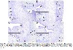
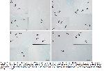
    AB (+), SO (+), and TB (+) uMCs were detected in rat uterine tissues using the AB/SO combined staining technique and toluidine blue staining. All uMCs that were TB (+), AB (+), or SO (+) were observed in proximity to the uterine glands and blood vessels within the stratum vasculare, specifically in the basal and deeper layers of the endometrial stroma. The combined AB/SO staining technique differentiated between uMCs staining blue, indicative of AB (+), and those exhibiting a red-pink color, characteristic of SO (+), in the uterine sections. The paroxetine+irisin treatment significantly increased the count of AB (+) uMCs compared to the sham vehicle group (Table 1, Figure 1, p<0.05).


    Büyütmek İçin Tıklayın
    Table 1: Effects of paroxetine and/or irisin on the MCs counts in rat uterine tissue after staining with alcian blue, safranin O, or toluidine blue


    Büyütmek İçin Tıklayın
    Figure 1: Representative photographs of uMCs in histological specimens from the uterus: (A) sham vehicle group, (B) paroxetine group, (C) irisin group, and (D) paroxetine+irisin group. uMCs were detected using toluidine blue (arrow: mast cell; curved arrow: blood vessel; asterisk: uterine glands; thick arrow: epithelium). Small frames: x40 magnification: Other areas: x10 magnification. Range bar=10 μm.

    In all groups, uMCs exhibited metachromatic staining with toluidine blue. These cells were noted in diverse sizes and morphologies, predominantly as round, oval, and elongated forms. The metachromatic granules displayed homogeneous staining within the cytoplasm, making individual selection unfeasible. The nuclei of the uMCs were observed in both central and eccentric positions, with granules often covering them in most cells. Paroxetine treatment significantly increased the number of TB (+) uMCs compared to the sham vehicle group (Table 1, Figure 1, p<0.01). Similarly, the number of TB (+) uMCs significantly increased in the paroxetine+irisin group compared to the sham vehicle group (Table 1, Figure 1, p<0.05). However, there was no significant change in the number of SO (+) uMCs across any group. When assessing serum hormone levels, neither paroxetine nor irisin treatment altered serum 17β-estradiol or progesterone levels (Figure 3).


    Büyütmek İçin Tıklayın
    Figure 2: Representative photographs of uMCs in histological specimens from the uterus: (A) sham vehicle group, (B) paroxetine group, (C) irisin group, and (D) paroxetine+irisin group. uMCs were detected using the AB/SO combined staining method (arrow: alcian blue (+) mast cell; curved arrow: safranin O (+) mast cell). Small frames: x40 magnification: Other areas: x20 magnification. Range bar=10 μm.


    Büyütmek İçin Tıklayın
    Figure 3: Effects of paroxetine and/or irisin on the: (A) serum 17β-estradiol levels and (B) serum progesterone levels (n = 10).

  • Başa Dön
  • Özet
  • Giriş
  • Materyal ve Metot
  • Bulgular
  • Tartışma
  • Kaynaklar
  • Tartışma
    Similar to MCs located in various body regions, uMCs contain a range of mediators such as cytokines, growth factors, and chemokines within their cytoplasmic granules 6. These mediators have a pivotal role in inflammatory responses, which are critical for successful blastocyst implantation within the uterus and placentation 7. Beyond these functions, uMCs also have the ability to affect angiogenesis and control uterine contractions, both fundamental to parturition 6. The current study investigated the impact of paroxetine and/or irisin on MCs counts in rat uterine tissue after staining with AB, SO, or TB. For the first time, this study shows a significant increase in the TB-stained uMCs count in the paroxetine group. Interestingly, the paroxetine+irisin treatment resulted in an increase in the MCs count stained with AB or TB vs sham vehicle group. However, no significant changes were found in the serum levels of 17β-estradiol and progesterone following any of the treatments.

    Among women in their reproductive years, approximately one in seven uses antidepressant treatments. Of these treatments, SSRIs are the predominant class prescribed, as noted by Sylvester et al. 12. Paroxetine is one such SSRI, and it has been shown to increase serum 5-HT levels 24. There is almost no study in the literature that directly shows how paroxetine affects serum 5-HT levels and, consequently, the number of uMCs in rats. In one study, the potential in vivo relevance of MC migration induced by 5-HT was highlighted by the observed increase of MCs at locations where 5-HT was injected into the mouse dermis 25. In the current study, paroxetine treatment significantly increased the count of TB-stained uMCs in the paroxetine group compared to the sham vehicle group. This indicates that the increase in 5-HT levels due to paroxetine may affect the migration of MCs from the periphery to the uterus.

    The number of mast cells can vary due to numerous factors. One of the primary factors is the influence of female reproductive hormones—namely, estrogen and progesterone—on MC recruitment to the uterus 26. There are very few studies indicating that estrogen and progesterone directly affect the number of MCs in the rat uterus. In a study conducted by Jensen et al. (2010), it was reported that treatment with estradiol led to a notable increase in the number of uMCs in ovariectomized mice. In contrast, the application of progesterone didn't result in any significant variations in their count. However, the combined administration of both estradiol and progesterone caused substantial changes in the MC count 27. Given that the serum levels of estradiol and progesterone remained unchanged in our study, the unaltered uMCs count due to irisin treatment may be considered normal. To the best of our knowledge, there is almost no study in the literature on the effects of irisin on the count of uMCs stained with AB, SO, or TB, showing subtypes of uMCs. For the first time, this study demonstrated that the irisin treatment does not affect the number of uMC subtypes. However, there was a significant increase in the count of TB (+) uMCs in the paroxetine+irisin group compared to the sham vehicle group. Since irisin has no direct effect on uMCs in rats, as previously explained, this increase might be attributed to the effects of 5-HT induced by paroxetine in this group.

    As mentioned earlier, for many years, the AB/safranin dye has been mainly used to differentiate between CTMCs and MMCs based on its unique staining characteristics 10. Various studies indicate that, when using this staining technique, MMC's granules appear blue with AB (+), whereas CTMC's granules exhibit a red color with SO (+) 28. The presence of heparin in CTMC results in a red stain when treated with safranin. On the other hand, MMC, which doesn't contain heparin, presents a blue coloration after being stained with AB 29. In the present study, AB (+) uMCs count significantly increased in paroxetine+irisin group vs sham vehicle group. Since MC maturation takes place in peripheral tissues, the variation in MCs across different tissues likely stems from the conditions of the microenvironment influencing gene expression and phenotypic growth. Consequently, it's believed that the diversity of MCs dynamically changes based on factors in their surrounding microenvironment, such as cytokine levels, hormones, reactive radical species, and interactions with neighboring cells 30. Hence, the changes in MMCs observed in the current study might be attributed to the effects of paroxetine rather than irisin, as previously described, on the microenvironmental conditions of the uterus. This notion requires deeper investigation through extensive preclinical studies focused on uMCs, especially on MMCs in uterus, to fully understand the underlying mechanisms affected by paroxetine. Based on the results of our study, and in light of previously published data, the increase in the number of MMCs stained with AB in this rat model suggests that 5-HT, induced by paroxetine, might play a role in the biology of these uMC subtypes in rats.

    In conclusion, this study provides novel insights into the influence of paroxetine and/or irisin on uMC counts within rat uterine tissue. It emphasizes the increase of TB-stained uMCs count following paroxetine treatment. Despite the widespread use of SSRIs among women, the direct relationship between paroxetine and its impact on serum 5-HT levels, which subsequently affects uMCs, remains uncharted. A notable change in the count of AB (+) uMCs in the paroxetine+irisin group hints at the potential role of paroxetine-induced 5-HT in modifying the uterine microenvironment. This could influence uMC maturation and numbers. The limitation of our study is the inability to more thoroughly investigate the uterine microenvironment using more detailed methods. There is a need for more comprehensive research to unravel the intricate mechanisms behind these observations, especially concerning the role of 5-HT in uMC biology.

  • Başa Dön
  • Özet
  • Giriş
  • Materyal ve Metot
  • Bulgular
  • Tartışma
  • Kaynaklar
  • Kaynaklar

    1) Tutuncu S, Ertugrul T. Immunohistochemical expression of tryptase-chymase and mast cell heterogeneity in capsaicin-treated rat ovaries. Int J Vet Anim Res 2019; 2: 25-31.

    2) Aydın Y, Tuncel N, Gurer F, et al. Ovarian, uterine and brain mast cells in female rats: Cyclic changes and contribution to tissue histamine. Comp Biochem Physiol Part A Mol Integr Physiol 1998; 120: 255-262.

    3) Herr N, Bode C, Duerschmied D. The effects of serotonin in immune cells. Front Cardiovasc Med 2017; 4: 48.

    4) Karaca T, Yoruk M, Uslu S. Distribution and quantitative patterns of mast cells in ovary and uterus of rat. Arch Med Vet 2007; 39: 135-139.

    5) Binda MM, Donnez J, Dolmans MM. Targeting mast cells: A new way to treat endometriosis. Expert Opin Ther Targets 2017; 21: 67-75.

    6) Meyer N, Zenclussen AC. Mast cells-good guys with a bad image? Am J Reprod Immunol 2018; 80: e13002.

    7) Mor G, Cardenas I, Abrahams V, et al. Inflammation and pregnancy: The role of the immune system at the implantation site: Inflammation and pregnancy. Ann NY Acad Sci 2011; 1221: 80-87.

    8) Bancroft JD, Gamble M. Theory and practice of histological techniques. 5th Edition, Edinburgh: Churchill Livingstone Pub, 2002.

    9) Lin T, Befus AD. Mast Cells in Mucosal Defensis and Pathogenesis. In: Mestecky J, Lamm ME, Strober W, et al (Editors). Mucosal Immunity. United States of America: Elsevier Academic Press, 2002: 703-710.

    10) Enerbäck L. Mast cells in rat gastrointestinal mucosa: Effects of fixation. APMIS 1966; 66: 289-302.

    11) Chen W, Alley MR, Manktelow BW, et al. Mast cells in the bovine lower respiratory tract: Morphology, density and distribution. Brit Vet J 1990; 146: 425-436.

    12) Sylvester C, Menke M, Gopalan P. Selective serotonin reuptake inhibitors and fertility: Considerations for couples trying to conceive. Harv Rev Psychiatry 2019; 27: 108-118.

    13) Rayen I, Steinbusch HWM, Charlier TD, et al. Developmental fluoxetine exposure facilitates sexual behavior in female offspring. Psychopharmacology 2014; 231: 123-133.

    14) Wu H, Denna TH, Storkersen JN, et al. Beyond a neurotransmitter: The role of serotonin in inflammation and immunity. Pharmacol Res 2019; 140: 100-114.

    15) Tran BX, Ha GH, Vu GT, et al. Indices of change, expectations, and popularity of biological treatments for major depressive disorder between 1988 and 2017: A scientometric analysis. IJERPH 2019; 16: 2255.

    16) Ulker N, Yardimci A, Kaya Tektemur N, et al. Chronic exposure to paroxetine or bupropion modulates the pubertal maturation and the reproductive system in female rats. Reprod Biol 2020; 20: 154-163.

    17) Panati K, Suneetha Y, Narala VR. Irisin/FNDC5 – An updated review. Eur Rev Med Pharmacol 2016; 20: 689-697.

    18) Canpolat S, Ulker N, Yardimci A, et al. Irisin ameliorates male sexual dysfunction in paroxetine-treated male rats. Psychoneuroendocrinology 2022; 136: 105597.

    19) Yardimci A, Ulker N, Bulmus O, et al. Irisin improves high-fat diet-induced sexual dysfunction in obese male rats. Neuroendocrinology 2022; 112: 1087-1103.

    20) Li C, Zhou L, Xie Y, et al. Effect of irisin on endometrial receptivity of rats with polycystic ovary syndrome. Gynecol Endocrinol 2019; 35: 395-400.

    21) Kaya Sezginer E, Kirlangic OF, Eskin Tanriverdi MD, et al. Analysis of changes in serum levels and gene expression profiles of novel adipocytokines (Omentin, vaspin, irisin and visfatin) and their correlation with serum C-reactive protein levels in women diagnosed with endometriosis. Turk J Pharm Sci 2022; 19: 48-53.

    22) De Jong TR, Pattij T, Veening JG, et al. Effects of chronic selective serotonin reuptake inhibitors on 8-OH-DPAT-induced facilitation of ejaculation in rats: Comparison of fluvoxamine and paroxetine. Psychopharmacology 2005; 179: 509-15.

    23) Ulker N, Yardimci A, Kaya Tektemur N, et al. Irisin may have a role in pubertal development and regulation of reproductive function in rats. Reproduction 2020; 160: 281-292.

    24) Li F, Zhang M, Xu D, et al. Co-administration of paroxetine and pravastatin causes deregulation of glucose homeostasis in diabetic rats via enhanced paroxetine exposure. Acta Pharmacol Sin 2014; 35: 792-805.

    25) Kushnir-Sukhov NM, Gilfillan AM, Coleman JW, et al. 5-Hydroxytryptamine induces mast cell adhesion and migration. J Immunol 2006; 177: 6422-6432.

    26) Zierau O, Zenclussen AC, Jensen F. Role of female sex hormones, estradiol and progesterone, in mast cell behavior. Front Immun 2012; 3: 169.

    27) Jensen F, Woudwyk M, Teles A, et al. Estradiol and progesterone regulate the migration of mast cells from the periphery to the uterus and ınduce their maturation and degranulation. PLoS ONE 2010; 5: e14409.

    28) Uslu S, Yörük M. Hindilerde sindirim sisteminde mast hücrelerinin dağılımı ve heterojenitesi üzerine morfolojik ve histometrik araştırmalar. Yüzüncü Yıl Üniversitesi Veteriner Fakültesi Dergisi 2008; 19: 47-51.

    29) Stephen JG. New Insights into “The Riddle of the Mast Cells”: Microenvironmental Regulation of Mast Cell Development and Phenotypic Heterogeneity. In: Rubin E, Damjanov I (Editors). Pathology Reviews. 1st Edition, New Jersey: Humana Press, 1990: 49-77.

    30) Moon TC, St Laurent CD, Morris KE, et al. Advances in mast cell biology: New understanding of heterogeneity and function. Mucosal Immunol 2010; 3: 111-128.

  • Başa Dön
  • Özet
  • Giriş
  • Materyal ve Metot
  • Bulgular
  • Tartışma
  • Kaynaklar
  • [ Başa Dön ] [ Özet ] [ PDF ] [ Benzer Makaleler ] [ Yazara E-Posta ] [ Editöre E-Posta ]
    [ Ana Sayfa | Editörler | Danışma Kurulu | Dergi Hakkında | İçindekiler | Arşiv | Yayın Arama | Yazarlara Bilgi | E-Posta ]