[ Ana Sayfa | Editörler | Danışma Kurulu | Dergi Hakkında | İçindekiler | Arşiv | Yayın Arama | Yazarlara Bilgi | E-Posta ]
Fırat Üniversitesi Sağlık Bilimleri Tıp Dergisi
2018, Cilt 32, Sayı 3, Sayfa(lar) 147-151
[ Özet ] [ PDF ] [ Benzer Makaleler ] [ Yazara E-Posta ] [ Editöre E-Posta ]
Lateral Humeral Kondil Kırıklarında Kırık Tipinin Klinik ve Radyolojik Sonuçlar Üzerine Etkisi
Murat GÜRGER
Fırat University, Faculty of Medicine, Department of Orthopedics and Traumatology, Elazığ, TURKEY
Anahtar Kelimeler: Çocuk, dirsek, lateral humeral kondil kırığı, tedavi
Özet
Amaç: Bu çalışmanın amacı lateral humeral kondil kırıklarında kırık tipinin klinik ve radyolojik sonuçlar üzerine olan etkisini araştırmaktır.

Gereç ve Yöntem: En az iki yıl takibi olan ve ek yaralanması olmayan 84 hasta bu retrospektif çalışmaya dahil edildi. Yetmiş iki hastaya cerrahi tedavi, 12 hastaya ise cerrahi olmayan tedavi yöntemleri uygulandı. Ortalama takip süresi 72.8 aydı. Kırıkları sınıflandırmak için Jacop sınıflandırma sistemi kullanıldı ve klinik sonuçlar ise Dhillon skorlama sistemine göre yapıldı.

Bulgular: Çalışmadaki hastaların 66'sı erkek ve 18'i kızdı. Ortalama yaş 5.5 yıl idi. Otuz altı tip I, 21 tip II ve 27 tip III kırık vardı. Lateral spur 30 hastada tespit edildi. Taşıma açısı ortalama 6.5 derece idi. Üç hastada valgus deformitesi, altı hastada varus deformitesi vardı. Altmış dokuz hastada mükemmel, 15 hastada ise iyi fonksiyonel sonuç elde edildi. Farklı kırık tiplerindeki fonksiyonel sonuçlar karşılaştırıldığında, istatistiksel olarak anlamlı bir fark bulunmadı (P>0.05). Farklı tedavi yöntemlerindeki (konservatif, kapalı redüksiyon perkutan tespit ve açık redüksiyon k teli ile tespit) klinik ve radyolojik sonuçlar karşılaştırıldığında, istatistiksel olarak anlamlı bir fark yoktu (P>0.05).

Sonuç: Bu çalışmada, başlangıçtaki kırık tipi ve klinik sonuçlar arasında anlamlı bir ilişki olmadığı ayrıca cerrahi tedavide, redüksiyonun yeterliliği ve stabilitenin kırık tipinden daha önemli olduğu sonucuna varılmıştır.

  • Başa Dön
  • Özet
  • Giriş
  • Materyal ve Metot
  • Bulgular
  • Tartışma
  • Kaynaklar
  • Giriş
    Lateral humeral condyle fractures (LHCF) rank as the second most frequent pediatric elbow fractures 1. These fractures constitute approximately 10% to 20% of all childhood elbow fractures 2. The classification system proposed by Milch and Jacob are generally used to classify pediatric LHCF 3-5. In Milch type 1 fracture, the fracture line extends lateral to capitello-trochlear groove while in Milch type 2 fractures, the fracture line extends medial to capitello-trochlear groove 4. In Jacob type I fracture, there is a displacement of less than 2 mm; in Jacob type II fractures, there is a displacement of more than 2 mm with an intact cartilaginous hinge and in Jacob type III fractures, there is a displaced fracture and the capitellum is rotated 5. The Milch classification system is widely used but is insufficient to determine the treatment method. The Jacob classification is more useful clinically and helps to choose a treatment method 6. The main purpose of classification systems is to assist physicians in determining the best treatment method and in predicting the prognosis but the classification of lateral condylar fractures is insufficient for predicting clinical and radiological outcomes 3. Here, to investigate the relationship between the initial fracture classification and clinical and radiological outcomes in patients with LHCF were aimed.
  • Başa Dön
  • Özet
  • Giriş
  • Materyal ve Metot
  • Bulgular
  • Tartışma
  • Kaynaklar
  • Materyal ve Metot
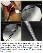
    The medical records of 84 patients, with at least a two-year follow-up and no additional injury were retrospectively analysed. The average follow-up period was 72.8 months (range, 24–132 months). Sixty-six (78.6%) patients were male and 18 (21.4%) patients were female. The average age was 5.5 (range: 2–16) years old. The right elbow was affected in 27 (32.1%) patients, and the left in 57 (67.9%) patients. The preoperative x-rays of the patients were examined, and the fractures were classified using the Jacob classification system. LHCF was determined as type I in 36 (42.9%) patients, type II in 21 (25%) patients and type III in 27 (32.1%) patients. The average period between diagnosis and operation was 3.4 (±4.4) days. Twelve patients (14.3%) had been treated with cast immobilisation (non-operative), 27 patients (31.1%) with closed reduction and percutaneous fixation (CRPF) and 45 patients (53.6%) with open reduction and internal fixation (ORIF). To decide which treatment method to use, the varus stress radiography was taken under sedation limiting the force for cases with a displacement less than 2 mm. The non-operative treatment method was applied for the fractures not displaced (<2 mm) under stress, CRPF surgical treatment for fractures with a displacement over 2 mm for which closed reduction can be achieved, and the ORIF surgical treatment when closed reduction cannot be achieved. The cast immobilisation had been applied for an average of 5.8 weeks (range: 5–7) in non-operatively treated patients. The Kocher (lateral) approach had been used for all patients having undergone open reduction. The fracture fixation had been performed with two Kirschner wires (Figure 1). Kirschner wires had been removed in average after 5.5 weeks (range: 4–7 weeks). The lateral spur, presence of cubitus varus or valgus deformity, and elbow motion had been evaluated during the last clinical examinations of the patients. The non-union, fishtail deformity, cubitus varus or valgus, carrying angle and lateral spurring had also been evaluated in the final controls of the patient as last radiological evaluations. The carrying angle was determined by measuring the angle between the line connecting the humerus metaphysis and diaphysis mid points and the line connecting the midpoint at the level of the bicipital tuberosity with the proximal edge of the ulna 7. The clinical results were evaluated using the scoring system defined by Dhillon et al, 8 based on pain, range of motion, neurological status and carrying angle, and a score was given as excellent, good, fair and poor (Table 1).


    Büyütmek İçin Tıklayın
    Figure 1: A 5-year old male patient with right LHCF. A) Intraoperative image (arrow, fracture line). B) and C) Anteroposterior and lateral fluoroscopy images. D) Postoperative control images in the 8th year, (arrow, lateral spurring).


    Büyütmek İçin Tıklayın
    Table 1: Dhillon scoring system

    The SPSS 21 (IBM Corp., Armonk, NY, USA) packaged programme was used to make the analysis of the data obtained in the study. The numerical data were expressed as average ± standard deviation and the categorical data were expressed as percentage. Chi-Square test or Fisher’s exact test were used in the analysis of the categorical data. Values with p < 0.05 were accepted as statistically significant.

  • Başa Dön
  • Özet
  • Giriş
  • Materyal ve Metot
  • Bulgular
  • Tartışma
  • Kaynaklar
  • Bulgular
    Twelve (14.3%) type I fractures were non-operatively treated; 24 (28.6%) type I and 3 (3.6%) type II fractures were treated with CRPF; 18 (21.4%) type II and all 27 (32.1%) type III fractures were treated with ORIF. Lateral spurring, delayed union and superficial infection were observed in 30 (35.7%), 3 (3.6%) and 3 (3.6%) patients, respectively. None of the patients presented non-union, avascular necrosis (AVN) or fishtail deformity. The carrying angle was in average 6.5 (±5.5) degrees in the final patient assessments. When compared with the contralateral side, the valgus deformity was >5 in 3 patients and the varus deformity was > 5 in 6 patients. During the final examinations, the number of patients with excellent results was 69 (82.1%), and good results was 15 (17.9%) using the functional grading from the Dhillon criteria (Table 2). Using the overall grading, excellent results were obtained in 42 (50%) patients and good results in 42 (50%) patients (Table 3). None of the patients treated non-operatively developed any complication as assessed in follow-up visits. When the functional grading in the different fracture types was compared, no statistically significant difference was found (P>0.05). However, when the overall grading was compared, a statistically significant difference was found (P<0.01). Similarly, when the lateral spurring was analysed in the different fracture types, there was a statistically significant difference (P<0.01). Especially, the difference was significant between type I and type III fractures (P<0.01) (Table 4).


    Büyütmek İçin Tıklayın
    Table 2: Relationship between the fracture type and functional result rating according to Dhillon’s criteria


    Büyütmek İçin Tıklayın
    Table 3: Relationship between the fracture type and overall result rating according to Dhillon’s criteria


    Büyütmek İçin Tıklayın
    Table 4: Lateral spurring occurrence in the different fracture types

    When the clinical, radiological results and lateral spurring were compared with the different treatment methods (non-operative, CRPF and ORIF), there was not a statistically significant difference (P>0.05).

  • Başa Dön
  • Özet
  • Giriş
  • Materyal ve Metot
  • Bulgular
  • Tartışma
  • Kaynaklar
  • Tartışma
    The most commonly cited classification system for pediatric lateral condyle fractures use anatomic configuration (Milch) and displacement (Jacob and Finnbogason) for descriptive and prognostic purposes 3. However, trochlear ossification occurs in children 9–10 years old and the actual fracture configuration and ultimate displacement cannot be predicted at this age 9. For this reason, the fracture displacement cannot be sufficiently evaluated by standard radiography especially in children younger than 8 years of age 10. Song et al. 11 determined that the internal oblique radiography is more valuable in LHCF. In addition, alternative imaging methods may be used for determining the displacement amount and stability, and MRI provides detailed images for evaluation of the growth plate and cartilage bridge. However, the disadvantages of this method include the necessity of anaesthesia and the high cost 12. Marzo et al. 13 have used arthrography to evaluate the fracture character and displacement range. However, the routine usage of this method is limited. Moreover, ultrasonography may be also used to distinguish stable from unstable fractures 14, the reliability of this method depending on the user’s experience. Chapman et al. 15 used multi-detector computed tomography to determine the fracture’s character. The disadvantage of this method is children are subjected to extra radiation. In this study, the varus stress test was applied gently under sedation, the amount of displacement was evaluated under an C-arm fluoroscopy and the treatment method was based on this examination. Existing classification systems are sometimes deemed inadequate in determining treatment modalities and predicting clinical outcomes 3. The effect of the fracture type according to Jacob’s classification on functional and radiological outcomes were investigated. There was no statistically significant difference between the fracture types when comparing the functional results according to the Dhillon criteria 8 in each type of fracture.

    A very limited number of radiographic criteria have been defined to evaluate a potential displacement and the necessity of an internal fixation during the follow-up of fractures of less than 2 mm 16. Although some orthopedists advocate conservative treatment for minimally displaced (<2 mm) fractures, others suggest open surgical treatment for these cases 17. Among the 51 LHCFs with a displacement less than 2 mm non-operatively treated by Pirker et al. 16, five patients had developed a displacement within the first week as assessed at follow-up visit, suggesting the requirement for an extensive follow-up for non-displaced fractures treated non-operatively.

    Surgical fixations are recommended for LHCF treatment involving a displacement over 2 mm. Among surgical techniques related to LHCF, good results are reported between 79% and 96% in various studies, and postoperative complications occur in 0% to 32% of cases 3. CRPF could potentially prevent these complications, even in cases of fractures devoid of cartilage continuity 3. Song et al. 18 have treated lateral condyle fractures with an average displacement of 13 mm and rotation using the CRPF method, and obtained successful results in all 18 patients.

    Lateral spurring is a significant radiological finding seen in children followed-up for LHCF 19. Pribaz et al. 19 observed that lateral spur had occurred at different degrees in 73% of their 212 patients treated with different methods. They showed that spur development and size are positively correlated with the fracture displacement at diagnosis. In addition, in the same study, spur occurred more often in cases surgically treated than in those non-operatively treated. Lateral spurs developed in 30 (35.7%) of our cases.

    There was a significant difference between lateral spur occurrence and initial fracture. This difference was especially significant between type I and type III fractures. However, lateral spur occurrence did not negatively affect our clinical results.

    Cubitus varus is another complication of LHCF 20. Previous studies have shown that the cubitus varus deformity is not related with the treatment method 20. This deformity is generally not symptomatic and surgical correction is rarely required 1. A cubitus varus deformity developed in 6 patients (7.1%) in this study, and these patients did not require corrective osteotomy.

    The distal humerus fishtail deformity describes an anatomic finding that originates from the osteonecrosis of the humeral trochlea, but may also result from a central physeal arrest. Generally, this deformity does not imply a functional problem 1. No fishtail deformity developed in any of our cases.

    Avascular necrosis (AVN) is a less often encountered complication and is likely related to the surgery 1. However, some authors defend that a surgical approach will not cause AVN and the principal causative reason is excessive dissection 21. Kocher (lateral) approach was used in our cases and no AVN developed in any of our cases.

    In conclusion, this study demonstrated no significant relationship between initial fracture displacement, fracture type and clinical outcome. However, the amount of initial displacement may partially negatively affect the radiological result, but radiological changes in the lateral condyle fractures usually do not adversely affect the functional outcomes. Good results may be obtained with the appropriate treatment in the lateral condyle fractures. Cases treated non-operatively should be comprehensively followed-up. In the surgical treatment, sufficiency and stability of the reduction are more important than fracture type.

  • Başa Dön
  • Özet
  • Giriş
  • Materyal ve Metot
  • Bulgular
  • Tartışma
  • Kaynaklar
  • Kaynaklar

    1) Shaerf DA, Vanhegan IS, Dattani R. Diagnosis, management and complications of distal humerus lateral condyle fractures in children. Shoulder & Elbow 2017; 10:114-120.

    2) Bakarman KA, Alsiddiky AM, Alzain KO, et al. Humeral lateral condyle fractures in children: redefining the criteria for displacement. J Pediatr Orthop B 2016; 25: 429-433.

    3) Weiss JM, Graves S, Yang S, et al. A new classification system predictive of complications in surgically treated pediatric humeral lateral condyle fractures. J Pediatr Orthop 2009; 29: 602-605.

    4) Milch H. Fractures and fracture dislocations of the humeral condyles. J Trauma 1964; 4: 592-607.

    5) Jakob R, Fowles JV, Rang M, Kassab MT. Observations concerning fractures of the lateral humeral condyle in children. J Bone Joint Surg Br 1975; 57: 430-436.

    6) Zale C, Winthrop ZA, Hennrikus W. Rate of displacement for Jakob Type 1 lateral condyle fractures treated with a cast. J Child Orthop 2018; 12: 117-122.

    7) Beals RK. The normal carrying angle of the elbow. A radiographic study of 422 patients. Clin Orthop Relat Res 1976; 119: 194-196.

    8) Dhillon KS, Sengupta S, Singh BJ. Delayed management of fracture of the lateral humeral condyle in children. Acta Orthop Scand 1988; 59: 419-424.

    9) Mirsky EC, Karas EH, Weiner LS. Lateral condyle fractures in children: Evaluation of classification and treatment. J Orthop Trauma 1997; 11: 117-120.

    10) Knapik DM, Gilmore A, Liu RW. Conservative management of minimally displaced (11) Song KS, Kang CH, Min BW, Bae KC, Cho CH. Internal oblique radiographs for diagnosis of nondisplaced or minimally displaced lateral condylar fractures of the humerus in children. J Bone Joint Surg Am 2007; 89: 58-63.

    12) Horn BD, Herman MJ, Crisci K, Pizzutillo PD, MacEwen GD. Fractures of the lateral humeral condyle: Role of the cartilage hinge in fracture stability. J Pediatr Orthop 2002; 22: 8-11.

    13) Marzo JM, d'Amato C, Strong M, Gillespie R. Usefulness and accuracy of arthrography in management of lateral humeral condyle fractures in children. J Pediatr Orthop 1990; 10: 317-321.

    14) Vocke-Hell AK, Schmid A. Sonographic differentiation of stable and unstable lateral condyle fractures of the humerus in children. J Pediatr Orthop B 2001; 10: 138-141.

    15) Chapman VM, Grottkau BE, Albright M, Salamipour H, Jaramillo D. Multidetector computed tomography of pediatric lateral condylar fractures. Journal of Computer Assisted Tomography 2005; 29: 842-846.

    16) Pirker ME, Weinberg AM, Höllwarth ME, Haberlik A. Subsequent displacement of initially nondisplaced and minimally displaced fractures of the lateral humeral condyle in children. Journal of Trauma and Acute Care Surgery 2005; 58: 1202-1207.

    17) Rizk AS, Essawy OM. Percutaneous fixation of pediatric humeral lateral condyle fractures. The Egyptian Orthopaedic Journal 2017; 52: 263-272.

    18) Song KS, Kang CH, Min BW, et al. Closed reduction and internal fixation of displaced unstable lateral condylar fractures of the humerus in children. J Bone Joint Surg Am 2008; 90: 2673-2681.

    19) Pribaz JR, Bernthal NM, Wong TC, Silva M. Lateral spurring (overgrowth) after pediatric lateral condyle fractures. J Pediatr Orthop 2012; 32: 456-460.

    20) Koh KH, Seo SW, Kim KM, Shim JS. Clinical and radiographic results of lateral condylar fracture of distal humerus in children. J Pediatr Orthop 2010; 30: 425-429.

    21) Conaway WK, Hennrikus WL, Ravanbakhsh S, Winthrop Z, Mahajan J. Surgical treatment of displaced pediatric lateral condyle fractures of the humerus by the posterior approach. J Pediatr Orthop B 2018; 27: 128-133.

  • Başa Dön
  • Özet
  • Giriş
  • Materyal ve Metot
  • Bulgular
  • Tartışma
  • Kaynaklar
  • [ Başa Dön ] [ Özet ] [ PDF ] [ Benzer Makaleler ] [ Yazara E-Posta ] [ Editöre E-Posta ]
    [ Ana Sayfa | Editörler | Danışma Kurulu | Dergi Hakkında | İçindekiler | Arşiv | Yayın Arama | Yazarlara Bilgi | E-Posta ]