[ Ana Sayfa | Editörler | Danışma Kurulu | Dergi Hakkında | İçindekiler | Arşiv | Yayın Arama | Yazarlara Bilgi | E-Posta ]
Fırat Üniversitesi Sağlık Bilimleri Tıp Dergisi
2021, Cilt 35, Sayı 2, Sayfa(lar) 092-096
[ Özet ] [ PDF ] [ Benzer Makaleler ] [ Yazara E-Posta ] [ Editöre E-Posta ]
Tüberoskleroz Kompleksi Olan Çocuklarda Derin Gri Cevherin Difüzyon Ağırlıklı Görüntüleme İle Değerlendirilmesi
Yeşim EROĞLU, Mustafa KOÇ
Firat University, Faculty of Medicine, Department of Radiology, Elazig, TURKIYE
Anahtar Kelimeler: Tüberoskleroz kompleksi, difüzyon ağırlıklı görüntüleme, görünür difüzyon katsayısı, çocuk
Özet
Amaç: Bu çalışmanın amacı tüberoskleroz kompleksi tanılı çocuklarda derin gri cevher yapılarının difüzyon ağırlıklı manyetik rezonans görüntüleme değerlerini kontrol grubu ile karşılaştırmaktır.

Gereç ve Yöntem: Ocak 2015 ile Aralık 2020 tarihleri arasında, difüzyon ağırlıklı manyetik rezonans görüntüleme yapılan tüberoskleroz kompleksi tanılı 14 çocuk hasta (8 erkek, 6 kız; ortalama yaş 9.50±4.16) ile yaş ve cinsiyet eşleştirmeli 28 kontrol hastası retrospektif olarak incelendi. Bilateral talamus ve bazal ganglionlara simetrik dairesel region of interest yerleştirilerek görünür difüzyon katsayısı değerleri ölçüldü.

Bulgular: Tüberoskleroz kompleksi tanılı hastalarda bilateral nükleus kaudatusta kontrol grubuna kıyasla görünür difüzyon katsayısı değerlerinde anlamlı artış saptandı (P<0.001).

Sonuç: Bu hastalarda olası derin gri cevher yapılarının etkilenmesi mevcut klinik bulgular ve gelişebilecek nörolojik bozukluklar için fizyopatolojiyi açıklayıcı olabilir.

  • Başa Dön
  • Özet
  • Giriş
  • Materyal ve Metot
  • Bulgular
  • Tartışma
  • Kaynaklar
  • Giriş
    Tuberous sclerosis complex (TSC) is an inherited autosomal dominant neurocutaneous syndrome. Epilepsy, mental retardation and cognitive inefficiency are the main clinical findings. Epilepsy is observed in 90% of the patients. Epilepsy starts in early infancy and its frequency and severity gradually increase. Drug-resistant epilepsy, which decreases the quality of life of the patient, is observed in the later periods 1. It is a multisystemic disease in which skin, central nervous system, eye, kidney, heart, lung and bone involvement may be observed 2. Cortical-subcortical tubers, subependymal nodules, subependymal giant cell astrocytomas and white matter abnormalities are the main central nervous system anomalies.

    Diffusion-weighted magnetic resonance imaging (DW-MRI) is an advanced MRI imaging method that shows functional data based on microscopic movements of water molecules in the tissue 3. In the literature; it was revealed that there were abnormalities at the cellular level in the cerebral parenchyma, which appeared normal in conventional MRI, and its significant contributions to the pathogenesis of the disease were reported with DW-MRI 3,4.

    The aim of this study is to research the presence of microstructural changes in the basal ganglia and thalamus, which are normal in conventional brain MRI, in children with TSC using DW-MRI.

  • Başa Dön
  • Özet
  • Giriş
  • Materyal ve Metot
  • Bulgular
  • Tartışma
  • Kaynaklar
  • Materyal ve Metot
    Research and Publication Ethics: Fırat University Non-Interventional Research Ethics Committee approval was obtained for the study (18.02.2021 / 2021/03 - 07).

    Patient and Control Group: The study included 14 pediatric patients diagnosed with TSC who had brain MRI in our hospital between January 2015 and December 2020. The control group consisted of 28 patients who underwent brain MRI due to headache, revealed normal results and were matched for age and gender. Physical and neurological examinations of the patients in the control group were normal.

    MRI Protocol: Brain MRI of the patients were obtained with 1.5 and 3 Tesla MRI devices (Philips Healthcare, Ingenia). MRI protocol consisted of sagittal T1-weighted (W) and T2-W and sagittal fluid attenuated inversion recovery (FLAIR), sagittal double inversion recovery (DIR) and their reconstruction images, susceptibility weighted imaging (SWI), diffusion-weighted imaging (DWI), apparent diffusion coefficient (ADC) images. DWI (b=0-1000 mm2/ s) was obtained in the axial plane (FOV=230x230 mm, matrix=256x256 mm, section thickness=5 mm).

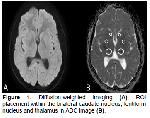
    Imaging Analysis: Brain MRI of the patients were evaluated in terms of cortical-subcortical tubers, subependymal nodules, subependymal giant cell astrocytoma, and white matter abnormalities. Tubers were recorded as cortical-subcortical localized, hypointense lesions in T1-W image, and hyperintense lesions in T2-W and FLAIR/DIR images 5. Subependymal nodules were described as focal subependymal hamartomas that extend along the ventricular surface and are often calcified 6. Subependymal giant cell astrocytomas were described as lesions that were hypointense on T1-W images and hyperintense on T2-W images with contrast enhancement 6. White matter abnormalities were described as hypointense in T1-W images, hyperintense in T2-W and FLAIR images, linear radial bands extending from the periventricular white matter to the cortex 7,8. Afterwards, measurements were taken by placing symmetrical circular region of interest (ROI) on the bilateral thalamus and basal ganglia in both TSC patients and the control group on ADC images (Figure 1). ROI size was chosen as 0.25 cm2 in thalamus, 0.10 cm2 in the caudate nucleus, and as 0.15 cm2 in the lentiform nucleus.


    Büyütmek İçin Tıklayın
    Figure 1: Diffusion-weighted imaging (A), ROI placement within the bilateral caudate nucleus, lentiform nucleus and thalamus in ADC image (B).

    Statistical Analysis: SPSS 22 package program was used for the statistical analysis of the study. After recording the data of the patients included in the study, Mann Whitney U test was used to compare ADC values. P<0.05 value was considered statistically significant.

  • Başa Dön
  • Özet
  • Giriş
  • Materyal ve Metot
  • Bulgular
  • Tartışma
  • Kaynaklar
  • Bulgular
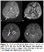
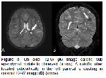
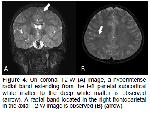
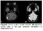
    There were 8 male and 6 female patients diagnosed with TSC in the study. The ages of the cases ranged from 4-16 years, and the mean age was found as 9.50±4.16. Cortical-subcortical tubers and subependymal nodules were existing in MRI of all patients (Figure 2). Subependymal nodules in 11 (78.57%) patients and tubers in 1 (7.14%) were calcified (Figure 3). Linear radial bands were existing in the white matter in 2 patients (14.28%) (Figure 4). There is the cerebellar calcific tuber in 1 (7.14%) of the patients (Figure 5). Subependymal giant cell astrocytoma was not observed in any of the patients. In this study, a significant increase in ADC values of the bilateral nucleus caudate (right=0.79±0.06; left=0.79±0.05) compared to the control group (right=0.71±0.04; left=0.71±0.04) in patients with TSC (P<0.001). ADC values were higher in the bilateral lentiform nucleus and thalamus compared to the control group, and no statistically significant difference was found (P>0.05). Besides, no significant difference was found in the ADC values of the mutual caudate nucleus, lentiform nucleus and thalamus of patients diagnosed with TSC (P>0.05). The comparison of ADC values of patients diagnosed with TSC and the control group is shown in Table 1. The comparison of ADC values of mutual deep gray matter structures of patients diagnosed with TSC is shown in Table 2.


    Büyütmek İçin Tıklayın
    Figure 2: Cortical-subcortical hyperintense tubers in axial T2-W (A) and FLAIR (B) images are observed. Hypointense, calcific subependymal nodules in the axial T2-W image are observed (C, D) (arrows).


    Büyütmek İçin Tıklayın
    Figure 3: On axial T2-W (A) image calcific sub ependymal nodule is observed (arrow). A calcific tuber located subcortically in the left parietal is existing in coronal T2-W image (B) (arrow).


    Büyütmek İçin Tıklayın
    Figure 4: On coronal T2-W (A) image, a hyperintense radial band extending from the left parietal subcortical white matter to the deep white matter is observed (arrow). A radial band located in the right frontoparietal in the axial T2-W image is observed (B) (arrow).


    Büyütmek İçin Tıklayın
    Figure 5: On the axial T2-W (A) and SWI (B) sequence, calcific tuber in the right cerebellar hemisphere is observed (arrow).


    Büyütmek İçin Tıklayın
    Table 1: Comparison of ADC values of TSC patients and control group (10-3 mm2/s)


    Büyütmek İçin Tıklayın
    Table 2: Comparison of ADC values of bilateral deep gray matter structures of TSC patients (10-3 mm2/s)

  • Başa Dön
  • Özet
  • Giriş
  • Materyal ve Metot
  • Bulgular
  • Tartışma
  • Kaynaklar
  • Tartışma
    Tuberous sclerosis complex is a congenital neurocutaneous syndrome also called tuberous sclerosis or Bourneville disease. Spontaneous mutations are observed in 50-90% of the patients, and others show autosomal dominant inheritance. Mutations in TSC1 and TSC2 genes were found in most of these patients 9,10. Its incidence has been reported as 1: 6000-10000 11. The main clinical findings are epilepsy, mental retardation, cognitive inefficiency and focal neurological deficits. A number of diagnostic criteria for the diagnosis of tuberous sclerosis patients have been created and were updated by the International Tuberous Sclerosis Complex Consensus Group in 2012 12. It is a disease that affects many organ systems such as skin, brain, eye, kidney, heart, lung and bone, and common central nervous system anomalies are observed 2. The main of central nervous system anomalies are cortical-subcortical tubers, subependymal nodules, subependymal giant cell astrocytoma and white matter abnormalities. Cortical or subcortical tubers are found in 80-95% of children diagnosed with TSC 13. Their number, size and localization differ. Most of the tubers are supratentorial and are often located in the frontal lobe 14.

    MRI is a superior imaging method in determining central nervous system findings in patients with TSC, as it provides high soft tissue resolution. Diffusion-weighted MR imaging shows functional data based on microscopic movements of water molecules within the tissue 3. Microscopic structural changes affect the diffusivity of the tissue by preventing the random movement of water molecules. Disruption of the integrity of the cell membrane and axons or loss of myelin may result in increased ADC values by facilitating the movement of water molecules. It has been reported that diffusion-weighted imaging has significant contributions to conventional MRI by showing abnormalities in the cerebral parenchyma that is observed as normal 3. Therefore, diffusion-weighted imaging has been used in neurological diseases on the purpose of research cerebral parenchymal involvement. Alkan et al. 15 found that ADC values of the hippocampus and thalamus increased in patients diagnosed with neurofibromatosis type 1 compared to the control group. Besides, Eastwood et al. 16 demonstrated that ADC values of the basal ganglia in patients diagnosed with neurofibromatosis type 1 have increased compared to the control group. Görkem et al. 17 found increased ADC values in ipsilateral white matter and deep gray matter in patients diagnosed with isolated unilateral polymicrogyria compared to the control group. Garaci et al. 4 compared the ADC values of perilesional and contralateral normal appearing white matter in patients diagnosed with TSC with the control group and found that the supratentorial white matter values in patients with TSC were higher than the control group. Jansen et al. 18 compared ADC values of cerebral tubers with normal brain tissue symmetrically in patients with TSC and found that ADC values in tubers were significantly higher. Firat et al. 19 compared the ADC values of the normal appearing white matter and basal ganglia of 6 patients diagnosed tuberous sclerosis with the control group and found no significant difference. The ADC values of the bilateral caudate nucleus were found significantly higher than the control group in this study. Increased ADC values may indicate the presence of abnormal myelinization in these areas while indicating increased water content and decreased cell density.

    Diffusion tensor imaging (DTI) is superior to diffusion-weighted imaging in evaluating the microanatomical structure of the cerebral parenchyma by providing information about the diffusion direction and size of water molecules. There are some studies using brain diffusion tensor imaging in patients diagnosed with tuberous sclerosis 20-22.

    Because the limitations of this study was a study from single center, the patient population was low. The using of advanced MRI techniques such as DTI can provide more useful information because it provides more comprehensive information, but since this study is retrospective, the existing images were evaluated.

    It has been shown that there are abnormalities in the caudate nuclei at the cellular level in patients diagnosed with TSC in this study. Affecting possible deep gray matter structures in these patients may explain the physiopathology for current clinical findings and neurological disorders that may develop.

  • Başa Dön
  • Özet
  • Giriş
  • Materyal ve Metot
  • Bulgular
  • Tartışma
  • Kaynaklar
  • Kaynaklar

    1) Curatolo P, Bombardieri R, Verdecchia M, Seri S. Intractable seizures in tuberous sclerosis complex: From molecular pathogenesis to rationale for treatment. J Child Neurol 2005; 20: 318-325.

    2) Tsao H, Luo S. Neurofibromatosis and tuberous sclerosis. In: Bolognia JL, Jorizzo JL, Rapini RP, (Editors). Dermatology. 3st Editon, Scotland: Elsevier Saunders; 2012: 925-941.

    3) Arulrajah S, Ertan G, Jordan L, et al. Magnetic resonance imaging and diffusion-weighted imaging of normal-appearing white matter in children and young adults with tuberous sclerosis complex. Neuroradiology 2009; 51: 781-786.

    4) Garaci FG, Floris R, Bozzao A, et al. Increased brain apparent diffusion coefficient in tuberous sclerosis. Radiology 2004; 232:461-465.

    5) Altman NR, Purser RK, Post MJ. Tuberous sclerosis: Characteristics at CT and MR imaging. Radiology 1988; 167: 527-532.

    6) Baskin Jr HJ. The pathogenesis and imaging of the tuberous sclerosis complex. Pediatr Radiol 2008; 38:936-952.

    7) Bernauer TA. The radial bands sign. Radiology 1999; 212: 761-762.

    8) Braffman BH, Bilaniuk LT, Naidich TP, et al. MR imaging of tuberous sclerosis: pathogenesis of this phakomatosis, use of gadopentetate dimeglumine, and literature review. Radiology 1992; 183: 227-238.

    9) Logue LG, Acker RE, Sienko AE. Best cases from the AFIP: Angiomyolipomas in tuberous sclerosis. Radiographics 2003; 23: 241-246.

    10) AlRayahi J, Zapotocky M, Ramaswamy V, et al. Pediatric brain tumor genetics: What radiologists need to know. Radio Graphics 2018; 38: 2102-2122.

    11) Umeoka S, Koyama T, Miki Y, et al. Pictorial review of tuberous sclerosis in various organs. Radiographics 2008; 28: e32.

    12) Northrup H, Krueger DA, Vries PJD, Prakash A. Tuberous sclerosis complex diagnostic criteria update: recommendations of the 2012 international tuberous sclerosis complex consensus conference. Pediatr Neurol 2013; 49:243-254.

    13) Jurkiewicz E, Jozwiak S, Bekiesinska-Figatowska M, et al. Cerebellar lesions in children with tuberous sclerosis. Neuroradiol J 2006; 19: 577-582.

    14) Kalantari BN, Salamon N. Neuroimaging of tuberous sclerosis: spectrum of pathologic findings and frontiers in imaging. AJR Am J Roentgenol 2008; 190: W304-309.

    15) Alkan A, Sigirci A, Kutlu R, et al. Neurofibromatosis type 1: diffusion weighted imaging findings of brain. Eur J Radiol 2005; 56: 229-231.

    16) Eastwood JD, Fiorella DJ, MacFall JF, et al. Increased brain apparent diffusion coefficient in children with neurofibromatosis type 1. Radiology 2001; 219: 354-358.

    17) Görkem SB, Doganay S, Gumus K, et al. The Role of Diffusion-Weighted Imaging in the Evaluation of the Whole Brain in Isolated Unilateral Polymicrogyria. J Child Neurol 2016; 31:1575-1578.

    18) Jansen FE, Braun KP, van Nieuwenhuizen O, et al. Diffusion weighted magnetic resonance imaging and identification of the epileptogenic tuber in patients with tuberous sclerosis. Arch Neurol 2003; 60:1580-1584.

    19) Firat AK, Karakaş HM, Erdem G, Yakinci C, Biçak U. Diffusion weighted MR findings of brain involvement in tuberous sclerosis. Diagn Interv Radiol 2006; 12: 57-60.

    20) Piao C, Yu A, Li K, et al. Cerebral diffusion tensor imaging in tuberous sclerosis. Eur J Radiol 2009; 71: 249-252.

    21) Peng SSF, Lee WT, Wang YH, Huang KM. Cerebral diffusion tensor images in children with tuberous sclerosis: A preliminary report. Pediatr Radiol 2004; 34: 387-392.

    22) Karadag D, Mentzel HJ, Güllmar D, et al. Diffusion tensor imaging in children and adolescents with tuberous sclerosis. Pediatr Radiol 2005; 35: 980-983.

  • Başa Dön
  • Özet
  • Giriş
  • Materyal ve Metot
  • Bulgular
  • Tartışma
  • Kaynaklar
  • [ Başa Dön ] [ Özet ] [ PDF ] [ Benzer Makaleler ] [ Yazara E-Posta ] [ Editöre E-Posta ]
    [ Ana Sayfa | Editörler | Danışma Kurulu | Dergi Hakkında | İçindekiler | Arşiv | Yayın Arama | Yazarlara Bilgi | E-Posta ]