[ Ana Sayfa | Editörler | Danışma Kurulu | Dergi Hakkında | İçindekiler | Arşiv | Yayın Arama | Yazarlara Bilgi | E-Posta ]
Fırat Üniversitesi Sağlık Bilimleri Tıp Dergisi
2021, Cilt 35, Sayı 3, Sayfa(lar) 172-177
[ Özet ] [ PDF ] [ Benzer Makaleler ] [ Yazara E-Posta ] [ Editöre E-Posta ]
Türkiye’nin Karadeniz Bölgesi’nde Ailesel Akdeniz Ateşi Hastalarında MEFV Geni Mutasyon ve Genotip Dağılımı
Özlem SEZER1, Aslıhan SANRI2
1Samsun Training and Research Hospital, University of Health Sciences, Department of Medical Genetics, Samsun, TURKIYE
2Samsun Training and Research Hospital, University of Health Sciences, Department of Pediatric Genetics, Samsun, TURKIYE
Anahtar Kelimeler: Ailesel Akdeniz Ateşi, MEFV mutasyonları, moleküler test
Özet
Amaç: Ailesel Akdeniz Ateşi (AAA), peritonit, plörit ve artritin eşlik ettiği tekrarlayan ateşli ataklarla karakterize en sık kalıtsal otoenflamatuar hastalıktır. Bu çalışma, tek bir merkezden büyük bir hasta grubundaki MEFV gen mutasyonlarının dağılımını değerlendirmeyi ve Türkiye'nin farklı bölgelerindeki diğer çalışmalarla karşılaştırmayı amaçlamaktadır.

Gereç ve Yöntem: Bu çalışmada, FMF şüphesi ile şehir devlet hastanesinin pediatrik genetik ve tıbbi genetik polikliniklerine başvuran 1113 hastanın demografik ve klinik bilgileri, MEFV mutasyon raporları ve moleküler test yöntemleri retrospektif olarak incelendi.

Bulgular: 854 hastada (% 76.7) MEFV'de en az bir mutasyon bulunurken, 259 hastada (% 23.3) mutasyon saptanmadı. 854 hastadan 394'ü (% 46.1) heterozigot, 63'ü (% 7.4) homozigot, 240'ı (% 28.1) bileşik heterozigot ve 157'si (% 18.4) kompleks genotipe sahipti. 31 farklı mutasyon ve toplam 1507 mutasyon tespit edildi. En yaygın beş mutasyon R202Q, M694V, E148Q, M608I ve P369S idi. 777 hastaya (% 91) MEFV dizi analizi uygulanırken, 77 hasta (% 9) strip yöntemi ile analiz edildi. Strip testi yapılan hastalarda en sık görülen mutasyon M694V (% 45.9) iken, MEFV ekzon 2,3,5, ve 10 dizi analizi yapılan hastalarda en sık görülen mutasyon R202Q (% 38.3) olarak saptandı.

Sonuç: Çalışmamızda Türkiye'de yapılmış olan önceki çalışmalardan farklı sonuçlar ortaya çıkmıştır. Çalışmamızda mutasyon tespit oranı yüksekti ve P369S en yaygın mutasyonlardan biriydi. MEFV varyantlarını tespit etmek için kullanılan yönteme göre, mutasyon frekansları çalışmalar arasında farklılık gösterebilmektedir.

  • Başa Dön
  • Özet
  • Giriş
  • Materyal ve Metot
  • Bulgular
  • Tartışma
  • Kaynaklar
  • Giriş
    Familial Mediterranean Fever (FMF) is a hereditary autoinflammatory disease characterized by recurrent, febrile episodes accompanied by peritonitis, pleuritis, arthritis, and/or erysipelas-like erythema 1. The frequency of attacks varies from once every week to several times a year. A typical attack of FMF lasts 0.5–3 days, and between attacks patients are generally asymptomatic 2. One of the devastating outcomes of FMF is the development of AA amyloidosis, which mostly affects the kidneys but may involve other organs 3. Life-long prophylactic colchicine is the main treatment of FMF, which reduces the number of acute attacks and prevents the development of amyloidosis 4,5.

    FMF is the most common hereditary autoinflammatory disease worldwide and affects about 70 million people 6,7. FMF is prevalent among populations surrounding the Mediterranean Sea, most commonly affects Jews, Turks, Armenians, and Arabs, however, in recent years, more cases have been reported in countries not related or close to this region. Turkey is one of the countries with the highest number of FMF patients, and a prevalence of about 1:1000. The carrier frequency among Turks is 1:5 8,9.

    FMF is an autosomal recessive disease caused by mutations in the MEFV gene encoding the pyrin protein 1. Pyrin is expressed in neutrophils, monocytes, dendritic cells and in peritoneal, pleural, synovial, and dermal fibroblasts. Mutated pyrin protein causes an exaggerated inflammatory response as a result of uncontrolled production of interleukin (IL)-1ß 10. MEFV has 10 exons and the majority of the disease-associated variants are encoded by exon 10. To date, more than 333 sequence variants have been defined, nearly all of which are missense substitutions 11. Although many variants are considered to be associated with the FMF phenotype, the majority of cases are caused by M694V, M608I, V726A, and M694I mutations clustered on exon 10, the prevalence of which varies according to the population studied 6.

    Several sets of diagnostic criteria have been proposed. Diagnosis of FMF is based on Tel-Hashomer criteria which depends on clinical manifestations 12. In atypical clinical cases, genetic analysis of MEFV is needed for confirming the FMF diagnosis.

    The present study aims to evaluate the distribution of MEFV gene mutations in a large patient group from a single center in the Black Sea Region of Turkey and compares with other studies from different regions of Turkey.

  • Başa Dön
  • Özet
  • Giriş
  • Materyal ve Metot
  • Bulgular
  • Tartışma
  • Kaynaklar
  • Materyal ve Metot
    Research and Publication Ethics: The Non-Invasive Clinical Research Ethics Committee of our hospital approved the study (protocol code: GOKA/2019/3/9).

    The molecular genetic testing of 1113 patients admitted to pediatric genetic and medical genetic outpatient clinics of a tertiary medical center in the Middle Black Sea Region, north of Turkey, with the suspicion of FMF according to Tel-Hashomer criteria between January 1, 2015, and December 31, 2019, were investigated retrospectively. Demographic and clinical informations, MEFV mutation reports, and molecular testing methods were evaluated from patients’ clinical genetic files retrospectively. The patients at least one mutation detected in the FMF genetic testing were included.

  • Başa Dön
  • Özet
  • Giriş
  • Materyal ve Metot
  • Bulgular
  • Tartışma
  • Kaynaklar
  • Bulgular
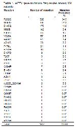
    From all 1113 patients, 854 patients (76.7%) had at least one mutation in MEFV, whereas 259 patients (23.3%) had no mutation. Among 854 patients, 510 were female (59.7%) and 344 were male (40.3%), with the mean age 22.3±16.7 years (min:1 years, max:85 years). The mean age of the complaint onset was 11.6±9.4 years. Among the mutation positive patients, 674 (78.9%) had abdominal pain, 495 (58%) had a fever, 514(63.6%) had arthralgia/arthritis and 93 (10.8%) had chest pain. The consanguinity rate was 31%. A family history of FMF was present in 425 (49.8%) patients. Of those 854 patients, 394 (46.1%) were heterozygous, 63 (7.4%) were homozygous, 240 (28.1%) were compound heterozygous and 157 (18.4%) had complex genotype. 31 different mutations and total 1507 mutations were detected in the patient group. The most common nine mutations were R202Q, M694V, E148Q, M608I, P369S, V726A, R408Q, A744S, and F479L respectively (Table 1).


    Büyütmek İçin Tıklayın
    Table 1: MEFV gene mutations frequencies among 854 patients

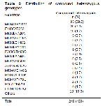
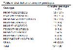
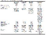
    These nine mutations were 94.6% (1425/1507) of total mutations. The most common six heterozygous mutations were R202Q, M694V, E148Q, M608I, V726A, A744S (Table 2). The most common homozygous genotypes were R202Q, M694V, M608I, E148Q, and V726A, respectively (Table 2). The three most common compound heterozygous genotypes were M694V/R202Q, E148Q/R202Q, and M608I/V726A (Table 3). Among 157 patients with complex genotype, 124 (79%) had three, 30 (19.1%) had four and 3 (1.9%) had five mutations. The most common complex genotype was M694V/R202Q/R202Q (Table 4). 777 (91%) patients underwent MEFV sequencing and the remaining 77 patients (9%) were analyzed for common known MEFV mutations (M694V, M694I, M608I(G/C), M608I(G/A), E148Q, P369S, V726A, A744S, I692del, K695R, R761H, and F479L) with strip assay technique. Exon 2,3,5 and 10 of the MEFV sequencing was the most common molecular testing method in the patient group (716 patients, 83.8%). Exon 10 and whole gene sequenced in 27 (3.2%) and 34 (4%) of the patients respectively. The most common mutation was M694V in the patients underwent strips assay and exon 10 of MEFV sequencing (45.9% and 48.6% respectively), whereas in the exon 2,3,5,10 of MEFV and whole MEFV sequenced patients the most common mutation was R202Q (38.3% and 35.1% respectively) (Table 5).


    Büyütmek İçin Tıklayın
    Table 2: Distribution of heterozygous and homozygous genotypes


    Büyütmek İçin Tıklayın
    Table 3: Distribution of compound heterozygous genotypes


    Büyütmek İçin Tıklayın
    Table 4: Distribution of complex genotypes


    Büyütmek İçin Tıklayın
    Table 5: Comparison of methods used for mutation analysis

  • Başa Dön
  • Özet
  • Giriş
  • Materyal ve Metot
  • Bulgular
  • Tartışma
  • Kaynaklar
  • Tartışma
    Here, we reported the MEFV mutations reports of 1113 patients with clinical suspicion of FMF living in the northern Anatolia. We detected at least one mutation in 854 (76.7%) patients, and 31 different mutations were detected. 259 patients (23.3%) had no mutation. In other studies from Turkey, the prevalence of patients without mutations was in the range of 38.2-65.9% 11,13,14. The lower number of the patients without mutation in our study may be due to the high rate of consanguinity (31%) and we conducted a regional study from a single center.

    FMF is a monogenic disorder inherited in the autosomal recessive manner. Although FMF is inherited autosomal recessively, some recent studies suggested that heterozygous people might manifest a spectrum of clinical features of mild and late-onset FMF. In several studies, as many as 30% of patients with FMF have only a single mutation in MEFV, even after whole exons sequencing. Partial penetrance and variable expressivity were suggested to explain the clinical features of FMF in heterozygous subjects 15,16. 46.1% of our patients with the clinical signs of FMF had heterozygous MEFV mutation.

    In previous regional and country-wide studies from Turkey, R202Q, M694V, E148Q, M608I, V726A, and M694I were reported as the most common mutations 17,18. In this study, the most frequently observed mutations were R202Q, M694V, E148Q, M608I, P369S, and V726A. None of our patients had M694I mutation.

    R202Q was the most common mutation (34.5%) in our study. R202Q was found in 5-34% of the Turkish population. Sayin et al. 19, Coskun et al. 20, Gumus et al. 11, Celep et al. 21 found the R202Q as the most common mutation. In some reports, it is still listed as a polymorphism and the clinical outcome of this alteration is not well defined. However, there were other studies accepted R202Q as a mutation with clinical significance. It was emphasized that homozygous or compound heterozygous R202Q mutations could cause the FMF phenotype 22. Arpacı et al. reported that R202Q, the clinical findings of the patients with R202Q mutation were similar to the diagnostic clinical findings of FMF reported in the literature, and all patients responded to colchicine treatment. They stated that R202Q may be a mutation rather than polymorphism and R202Q may be a risk factor in the development of the FMF clinic 23.

    The G allele of R202Q was found in linkage disequilibrium (LD) with M694V, it means that certain alleles of each gene are inherited together more often than that would be accepted by chance 24. We found that in 179 alleles M694V and R202Q were in LD. Celep et. al showed LD between M694V and R202Q in 43 alleles 21. Kılınç et al. emphasized the frequent accompanying presence of M694V with R202Q and its’ clinical impact in their study 17.

    M694V was the second most common mutation (24.3%) in our study. In the majority of the reports from Turkey, M694V was the most common mutation. Dogan et al. 25 Oztuzcu et al. 26, Barut et al. 27 reported M694V as a first common mutation with the frequency of 42.8%, 41.7%, and 41.1% respectively, whereas, in the study of Evliyaoglu et al. 28 M694V frequency was 3.2%.

    In our study, E148Q was the third most common mutation (11.6%). It was reported as a third common mutation in Turkey with a frequency of 6.8% 8,19. It is found in 3-18% of the major ethnicities where FMF is common 2. The clinical significance of E148Q mutation is not well defined. It is considered as a polymorphism, and usually related with mild FMF phenotype 24.

    P369S was the fifth common mutation (4%) in our patient group. This was the one of the major difference of our study from previous ones. Celep et al. 21 reported the allele frequency of P369S as 1.1% from the northern Anatolia. The allele frequency of P369S was 2.5% in the study of Gumus et al. 11.

    M608I and V726A mutations were two of the common mutations in Turkish population 11. M608I frequency was reported in the range of 14.1-2.4% from previous studies 21. We found M608I as a fourth common mutation (9.4%). V726A frequency was reported in the range of 16.3-1.8 % from previous studies 21. V726A was the sixth common mutation (3.8%) in our study.

    The majority of MEFV gene mutations responsible for the disease are missense mutations, whereas nonsense or deletion type mutations are rare 2. 29 (93.5%) of total 31 different mutations were missense mutations. Only 1 mutation was deletion type and 1 was a nonsense mutation.

    When interpreting the mutation results of the patients, it is important to consider the method used for mutation detection. A number of methods are used in the diagnosis of FMF 17. In some studies, the patients were screened for the most common known mutations responsible for the majority of FMF, whereas some used sequencing of the gene. Some centers prefer sequencing only the exons coding the majority of disease-causing mutations, some others perform whole gene sequencing. Although the patient number was not homogenous, we saw that the method of exon 2,3,5 and 10 sequencing and whole gene sequencing revealed a more different number of mutations, more compound heterozygous and complex genotypes than the strip assay method. We also noticed that the most common mutations varied according to the method used to identify mutations. Gene sequencing either by the Sanger method or next-generation sequencing has the advantage to identify rare or novel variants, whereas interpreting the complex genotypes and novel variants may be difficult, and segregation analysis of the family is needed often.

    In conclusion, our study revealed different results from the previous studies from Turkey. We had a high mutation detection rate in patients. P369S was the one of the commonest mutation in our study. According to the method used to detect MEFV gene variants, the frequencies of the mutations may vary in between the studies. The method should be in consider when giving genetic counseling to the families. We believe that our study may add some contribution to the mutational data of FMF.

  • Başa Dön
  • Özet
  • Giriş
  • Materyal ve Metot
  • Bulgular
  • Tartışma
  • Kaynaklar
  • Kaynaklar

    1) Sohar E, Gafni J, Pras M, Heller H. Familial Mediterranean fever. A survey of 470 cases and review of the literature. Am J Med 1967; 2: 227-253.

    2) Padeh S, Berkun Y. Familial Mediterranean fever. Curr Opin Rheumatol 2016; 5: 523-529.

    3) Van der Hilst JC, Simon A, Drenth JP. Hereditary periodic fever and reactive amyloidosis. Clin Exp Med 2005; 3: 87-98.

    4) Saatci U, Ozen S, Ozdemir S, et al. Familial Mediterranean fever in children: report of a large series and discussion of the risk and prognostic factors of amyloidosis. Eur J Pediatr 1997; 8: 619-623.

    5) Zemer D, Pras M, Sohar E, et al. Colchicine in the prevention and treatment of the amyloidosis of familial Mediterranean fever. N Engl J Med 1986; 16: 1001-1005.

    6) Ben-Chetrit E, Touitou I. Familial Mediterranean fever in the world. Arthritis Rheum 2009; 10: 1447-1453.

    7) Assouad E, El Hage S, Safi S, et al. Familial Mediterranean fever research activity in the Arab world: the need for regional and international collaborations. East Mediterr Health J 2021; 10: 984-992.

    8) Turkish FMF Study Group. Familial Mediterranean fever (FMF) in Turkey: results of a nationwide multicenter study. Medicine (Baltimore) 2005; 1: 1-11.

    9) Yılmaz E, Ozen S, Balcı B, et al. Mutation frequency of familial Mediterranean fever and evidence for a high carrier rate in the Turkish population. Eur J Hum Genet 2001; 7: 553-555.

    10) Chae JJ, Wood G, Richard K, et al. The familial Mediterranean fever protein, pyrin, is cleaved by caspase-1 and activates NF-kappa B through its N-terminal fragment. Blood 2008; 5: 1794-1803.

    11) Gumus E. The Frequency of MEFV Gene Mutations and Genotypes in Sanliurfa Province, South-Eastern Region of Turkey, after the Syrian Civil War by Using Next Generation Sequencing and Report of a Novel Exon 4 Mutation (I423T). J Clin Med 2018; 5: 105.

    12) Livneh A, Langevitz P, Zemer D, et al. Criteria for the diagnosis of familial Mediterranean fever. Arthritis Rheum.1997; 10: 1879-1885.

    13) Gunesacar R, Celik MM, Arica V, et al. Frequency of MEFV gene mutations in Hatay province, Mediterranean region of Turkey and report of a novel missense mutation (I247V). Gene 2014; 2: 195-199.

    14) Ece A, Çakmak E, Uluca Ü, et al. The MEFV mutations and their clinical correlations in children with familial Mediterranean fever in southeast Turkey. Rheumatol Int 2014; 2: 207-212. 15. Ozen S. Changing concepts in familial Mediterranean fever: is it possible to have an autosomal-recessive disease with only one mutation? Arthritis Rheum 2009; 6: 1575-1577.

    16) Marek-Yagel D, Berkun Y, Padeh S, et al. Clinical disease among patients heterozygous for familial Mediterranean fever. Arthritis Rheum 2009; 6: 1862-1866.

    17) Kilinc M, Ganiyusufoglu E, Sager H, et al. The report of sequence analysis on familial Mediterranean fever gene (MEFV) in South- eastern Mediterranean region (Kahramanmaraş) of Turkey. Rheumatol Int 2016; 1: 25-31.

    18) Dundar M, Emirogullari EF, Kiraz A, Taheri S, Baskol M. Common familial Mediterranean fever gene mutations in a Turkish cohort. Mol Biol Rep 2011; 8: 5065-5069.

    19) Sayın Kocakap DB, Günel-Özcan A, Çabuk F, Ensari C. The frequency of familial Mediterranean fever gene mutations and genotypes at Kirikkale and comparison with the mean of regional MEFV mutation frequency of Turkey. Mol Biol Rep 2014; 3: 1419-1426.

    20) Coşkun S, Ustyol L, Bayram Y, et al. The spectrum of MEFV gene mutations and genotypes in Van province, the eastern region of Turkey, and report of a novel mutation (R361T). Gene 2015; 1: 128-131.

    21) Celep G, Durmaz ZH, Erdogan Y, et al. The Spectrum of MEFV Gene Mutations and Genotypes in the Middle Northern Region of Turkey. Eurasian J Med 2019; 3: 252-256.

    22) Ritis K, Giaglis S, Spathari N, et al. Non-isotopic RNAase cleavage assay for mutation detection in MEFV, the gene responsible for familial Mediterranean fever, in a cohort of Greek patients. Ann Rheum Dis 2004; 4: 438-443.

    23) Arpacı A, Doğan S, Erdoğan HF, et al. Presentation of a new mutation in FMF and evaluating the frequency of distribution of the MEFV gene mutation in our region with clinical findings. Mol Biol Rep 2021; 3: 2025-2033.

    24) Touitou I, Lesage S, McDermott M, et al.. Infevers: an evolving mutation database for auto-inflammatory syndromes. Human Mutat 2004; 3: 194-198.

    25) Dogan H, Faruk Bayrak O, Emet M, et al. Familial Medi-terranean fever gene mutations in north-eastern part of Anatolia with special respect to rare mutations. Gene 2015; 2: 170-175.

    26) Oztuzcu S, Ulaşlı M, Ergun S, et al. Screening of common and novel familial mediterranean fever mutations in south-east part of Turkey. Mol Biol Rep 2014; 4: 2601-2607.

    27) Barut K, Pamuk G, Adrovic A, et al. Comparison of Familial Mediterranean Fever and juvenile idiopathic arthritis patients according to family origin. Turk Pediatri Ars 2018; 1: 31-36.

    28) Evliyaoglu O, Bilici S, Yolbas I, Kelekci S, Sen V. Common MEFV gene mutations in children with FMF in Diyarbakır, Turkey. Dicle Tıp Dergisi (Dicle Medical Journal) 2009; 2: 80-84.

  • Başa Dön
  • Özet
  • Giriş
  • Materyal ve Metot
  • Bulgular
  • Tartışma
  • Kaynaklar
  • [ Başa Dön ] [ Özet ] [ PDF ] [ Benzer Makaleler ] [ Yazara E-Posta ] [ Editöre E-Posta ]
    [ Ana Sayfa | Editörler | Danışma Kurulu | Dergi Hakkında | İçindekiler | Arşiv | Yayın Arama | Yazarlara Bilgi | E-Posta ]