[ Ana Sayfa | Editörler | Danışma Kurulu | Dergi Hakkında | İçindekiler | Arşiv | Yayın Arama | Yazarlara Bilgi | E-Posta ]
Fırat Üniversitesi Sağlık Bilimleri Tıp Dergisi
2022, Cilt 36, Sayı 1, Sayfa(lar) 046-050
[ Özet ] [ PDF ] [ Benzer Makaleler ] [ Yazara E-Posta ] [ Editöre E-Posta ]
TGF-Β1 Covid-19 Hastalığının Şiddetini Öngörmede Önemli Bir Prognostik Faktör Olabilir mi?
Mustafa TİMURKAAN1, Hakan AYYILDIZ2, Mehmet KALAYCI3
1Fethi Sekin City Hospital, Internal Medicine Clinic, Elazığ, TÜRKİYE
2Fethi Sekin City Hospital, Biochemistry Laboratory, Elazığ, TÜRKİYE
3Akçakoca State Hospital, Biochemistry Laboratory, Düzce, TÜRKİYE
Anahtar Kelimeler: TGFβ1, Covid19, pandemi, fibroz
Özet
Amaç: Küresel bir salgına neden olan Covid-19, varyant formlarının da ortaya çıkmasıyla birlikte milyonlarca insanı etkilemeye devam etmektedir. Bu çalışmanın amacı, Covid-19 fizyopatolojisinde hastalık şiddetiyle ilişkili olabileceğini düşündüğümüz Transforming growth factor β1 (TGFβ1)’in, hastalar ve sağlıklı popülasyon düzeylerini karşılaştırıp prognostik ve prediktif bir belirteç olarak kullanılıp kullanılmayacağını araştırmaktır.

Gereç ve Yöntem: Bu çalışmada COVID-19 tanısı konmuş ve hastalık şiddetine göre sınıflandırılmış 43 hafif (Bilgisayarlı Tomography (BT) [-], Kadın=21-Erkek=22), 45 şiddetli (BT [+], Kadın=20-Erkek=25) Covid-19 hastası ve 88 Sağlıklı kontrol (Kadın=46-Erkek=42) grubundan oluşmuştur.

Bulgular: Covid-19’ da; TGFβ1, CRP ve D-Dimer düzeylerinde kontrol grubuna göre anlamlı düzeyde artış bulduk. Hasta grubunda TGFβ1 düzeyleri ile CRP düzeyleri arasında pozitif korelasyon bulduk.

Sonuç: Antiviral ilaç ve aşı seçenekleriyle pandemiyi önleme çalışmaları tüm Dünya’ da devam ederken, halen Covid-19 enfeksiyonuna yönelik nihai tedavi bulunabilmiş değildir. Covid-19 enfeksiyonu tanısı almış hastalarda TGFβ1 düzeylerinin, bu hastalık için anahtar molekül olabileceğini ve patofizyolojisinin anlaşılmasıyla birlikte, hastalığın şiddetinin seyri açısından erken tanıda yol gösterici olabileceğini düşünüyoruz.

  • Başa Dön
  • Özet
  • Giriş
  • Materyal ve Metot
  • Bulgular
  • Tartışma
  • Kaynaklar
  • Giriş
    Covid-19 virus (SARS-Cov-2), a new member of the coronavirus family whose origin is still not fully known, has alarmed the entire world, causing announcement of a global pandemic by the WHO (World Health Organization) in March 2020. As of the date of writing of the article, 473.092.602 people were infected, and 1.3% of infected patients died 1,2. Although vaccination efforts continue globally, it is obvious that there are currently no countries that do not have a single dose of the vaccine, as well as that vaccines cannot be adequately vaccinated, given both the production rates and costs.

    For Covid-19 disease, which has a limited treatment option in the current environment, the treatment option in severe patients is provided with ventilator support. In the studies on the pathophysiology of the disease, the viral load and inflammation, fibrosis, and apoptosis caused by this viral load are prominent. Pulmonary fibrosis also increases in direct proportion to the severity of the disease, and there is no treatment option in this regard yet 3,4.

    Transforming growth factor β (TGFβ); TGFβ1 is an important family of cytokines consisting of β2 and β3. The most common of these isoforms is TGFβ1. The TGFβ family is generally synthesized as precursor proteins. The C and N-terminal regions separate from each other when returning from the latent form to the active form. The C-terminal region is called Latency associated peptide, while the N-terminal domain is known as the active TGFβ. The Regulation of this activity is governed by highly complex processes 5. TGF-β1, a pro-fibrotic cytokine, has many functions such as immune function, angiogenesis, cell differentiation, and regulation of cell proliferation. It is also a key mediator in organ fibrosis as a result of excessive collagen production 6-10. TGFβ1, one of the molecules 11 that creates the movement of monocytes, lymphocytes, neutrophils, and fibroblasts to damaged tissues, has a strong chemotaxis effect.

    This study aims to determine whether TGFβ1, which is responsible for fibrosis, chemotaxis, and immune response and is a potent cytokine that is predicted to be related to disease severity in Covid-19 physiopathology, can be used as a prognostic and predictive marker by comparing its values in the patient group and healthy population.

  • Başa Dön
  • Özet
  • Giriş
  • Materyal ve Metot
  • Bulgular
  • Tartışma
  • Kaynaklar
  • Materyal ve Metot
    Research and Publication Ethics: The study was carried out in partnership with Fethi Sekin City Hospital Internal Medicine Clinic and Biochemistry Laboratory. Ethics Committee approval was obtained from Firat University Non-Interventional Research Ethics Commission. (24.06.2021, 2021/08-31)

    In the study, patients with Covid-19 were separated into two groups. The group with pneumonia with lung signs was classified as severe (n=45), and patients with Covid-19 without lung signs were classified as mild (n=43). In addition, a control group consisting of 88 healthy people was included in the study.

    Samples were taken from the people involved in the study to the tube containing aprotinin. Blood samples taken were centrifuged at 4100 rpm for 10 minutes. The plasmas were placed in small volume tubes to study TGFβ1 and these tubes were stored at -70°C until the study day.

    CRP measurements were made with Immage 800 (Beckman Coulter, Inc., Miami, FL, USA). D-Dimer levels were analyzed with the Beckman AU5800 autoanalyzer and the Complete Blood Count (CBC) was analyzed with the Beckman DxH800 (Beckman Coulter, Inc., Miami, FL, USA). Plasma TGFβ1 levels were studied using TGFβ1 Enzyme-Linked ImmunoSorbent Assay kit (FN-Test, Wuhan Fine Biotech Co., Ltd., catalog no: EH0287, Wuhan, China) following the operating procedures outlined in the kit catalog. Absorbance measurement was performed with the Chromate 4300 Microplate Reader (Awareness Technology, Inc., USA). The minimum detection limit of TGFβ1 was 18.75 pg/mL. The intra-inter assay CV% for plasma TGFβ1 were <8% and <10%, respectively.

    G Power programme (Heinrich-Heine-Universitat, Düsseldorf, Germany) was used to calculate the sample size in the study. According to the criteria suggested by Cohen, the effect size was identified as small (d=0.2), medium (d=0.5) and large (d=0.8). The sample size was calculated using the sub-limit (0.8) of the large effect size noted by Cohen 12. In this respect, the minimum sample size for each group was determined to be 42.

    SPSS21 Package Program was used for statistical analysis. Kolmogorov-Smirnov test was used to find out whether the variables showed a normal distribution. Qualitative data were evaluated by the Chi-square test. For categorical or continuous variables, to detect the difference between three or more groups, analyzed by Kruskal-Wallis test using Bonferroni correction then Dunnett's T3 post hoc test was used. To examine the relations between the parameters in the groups with each other, the Spearman correlation test is performed. p<0.05 levels were accepted as the minimum significance level.

  • Başa Dön
  • Özet
  • Giriş
  • Materyal ve Metot
  • Bulgular
  • Tartışma
  • Kaynaklar
  • Bulgular
    The Laboratory, clinical and demographic characteristics of our study are presented in Table 1.


    Büyütmek İçin Tıklayın
    Table 1: The demographic, clinical and laboratory characteristics of Covid-19 and control group.

    As can be seen in Table 1, although there is no significant difference between the groups in terms of gender, there is a significant difference in Covid19 patients with lung findings compared to the control group in terms of age (p= 0.035).

    D-Dimer levels were significantly higher in both patient groups than in the control group (p<0.001). Lymphocyte was significantly lower in both patient groups compared to the control group (p<0.001), the decrease in monocyte levels was significant in CT (+) Covid19 patients.

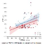
    A positive correlation (r= 0.308, p= 0.003) was found between TGFβ1 and CRP levels in the patient group. In addition, a positive correlation between CRP and D-dimer levels (r= 0.346, p= 0.001); a negative correlation with lymphocyte levels (r= -0.270, p= 0.011) was found.

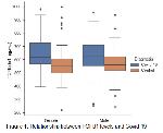
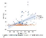
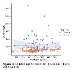
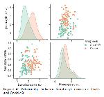
    Plasma TGFβ1, C-Reactive Protein, and D-Dimer levels were determined to have significantly increased in the Covid-19 group compared to the control group (Figure 1, 2 and 3). In addition, White Blood Cell, lymphocyte, monocyte, and platelet levels were significantly lower in Covid-19 patients compared to the control group. Lymphocyte-monocyte relationship is shown in Figure 4.


    Büyütmek İçin Tıklayın
    Figure 1: Relationship between TGFβ1 levels and Covid-19


    Büyütmek İçin Tıklayın
    Figure 2: Relationship between TGFβ1-CRP levels and Covid-19


    Büyütmek İçin Tıklayın
    Figure 3: Relationship between TGFβ1-D-Dimer levels and Covid-19


    Büyütmek İçin Tıklayın
    Figure 4: Relationship between lymphocyte-monocyte levels and Covid-19

    CRP levels were significantly higher in CT (+) and CT (-) Covid 19 patients compared to the control group (p<0.001). CRP levels in the CT (+) group were significantly higher than in the CT (-) group when the patient groups were compared among themselves (p= 0.004) (Figure 5).


    Büyütmek İçin Tıklayın
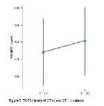
    Figure 5: TGFβ1 levels of CT (-) and CT (+) patients

    Although TGFβ1 levels were high in both patient groups compared to the control group, CT (+) was significantly higher in the patient group than in the control (p= 0.005).

    The relationship between TGFB1 and CRP levels between CT (+) and CT (-) patient groups is shown in Figure 6.


    Büyütmek İçin Tıklayın
    Figure 6: TGFβ1-CRP levels and severity of the disease

  • Başa Dön
  • Özet
  • Giriş
  • Materyal ve Metot
  • Bulgular
  • Tartışma
  • Kaynaklar
  • Tartışma
    In the study, it was concluded that TGFβ1 levels in 88 COVID-19 patients with dissimilar disease levels (mild, severe) were significantly different from TGFβ1 levels in the control group, and it was associated with WBC subgroups, particularly lymphocyte and monocyte.

    Lymphopenia is mostly observed during administration in Covid-19 patients, depending on the severity of the disease. Some studies 13,14 indicate that lymphocyte level may be a potential predictor for the prognosis of the disease. In our study, we found that there is a negative and significant relationship not only between the lymphocyte level but also between the monocyte level and Covid 19. It is believed that the source of this decrease in lymphocyte level is irregular cytokine production, apoptosis of lymphocytes, and autophagy 15,16.

    While lymphocyte levels are low regardless of Covid-19 disease severity, the fact that low monocyte levels are significantly lower in patients diagnosed with Covid-19 pneumonia especially with CT findings, compared to the control group, suggests that monocyte levels can be a rapid diagnostic tool to determine the severity of the disease.

    Moore et al. Covid-19 reported that high-level inflammatory cytokines may be associated with disease severity 17. Cytokine release syndrome is closely related to the clinic of COVID-19 patients and can have serious effects such as respiratory failure, ARDS (Acute Respiratory Distress Syndrome). High levels of TGFβ1 in ARDS play has a significant role in the pathophysiology of pulmonary fibrosis by altering epithelial permeability 18-21. In addition, experimental models have shown that TGF-β has effects such as cell death, glutathione consuming and degradation of epithelial unity 22-24. Based on these actions of TGF-β, it has been stated that this pluripotent cytokine may be a potential target for the treatment of Covid-19 25. In our study, we found that TGFβ1 levels were significantly higher in the Covid-19 of patients with Covid pnomony in CT than in the control group. These findings suggest that TGFβ1, a pro-fibrotic cytokine that may play an important role in disease severity, may occur as a result of changes in permeability at the cellular level caused by alveoli.

    CRP levels are positively correlated with disease seriousness in Covid-19 patients 26,27. CRP levels are high, which is not observed in any other infectious viral disease 28. In our study, we similarly found that CRP levels with TGFβ1 were positively associated with the severity of the disease. It has been reported that the excessive activation of the TGF-β has a central role in the formation of atrial fibrosis 29.

    It is considered that the CRP levels associated with disease severity in Covid-19 patients, which are higher than expected for viral infection, expresses the tissue factor in alveolar epithelial cells 30, myeloid cells 31, endothelial cells 32 and indirectly contributes to TGFβ1 production 33 through Signal Transducer and Activator of Transcription 3 (STAT-3), which is considered an important mediator of cell apoptosis. Thus, it changes TGFβ1 levels, which are associated with lung and atrial fibrosis pathophysiology. For this reason, TGFβ1 levels may also be associated with the pathophysiology of deaths from sudden cardiac diseases in heavy Covid-19 patients and in patients who have had a severe illness and have been discharged.

    In conclusion, the study indicated that TGFβ1 and monocyte levels can be used as a predictive marker of disease severity in Covid19 patients, and D-Dimer and lymphocyte levels can be diagnostic markers. A more detailed examination of the complex physiology of the TGFβ family will shed light on the reasons why some patients have mild symptoms while others have pneumonia cases that are severe enough to require ventilator support. It is considered that the results to be obtained may guide the early diagnosis in terms of the course of severe disease.

    Acknowledgments:Funding Details: No financial support for the research.

    Disclosure Statement: No conflicts of interest.

  • Başa Dön
  • Özet
  • Giriş
  • Materyal ve Metot
  • Bulgular
  • Tartışma
  • Kaynaklar
  • Kaynaklar

    1) WHO/Europe | Coronavirus disease (COVID-19) outbreak - About the virus (Accessed: 30.12.2021).

    2) COVID-19 Map - Johns Hopkins Coronavirus Resource Center (jhu.edu) (Accessed: 30.12.2021).

    3) Carlson FR, Bosukonda D, Keck PC, Carlson WD. Multiorgan Damage in Patients With COVID-19. JACC: Basic to Translational Science 2020; 5: 1145-1148.

    4) Gu J, Gong E, Zhang B, et al. Multiple organ infection and the pathogenesis of SARS. J Exp Med 2005; 202: 415-424.

    5) Batlle E, Massague J. Transforming Growth Factor-β Signaling in Immunity and Cancer. Immunity 2019; 50: 924-940.

    6) Zhang Y, Alexander PB, Wang XF. TGF-β Family Signaling in the Control of Cell Proliferation and Survival. Cold Spring Harb Perspect Biol 2017; 9: a022145.

    7) Ferrari G, Cook BD, Terushkin V, Pintucci G, Mignatti P. Transforming growth factor-beta 1 (TGF-beta1) induces angiogenesis through vascular endothelial growth factor (VEGF)-mediated apoptosis. J Cell Physiol 2009; 219: 449-458.

    8) Meng XM, Nikolic-Paterson DJ, Lan HY. TGF-β: the master regulator of fibrosis. Nat Rev Nephrol 2016; 12: 325-438.

    9) Travis MA, Sheppard D. TGF-β activation and function in immunity. Annu Rev Immunol 2014; 32: 51-82.

    10) Wang MK, Sun HQ, Xiang YC, Jiang F, Su YP, Zou ZM. Different roles of TGF-β in the multi-lineage differentiation of stem cells. World J Stem Cells 2012; 4: 28-34.

    11) Han G, Li F, Singh TP, Wolf P, Wang XJ. The Pro-inflammatory Role of TGFβ1: A Paradox? Int J Biol Sci 2012; 8: 228-235.

    12) Lakens D. Calculating and reporting effect sizes to facilitate cumulative science: a practical primer for t-tests and ANOVAs. Front Psychol 2013; 4: 863

    13) Chen G, Wu D, Guo W, et al. Clinical and immunological features of severe and moderate coronavirus disease 2019. J Clin Invest 2020; 130: 2620-2629.

    14) Yuan X, Huang W, Ye B, et al. Changes of hematological and immunological parameters in COVID-19 patients. Int J Hematol 2020; 12: 1-7.

    15) Huang C, Wang Y, Li X, et al. Clinical features of patients infected with 2019 novel coronavirus in Wuhan, China. Lancet 2020; 395: 497-506.

    16) Xiong Y, Liu Y, Cao L, et al. Transcriptomic characteristics of bronchoalveolar lavage fluid and peripheral blood mononuclear cells in COVID-19 patients. Emerg Microbes Infect 2020; 9: 761-770.

    17) Moore JB, June CH. Cytokine release syndrome in severe COVID-19. Science 2020; 368: 473-474.

    18) Liu Y, Tan W, Chen H, et al. Dynamic changes in lymphocyte subsets and parallel cytokine levels in patients with severe and critical COVID-19. BMC Infect Dis 2021; 21: 79.

    19) Frank JA, Matthay MA. TGF-beta and lung fluid balance in ARDS. Proc Natl Acad Sci 2014; 111: 885-886.

    20) Overgaard CE, Schlingmann B, Dorsainvil WS, et al. The relative balance of GM-CSF and TGF-beta1 regulates lung epithelial barrier function. Am J Physiol Lung Cell Mol Physiol 2015; 308: 1212-1223.

    21) Peters DM, Vadasz I, Wujak L, et al. TGF-beta directs trafficking of the epithelial sodium channel ENaC which has implications for ion and fluid transport in acute lung injury. Proc Natl Acad Sci 2014; 111: 374-383.

    22) Pittet JF, Griffiths MJD, Geiser T, et al. TGF-beta is a critical mediator of acute lung injury. J Clin Invest 2001; 107:1537-1544.

    23) Xu X, Zheng L, Yuan Q, et al. Transforming growth factor-β in stem cells and tissue homeostasis. Bone Res 2018; 6:2.

    24) Liu RM, Gaston Pravia KA. Oxidative stress and glutathione in TGF-beta-mediated fibrogenesis. Free Radic Biol Med 2010; 48: 1-15.

    25) Chen W. A potential treatment of COVID-19 with TGF-beta blockade. Int J Biol Sci 2020; 16: 1954-1955.

    26) Wang L. C-reactive protein levels in the early stage of COVID-19. Med Mal Infect 2020; 50: 332-334.

    27) Chen W, Zheng KI, Liu S, et al. Plasma CRP level is positively associated with the severity of COVID-19. Ann Clin Microbiol Antimicrob 2020; 19: 18.

    28) Kayser S, Kunze R, Sheriff A. Selective C-reactive protein apheresis for Covid-19 patients suffering from organ damage. Ther Apher Dial 2021; 25: 251-252.

    29) Gramley F, Lorenzen J, Koellensperger E, et al. Atrial fibrosis and atrial fibrillation: The role of the TGF-beta1 signaling pathway. Int J Cardiol 2010; 143: 405-413.

    30) Bastarache JA, Wang L, Geiser T, et al. The alveolar epithelium can initiate the extrinsic coagulation cascade through expression of tissue factor. Thorax 2007; 62: 608-616.

    31) Shaver CM, Grove BS, Clune JK, et al. Myeloid tissue factor does not modulate lung inflammation or permeability during experimental acute lung injury. Sci Rep 2016; 6: 22249.

    32) Devaraj S, Kumaresan PR, Jialal I. C-reactive protein induces release of both endothelial microparticles and circulating endothelial cells in vitro and in vivo: further evidence of endothelial dysfunction. Clin Chem 2011; 57: 1757-1761.

    33) Matsuyama T, Kubli SP, Yoshinaga SK, Pfeffer K, Mak TW. An aberrant STAT pathway is central to COVID-19. Cell Death Differ 2020; 27: 3209-3225.

  • Başa Dön
  • Özet
  • Giriş
  • Materyal ve Metot
  • Bulgular
  • Tartışma
  • Kaynaklar
  • [ Başa Dön ] [ Özet ] [ PDF ] [ Benzer Makaleler ] [ Yazara E-Posta ] [ Editöre E-Posta ]
    [ Ana Sayfa | Editörler | Danışma Kurulu | Dergi Hakkında | İçindekiler | Arşiv | Yayın Arama | Yazarlara Bilgi | E-Posta ]