[ Ana Sayfa | Editörler | Danışma Kurulu | Dergi Hakkında | İçindekiler | Arşiv | Yayın Arama | Yazarlara Bilgi | E-Posta ]
Fırat Üniversitesi Sağlık Bilimleri Tıp Dergisi
2022, Cilt 36, Sayı 2, Sayfa(lar) 117-124
[ Özet ] [ PDF ] [ Benzer Makaleler ] [ Yazara E-Posta ] [ Editöre E-Posta ]
Şiddetli COVID-19 Enfeksiyonunda Önemli Bir Biyomarker Olarak Artan Platelet Large Cell-Ratio Düzeyi
Pınar ÖNER1, Sait Fatih ÖNER2
1Elazığ Fethi Sekin City Hospital, Department of Microbiology, Elazığ, TÜRKİYE
2Elazığ Fethi Sekin City Hospital, Department of Anesthesiology and Reanimation, Elazığ, TÜRKİYE
Anahtar Kelimeler: COVID-19, SARS-CoV-2, trombosit büyük hücre oranı, trombosit hacim indeksleri
Özet
Amaç: Şiddetli COVID-19 enfeksiyonunun PLC-R düzeyleri üzerindeki etkilerini ortaya koymak amaçlandı.

Gereç ve Yöntem: COVID-19 tanısı konan 111 hasta çalışmaya dahil edildi. Nazofaringeal sürüntülerinde RT-PCR testleri ile SARS-CoV-2 pozitif tespit edilen hastalar çalışmaya dahil edildi. COVID-19 hastaları akciğer BT bulgularına göre üç gruba ayrıldı. Grup 1 (45 hasta) hafif, Grup 2 (34 hasta) orta ve Grup 3 (32 hasta) şiddetli olarak tanımlandı. Tüm hastaların PVI değerleri, CRP, D-dimer ve lipid profilleri dahil olmak üzere tam kan sayımı parametreleri analiz edildi. COVID-19 hasta grupları ile PLC-R değerleri arasındaki korelasyon SPSS programı ve ANFC yöntemleri kullanılarak gösterildi.

Bulgular: Bu çalışmanın en önemli bulgusu, şiddetli COVID-19 hastalarında PLC-R'nin orta ve hafif hastalara göre anlamlı derecede daha yüksek olmasıdır. Spearman's rho korelasyon analizi hastalığın şiddeti ile PLC-R ve WBC düzeylerinin arttığını, Htc ve Hb düzeylerinin ise azaldığını gösterdi. ROC analizi, PLC-R>%38.3'ün şiddetli COVID-19 hastalığını öngörmede %59.4 duyarlılığa ve %68,4 özgüllüğe sahip olduğunu gösterdi (AUC 0.672, %95 CI 0.560, 0.784; p=0.005, cut off=38.3). Grup 3'teki hastaların CRP, ferritin ve D-dimer değerlerinin Grup 1'deki hastalara göre anlamlı düzeyde yüksek olduğu, grup 3'teki hastaların demir değerlerinin ise grup 1'deki hastalara göre anlamlı düzeyde düşük olduğu görüldü.

Sonuç: PLC-R değerleri, akut trombotik olayları öngörmede değerli bir parametredir. Bu çalışmanın sonuçlarına göre PLC-R değerleri, COVID-19 enfeksiyonunun şiddetini tanımlamak için uygun bir biyobelirteç olarak kullanılabilir.

  • Başa Dön
  • Özet
  • Giriş
  • Materyal ve Metot
  • Bulgular
  • Tartışma
  • Kaynaklar
  • Giriş
    In December 2019, the World Health Organization reported a new beta-coronavirus named "SARS-CoV2" responsible for the COVID-19 outbreak 1. Since clinical symptoms and radiological findings are not specific for this disease, the diagnosis should be confirmed by nucleic acid-based polymerase chain reaction (PCR) 2. Individuals infected with SARS-CoV-2 present with different clinical pictures ranging from mild respiratory tract infection to acute respiratory distress syndrome, organ dysfunctions, and even death in critical cases 3. Timely clinical intervention and follow-up of the diagnosed patients are essential in terms of prognosis 4. Severe COVID-19 patients have an increased risk of thrombosis, including pulmonary embolism, venous thrombosis, and ischemic stroke 5. The high incidence of venous thromboembolism and the importance of anticoagulant thromboprophylaxis are noted in the guidelines and supported by sequential autopsy findings 6.

    It is known that platelets, which also have important roles in the immune response, play an important role in developing these thrombotic complications 7. Hematological tests used in diagnosing and treating many diseases, including COVID-19 infection, are used as the first choice in terms of low cost and ease of operation 8,9. Platelet volume indices (PVI) are parameters that are checked in hematological tests. PVI can be considered an indicator of increased platelet activity. PVI parameters include PLC-R, platelet large cell count (PLCC), mean platelet volume (MPV), mean platelet volume-to-platelet count ratio (MPV/P), platelet distribution width (PDW) and platelet distribution width-to-platelet count ratio (PDW/P) 10.

    PLC-R is a percentage-based measure of circulating larger platelets (>12 fL), measured by using the formula PLC-R=Large platelet cell counts (>12 fL)/Total platelet counts. It has been used to monitor platelet activity as well. The number of platelets greater than 12 fL is referred to as PLCC. Plt×PLC-R is multiplied to form PLCC 11.

    In this study, we aimed to represent the effects of the severity of COVID-19 infection on PLC-R.

  • Başa Dön
  • Özet
  • Giriş
  • Materyal ve Metot
  • Bulgular
  • Tartışma
  • Kaynaklar
  • Materyal ve Metot
    Research and Publication Ethics:The study was performed by Helsinki principles and approved by the local Ethics Committee (Presidency of T.C. Firat University Ethics Committee). Ethical approval number: 2021/07-37, date: 27.05.2021.

    Study Participants: This cross-sectional study was conducted in the Elazığ Fethi Sekin City Hospital, designated as the coronavirus pandemic hospital in the province by the ministry of health. A total of 111 SARS-CoV-2 infected patients with lung involvement treated in the COVID-19 wards and intensive care units between November 2020 and April 2021 were included in the study. Patients with a prior diagnosis of chronic illness or atherosclerotic risk factors were excepted from the study. Positive results for SARS-CoV-2 based on a typical RT-PCR test Bio-Rad CFX96 Real-Time PCR Detection System (Bio-Rad Laboratories, Inc., Hercules, CA, USA) performed on nasopharyngeal swabs were included in the study Groups.

    Patients with COVID-19 pneumonia were divided into three Groups according to their chest computed tomography (CT) features. Group 1 (45 patients) was defined as mild, Group 2 (34 patients) as moderate, Group 3 (32 patients) as severe. The distribution of ground-glass opacities and consolidations to the affected lung lobes was evaluated qualitatively and visually using CT images 12. Group 1 (mild): small, localized ground glass opacities beginning at the lung periphery (SpO2 96%-98%). Group 2 (moderate): opacities in more lung lobule, diffused, reticular "crazy-paving" patterns, and arterial stiffening, with small consolidations possible (SpO2 90%-95%). Group 3 (severe): heavy porous consolidations covering a large proportion of the lungs, with more noticeable septal thickenings (SpO2 90%). Laboratory parameters of the patients at the time of admission were examined.

    Laboratory Measurements: Each blood sample (6 mL for complete biochemistry, 5 mL for complete blood count) was drawn from the antecubital vein. Samples were collected and analyzed in vacuum tubes, including 15% K3-ethylenediaminetetraacetic acid anticoagulation tubes (Sarstedt, Essen, Belgium). Complete blood count (CBC) parameters were measured using a Sysmex XN-550 hematology analysis tool (Sysmex Europe GmbH, Sysmex Corporation, Hamburg, Germany). A 3 mL sample of venous blood obtained in diamine tetraacetic acid-containing anticoagulation tubes were used to assess PVI. After half an hour of bloodletting, the blood samples of two Groups by using an electronic blood counter were analyzed. Platelet shape changes were allowed to stabilize during the half-hour waiting period. The levels of D-dimer, glucose, urea, creatinine, total cholesterol, triglycerides, high-density lipoprotein cholesterol, and low-density lipoprotein cholesterol (LDL-c) were tested using the chemiluminescence method on a Beckman Coulter AU5800 (Beckman Coulter, USA) auto-analyzer. C-reactive protein (CRP) levels were measured by using a nephelometric method (image 800, Beckman Coulter, USA).

    Statistical Analysis: Power analysis was applied with the G*Power program to determine the adequacy of the sample size for PLC-R value comparison in COVID-19 patients. The sample size was found to be 58 patients in total, 29 patients for each Group (d: 0.879 ve Actual Power:0.951).

    Statistical analyses were performed using SPSS software, version 26.0 (SPSS Inc., Chicago, IL, USA) for Windows. The Kolmogorov-Smirnov test was used to evaluate whether the variables were normally distributed. Platelet, glucose, triglycerides, urea, creatinine, CRP, iron, transferrin saturation (TSAT), ferritin, and D-dimer didn't show normal distribution. These variables were analyzed by the Kruskal Wallis test and were presented as medians with 25th–75th percentiles. Tamhane's T2 test was used for post hoc comparisons. Other continuous variables were analyzed by one-way ANOVA and were presented as means with standard deviations (SD). Bonferronni test was used for post hoc comparisons. Correlation analyses were performed using Spearman's rho correlation test. Receiver operating characteristics (ROC) analysis was performed to evaluate the specificity and sensitivity of PLC-R levels in detecting severe COVID-19 disease. All P-values were two-tailed, and values <0.05 were considered to indicate statistical significance.

    In addition, all parameters were analyzed in detail with the adaptive neuro-fuzzy classifier (ANFC) method. ANFC systems can be used in pattern recognition, decision making, and many other fields. These systems depend on linguistic rules, which are ensured by experts or the rules, and are removed from a given training dataset by various clustering methods 13.

  • Başa Dön
  • Özet
  • Giriş
  • Materyal ve Metot
  • Bulgular
  • Tartışma
  • Kaynaklar
  • Bulgular
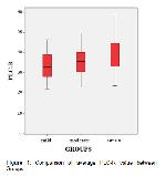
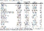
    This study performed laboratory analyses, among 111 SARS-CoV-2 infected patients, including 45 mild, 34 moderates, and 32 severe cases. Comparison of PLC-R average value comparison between mild, moderate, and severe Groups are demonstrated (Figure 1). It was observed that PLC-R values increased significantly in severe SARS CoV-2 patients (33.24%±6.24%, 35.73%±6.98% and 38.92%±7.64% in three Groups, respectively, p=0.003) (Table 1). There was no statistically significant difference in other PVI values between the three Groups (MPV (p=0.290), PDW (p=0.493), PLCC (p=0.080), total platelet counts (p=0.073) (Table 1). White blood cell (WBC), LDL-c, hemoglobin and hematocrit results showed significant values between Groups (p=0.002, p=0.002, p=0.018, p=0.007, respectively) (Table 1).


    Büyütmek İçin Tıklayın
    Figure 1: Comparison of average PLC-R value between Groups.


    Büyütmek İçin Tıklayın
    Table 1: Comparison of laboratory data between three Groups.

    CRP, ferritin, iron and D-dimer values differ significantly between Group 3 and Group 1 (p<0.001, p=0.003, p=0.031 and p<0.001, respectively). It was observed that the CRP, ferritin and D-dimer values of the patients in Group 3 were significantly higher than the patients in Group 1, and the iron values of the patients in Group 3 were significantly lower than the patients in Group 1. There was no significant difference between Groups in TSAT levels (p=0.399) (Table 1).

    According to Spearman's rho correlation analysis results, PLC-R levels (r=0.299, p=0.001) and WBC levels (r=0.332, p<0.001) were positively correlated with COVID-19 severity. Ht levels (r=-0.276, p=0.003) and Hb levels (r=-0.273, p=0.004) were negatively correlated with COVID-19 severity.

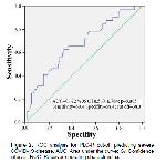
    The ROC curve analysis showed that PLC-R> 38.3% had 59.4% sensitivity and 68.4% specificity in predicting severe COVID-19 disease (AUC 0.672, %95 CI 0.560, 0.784; p=0.005, cut off=38.3) (Figure 2). There was no statistically significant difference between the Groups regarding age and other investigated laboratory parameters (Table 1).


    Büyütmek İçin Tıklayın
    Figure 2: ROC analysis for PLC-R cut-off predicting severe COVID-19 disease. AUC: Area under the curve; CI: Confidence interval; ROC: Receiver operating characteristics.

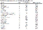
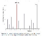
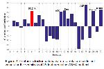
    As shown in Table 2, the increase in the p-value calculated by the ANFC method detects an increasing disease association. Thus, the relationship between PLC-R and COVID-19 is proven in Table 3. It makes sense if the correlation coefficient is positive and significant. For this reason, it is understood that PLC-R is an effective finding parameter among COVID-19 patients. In the meantime, we made two separate calculations of total linguistic hedge values and correlation coefficient with the ANFC method. The PLC-R value was found to be the only platelet parameter that significantly correlated with these two calculations (Figure 3 and 4).


    Büyütmek İçin Tıklayın
    Table 2: The representation of linguistic hedge (P) values


    Büyütmek İçin Tıklayın
    Table 3: The representation of p and correlation Coefficient values oth the ANFC method


    Büyütmek İçin Tıklayın
    Figure 3: Feature selection criteria with total linguistic hedge values for the sensitivity of 25 features in the ANFC method.


    Büyütmek İçin Tıklayın
    Figure 4: Feature selection criteria with a correlation coefficient values for the sensitivity of 25 features in the ANFC method.

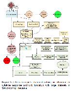
    With many different analysis results, the PLC-R value has been an essential parameter for COVID-19 patients. Also schematic representation of pathogenic inflammatory cytokine response and clot formation with large platelets in SARS-CoV-2 infections are demonstrated in Figure 5.


    Büyütmek İçin Tıklayın
    Figure 5: Schematic representation of pathogenic inflammatory cytokine response and clot formation with large platelets in SARS-CoV-2 infections.

  • Başa Dön
  • Özet
  • Giriş
  • Materyal ve Metot
  • Bulgular
  • Tartışma
  • Kaynaklar
  • Tartışma
    The critical result of our study is that PLC-R was remarkably higher in the severe COVID-19 patients. According to different types of correlation analysis results, PLC-R levels were positively correlated with COVID-19 severity.

    Inflammation caused by viral infections can cause dyslipidemia in patients. A previous study reported that the LDL-c levels of COVID-19 patients decreased 14. SARS-CoV-2 can damage, liver function and reduce LDL-c biosynthesis. SARS-CoV-2 causes acute inflammation by causing an increase in proinflammatory cytokine levels. It can disrupt the efflux and transport of lipids by changing liver functions in cytokines. Free radicals occurring in the host cell in viral infection can also disrupt lipid metabolism 15. SARS-CoV-2 infection can cause cholesterol molecules to leak into tissues such as alveolar spaces to form exudate by increasing vascular permeability. This causes the lipid level in the plasma to decrease 16. This study observed that LDL-c levels decreased in the severe Group of COVID-19 patients.

    The literature studies found severe impairment occurred in kidney function tests and blood electrolyte levels in severe COVID-19 patients. Acute kidney damage due to cell and tissue damage from SARS-CoV-2 and lack of oral intake of patients reveasl these pathological findings 17. According to our data, as the severity of COVID-19 infection increased, urea, Na, Ca levels increased meaningfully.

    An increase in non-specific inflammatory biomarkers such as CRP, D-dimer, and ferritin is observed in COVID-19 patients. A study of 653 COVID-19 patients reported that serum ferritin values in patients with severe COVID-19 infection were higher than patients with mild COVID-19 infection 18. Shah et al. reported no significant difference in serum ferritin levels and transferrin saturation between patients with non-severe and severe hypoxemia, but serum iron levels were significantly lower 19. In our study, in the Group with severe COVID-19 infection, it was observed that CRP and ferritin levels increased statistically, and iron levels were statistically decreased. There was no significant difference in transferrin saturation. High ferritin levels may indicate a robust inflammatory response, with SARS-CoV-2 entry into the human body and its effect on iron metabolism 20. Iron is an essential micronutrient for both humans and pathogens 21. During infection, humans' innate immune system response limits the amount of iron to deprive it of the pathogen, leading to anemia 22. Anemia reduces oxygen delivery to tissues and causes multi-organ failure. Studies show that Hb levels decrease in COVID-19 infection 23. We also obtained findings that support previous studies. Inflammatory changes caused by COVID-19 infection can affect erythropoiesis and cause a decrease in hemoglobin. SARS-CoV-2 binds to the beta chains of hemoglobin via surface glycoproteins, inhibiting both metabolism and hemoglobin denaturation. Hemoglobinopathy and iron dysmetabolism together with hypoxia can seriously compromise the O2 carrying capacity of erythrocytes and induce tissue changes due to hyperferritinemia 24. For these reasons, decreased hemoglobin levels for COVID-19 may be an indicator of disease progression.

    Viral and bacterial infections are generally associated with thrombocytopenia and changes in platelet morphology. Platelets are involved in hemostasis, coagulation, immune, and inflammatory responses 25. SARS-CoV-2 uses the spike protein to enter host cells by binding to angiotensin-converting enzyme 2 in the host cell membrane. Spike protein enhances thrombus formation. Coagulation factors are released, inflammatory cytokines are secreted, and leukocyte platelet clusters are formed 26. Fibrin deposition with coagulation activation and subsequent fibrinolysis increase D-dimer level, an essential intravascular coagulation indicator. Studies have proven that high D-dimer values at admission to the hospital are associated with poor prognosis 27. According to the results of our research, it was observed that D-dimer values increased statistically significantly in severe COVID-19 infection. Continued inflammation can rapidly lead to the development of cytokine storm and macrophage activation syndrome and hyperinflammatory response. Cytokine storm is a syndrome that releases proinflammatory cytokines that cause acute respiratory distress syndrome and multiple organ dysfunction syndromes in case of infection 28. Macrophage activation syndrome is a proinflammatory cascade associated with a high rate of thrombosis and death in sepsis 29. In COVID-19 disease, increasing cytokine levels and inflammation initiate platelet destruction by causing bone marrow involvement. Accumulation in the lungs causes more platelet consumption. The decrease in the platelet count leads to an increase in the production of young and immature platelets that are functionally more active and larger sized 30. Larger platelets have more granules and receptors, and they are metabolically and enzymatically more active than small platelets, produce more thromboxane A2, and tend to clot more quickly 31. Since the destruction in the bone marrow will be more significant in severe COVID-19 infection, the number of large platelets released into the circulation will increase with the release of more megakaryocytes 32. Mitophagy impairment occurs in patients with COVID-19 due to oxidative stress and hyperinflammation. Mitophagy protects platelets from oxidative stress and mitochondrial destruction by removing damaged mitochondria to prevent platelet apoptosis in healthy individuals. However, mitochondrial dysfunction in platelets in COVID-19 infection may affect platelet survival and apoptosis, potentially increasing the risk of thrombus formation. Studies have shown that apoptotic platelets can induce clotting ≥50 times faster than normal platelets 33. In the studies, patients with myocardial infection have reported that larger platelets contribute to the prothrombotic state 34. Microvascular and macrovascular thromboembolic complications in the lung, spleen, brain, intestine and peripheral vessels have been reported in COVID-19 35. It has been reported that stroke has been detected in young patients with covid-19, even without underlying coagulopathy before the infection 36. Large platelets may be one of the causes of these thrombotic events seen in COVID-19 infection.

    Platelet activity should be evaluated with PVI parameter values, not platelet counts. PVI parameters are measurements calculated by devices and can be easily detected in blood tests, typically as part of the CBC, not requiring advanced or expensive technology 11.

    The MPV and PDW parameters are widely and routinely used in clinics around the world. When platelet production decreases, young platelets grow, their volume increases, and they become more active. As a result, MPV and PDW levels increase 37. It has been reported that MPV and PDW values increase to higher levels in patients with sepsis, and PDW is a poor prognostic factor in severe sepsis 38. In a study by Güçlü et al., it was reported that mortality increased in parallel with the increase in MPV value in COVID-19 infection 39. However, we found that PLC-R was significantly higher in severe COVID-19 disease, although we did not see an increase in these values. In the meantime, a comprehensive calculation was made with the ANFC method, while PLC-R findings were found as a constant factor parameter, and it was brought to the literature.

    PVI in patients with COVID-19 infection is a simple, effortless, and economical test that should be used to predict the probability of acute thrombotic events. Larger platelets can easily be identified during routine complete blood count analysis, and such patients can benefit from preventive treatment. According to our data, it has been proven by many analysis methods that the PLC-R value can be used as a suitable biomarker that can describe the severity of COVID-19 infection.

  • Başa Dön
  • Özet
  • Giriş
  • Materyal ve Metot
  • Bulgular
  • Tartışma
  • Kaynaklar
  • Kaynaklar

    1) Huang C, Wang Y, Li X, et al. Clinical features of patients infected with 2019 novel coronavirus in Wuhan. China Lancet 2020; 395: 497-506.

    2) Zhou P, Yang XL, Wang XG, et al. A pneumonia outbreak associated with a new coronavirus of probable bat origin. Nature 2020; 579: 270-273.

    3) Gordon DE, Jung GM, Bouhaddou M, et al. A SARS-CoV-2 protein interaction map reveals targets for drug repurposing. Nature 2020; 583: 459-468.

    4) Taleghani N, Taghipour F. Diagnosis of COVID-19 for controlling the pandemic: A review of the state-of-the-art. Biosens Bioelectron 2021; 174: 112830.

    5) Llitjos JF, Leclerc M, Chochois C, et al. High incidence of venous thromboembolic events in anticoagulated severe COVID-19 patients. J Thromb Haemost 2020; 18: 1743-1746.

    6) Iba T, Levy JH, Connors JM, et al. The unique characteristics of COVID-19 coagulopathy. Crit Care 2020; 24: 360.

    7) Manne BK, Denorme F, Middleton EA, et al. Platelet gene expression and function in patients with COVID-19. Blood. 2020; 136: 1317-1329.

    8) Gong J, Ou J, Qiu X, et al. A Tool to Early Predict Severe 2019-Novel Coronavirus Pneumonia (COVID-19): A Multicenter Study using the Risk Nomogram in Wuhan and Guangdong, China. Clin Infect Dis 2020; 71: 833-840.

    9) Sharma D, Dayama A, Banerjee S, et al. To Study the Role of Absolute Lymphocyte Count and RDW in COVID 19 Patients and their Association with Appearance of Symptoms and Severity. J Assoc Physicians India 2020; 68: 39-42.

    10) Takeuchi H, Abe M, Takumi Y, et al. The prognostic impact of the platelet distribution width-to-platelet count ratio in patients with breast cancer. PLoS One 2017; 12: e0189166.

    11) Yılmaz M, Kayançiçek H, Gözel N, et al. Platelet Large Cell Ratio Levels in Smoking. F.U.Med. J. Health Sci. 2018; 32: 65-70.

    12) Ippolito D, Ragusi M, Gandola D, et al. Computed tomography semi-automated lung volume quantification in SARS-CoV-2-related pneumonia. Eur Radiol 2021; 31: 2726-2736.

    13) Cetisli B. Development of an adaptive neuro-fuzzy classifier using linguistic hedges: Part 1. Expert Systems with Applications 2010; 37: 6093-6101.

    14) Wei X, Zeng W, Su J, et al. Hypolipidemia is associated with the severity of COVID-19. J Clin Lipidol 2020; 14: 297-304.

    15) Zidar DA, Juchnowski S, Ferrari B, et al. Oxidized LDL levels are increased in HIV infection and may drive monocyte activation. J Acquir Immune Defic Syndr 2015; 69: 154-160.

    16) Tian S, Hu W, Niu L, et al. Pulmonary pathology of early phase 2019 novel coronavirus (COVID-19) pneumonia in two patients with lung cancer. J Thorac Oncol 2020; 15: 700-704.

    17) Cheng Y, Luo R, Wang K, et al. Kidney disease is associated with in-hospital death of patients with COVID-19. Kidney Int 2020; 97: 829-838.

    18) Henry BM, De OMHS, Benoit S, Plebani M, Lippi G. Hematologic, biochemical and immune biomarker abnormalities associated with severe illness and mortality in coronavirus disease 2019 (COVID-19): A meta-analysis. Clin Chem Lab Med 2020; 58: 1021-1028.

    19) Shah A, Frost JN, Aaron L, Donovan K, Drakesmith H. Collaborators Systemic hypoferremia and severity of hypoxemic respiratory failure in COVID-19. Crit Care 2020; 24: 320.

    20) Middeldorp S, Coppens M, van Haaps TF, et al. Incidence of venous thromboembolism in hospitalized patients with COVID-19. J Thromb Haemost 2020; 18: 1995-2002.

    21) Lodigiani C, Iapichino G, Carenzo L, et al. Venous and arterial thromboembolic complications in COVID-19 patients admitted to an academic hospital in Milan, Italy. Thromb Res 2020; 191: 9-14.

    22) Poissy J, Goutay J, Caplan M, et al. Pulmonary embolism in COVID-19 patients: awareness of an increased prevalence. Circulation 2020; 142: 184-186.

    23) Sun S, Cai X, Wang H, et al. Abnormalities of the peripheral blood system in patients with COVID-19 in Wenzhou, China Clin Chim Acta 2020; 507: 174-180.

    24) Cavezzi A, Troiani E, Corrao S. COVID-19: Hemoglobin, iron, and hypoxia beyond inflammation. A narrative review. Clin Pract 2020; 10: 1271.

    25) Qu R, Ling Y, Zhang YH, et al. Platelet-to-lymphocyte ratio is associated with prognosis in patients with coronavirus disease-19. J Med Virol 2020; 92: 1533-1541.

    26) Zhang S, Liu Y, Wang X, et al. SARS-CoV-2 binds platelet ACE2 to enhance thrombosis in COVID-19. J Hematol Oncol 2020; 13: 120.

    27) Hanff TC, Mohareb AM, Giri J, Cohen JB, Chirinos JA. Thrombosis in COVID-19. Am J Hematol 2020; 95: 1578-1589.

    28) England JT, Abdulla A, Biggs CM, et al. Weathering the COVID-19 storm: Lessons from hematologic cytokine syndromes. Blood Rev 2021; 45: 100707.

    29) Karakike E, Giamarellos-Bourboulis EJ. Macrophage activation-like syndrome: a distinct entity is leading to early death in sepsis. Front Immunol 2019; 10: 55.

    30) Gao Y, Li Y, Yu X, et al. The impact of various platelet indices as prognostic markers of septic shock. PLoS One 2014; 9: 103761.

    31) Thompson CB, Eaton KA, Princiotta SM, Rushin CA, Valeri CR. Size-dependent platelet subpopulation: Relationship of platelet volume to ultrastructure, enzymatic activity and function. Br J Haematol 1982; 50: 509–520.

    32) Geddis AE. Megakaryopoiesis. Semin Hematol 2010; 47: 212-219.

    33) Saleh J, Peyssonnaux C, Singh KK, Edeas M. Mitochondria and microbiota dysfunction in COVID-19 pathogenesis. Mitochondrion 2020; 54: 1-7.

    34) Pizzulli L, Yang A, Martin JF, Lüderitz B. Changes in platelet size and count in UA compared to stable angina or non-cardiac chest pain. Eur Heart J 1998; 19: 80-84.

    35) Vulliamy P, Jacob S, Davenport RA. Acute aorto-iliac and mesenteric arterial thromboses as presenting features of COVID-19. Br J Haematol 2020; 189: 1053-1054.

    36) Klok FA, Kruip MJHA, Van Der Meer NJM, et al. Confirmation of the highest cumulative incidence of thrombotic complications in critically ill ICU patients with COVID-19: an updated analysis. Thromb Res 2020; 191: 148-150.

    37) Gowda SB, Gosavi S, Rao AA, et al. Prognosis of COVID-19: Red cell distribution width, platelet distribution width, and c-reactive protein. Cureus 2021; 13: e13078.

    38) Güçlü E, Durmaz Y, Karabay O. Effect of severe sepsis on platelet count and their indices. Afr Health Sci 2013; 13: 333-338.

    39) Güçlü E, Kocayiğit H, Okan HD, et al. Effect of COVID-19 on platelet count and its indices. Rev Assoc Med Bras 2020; 66: 1122-1127.

  • Başa Dön
  • Özet
  • Giriş
  • Materyal ve Metot
  • Bulgular
  • Tartışma
  • Kaynaklar
  • [ Başa Dön ] [ Özet ] [ PDF ] [ Benzer Makaleler ] [ Yazara E-Posta ] [ Editöre E-Posta ]
    [ Ana Sayfa | Editörler | Danışma Kurulu | Dergi Hakkında | İçindekiler | Arşiv | Yayın Arama | Yazarlara Bilgi | E-Posta ]