[ Ana Sayfa | Editörler | Danışma Kurulu | Dergi Hakkında | İçindekiler | Arşiv | Yayın Arama | Yazarlara Bilgi | E-Posta ]
Fırat Üniversitesi Sağlık Bilimleri Tıp Dergisi
2023, Cilt 37, Sayı 1, Sayfa(lar) 036-042
[ Özet ] [ PDF ] [ Benzer Makaleler ] [ Yazara E-Posta ] [ Editöre E-Posta ]
Artmış Ürotensin II Düzeyleri, COVID-19 Şiddetini Öngörebilir mi?
Selda TELO1, Mutlu KULUÖZTÜRK2
1Fırat University, Faculty of Medicine, Department of Biochemistry, Elazığ, TÜRKİYE
2Fırat University, Faculty of Medicine, Department of Chest Disease, Elazığ, TÜRKİYE
Anahtar Kelimeler: COVID-19, ürotensin II, mortalite
Özet
Amaç: COVID tanısı almış hastalarda ürotensin II (UII) düzeyinin ve prognostik değerinin belirlenmesi amaçlanmıştır.

Gereç ve Yöntem: COVID-19 tanısı alan ardışık 90 hasta ve 30 sağlıklı birey değerlendirildi. COVID-19 hastaları başvuruda hafif-orta ve ağır hastalar olarak sınıflandırıldı. Tedavi öncesi tam kan sayımı, serum UII ve CRP düzeyleri belirlendi. Hastane mortalitesi, yoğun bakım ünitesine (YBÜ) transfer ve taburcu edilen hastalar kaydedildi.

Bulgular: 76 hastanın (%84,4) taburcu edildiği, 14 hastanın (%15,6) yoğun bakıma alındığı veya eksitus olduğu belirlendi. Ağır COVID-19 hastalarında, hafif-orta dereceli COVID-19 hastalarına göre mortalite ve yoğun bakıma transfer oranlarında artış bulundu. Şiddetli COVID-19 hastalarında serum UII seviyeleri, hafif-orta dereceli COVID-19 hastalarına ve sağlıklı kontrole göre istatistiksel olarak anlamlı şekilde arttı. Ayrıca, hafif-orta dereceli COVID-19 hastaları, sağlıklı kontrole göre daha yüksek UII seviyelerine sahipti. Yoğun bakım ünitesine transfer edilen veya eksitus olan COVID-19 hastalarında, taburcu olan COVID-19 hastalarına kıyasla artmış UII seviyeleri bulundu. Hastalığın ciddiyetini tahmin etmek için ROC analizi ile UII'nin cut-off değeri ≥5.26 alındığında, UII'nin ROC eğrisinde eğri altında kalan alanı (AUC) 0.688 (0,580-0.796; %95 CI; p<0.01) vardı. Ayrıca, UII'nin duyarlılığı %80 ve özgüllüğü %59'du.

Sonuç: Sonuçlarımız, artan UII düzeylerinin COVID-19 hastalarında hastalık şiddeti ile ilişkili olabileceğini göstermiştir. Bu hastalarda UII'nin ilk değerlendirmesi, olumsuz sonuçların tahmin edilmesine yardımcı olabilir.

  • Başa Dön
  • Özet
  • Giriş
  • Materyal ve Metot
  • Bulgular
  • Tartışma
  • Kaynaklar
  • Giriş
    Inflammatory and immune responses in COVID-19 infection are important for the treatment of the infection. Additionally, they may influence the expression of the clinical spectrum of COVID-19 disease. Inflammatory responses play a critical role in the progression of COVID-19 1. Accumulating evidence demonstrated that several inflammatory markers, such as procalcitonin (PCT), serum ferritin, C-reactive protein (CRP), and interleukin-6 (IL-6), were significantly associated with the high risks of the development of severe COVID-19 2-5. However, the role of inflammatory markers in monitoring the severity of COVID-19 is still controversial.

    Human urotensin II (UII) is an 11 amino acid cyclic peptide and is known as the most potent vasoconstrictor. It is expressed in the central nervous system as well as other tissues and circulates in human plasma. The net effect of UII on vascular tone is a balance between endothelium-independent vasoconstriction and endothelium-dependent vasodilatation 6. Increased plasma levels were observed in several diseases, such as renal failure, congestive heart failure, diabetes mellitus, and systemic hypertension 7-10. The remarkable relationship between UII and inflammation was also demonstrated in recent studies 11,12. In these studies, it was determined that UII was involved in the process of inflammatory injury while having a notable effect on the development of inflammatory diseases.

    In this study, we aimed to determine the level of UII and its prognostic value in patients with COVID-19. Furthermore, it was aimed to evaluate the correlation between UII and other laboratory parameters.

  • Başa Dön
  • Özet
  • Giriş
  • Materyal ve Metot
  • Bulgular
  • Tartışma
  • Kaynaklar
  • Materyal ve Metot
    Research and Publication Ethics: Ethical approval was obtained from the Ethics Review Committee of Firat University, Elazig, Türkiye. Ethical approval number: 2020/13-21.

    Study Design and Participants: Ninety consecutive patients with COVID-19 diagnosis, who were admitted to the Pandemic Clinic in the Faculty of Medicine at Firat University, were evaluated. During the period from August 2020 to March 2020, confirmed and hospitalized cases of COVID-19 were included in this study. Moreover, 30 healthy individuals, who matched the age and sex with the subjects in the sample group, were included in the study as the control group. All of the cases included in the study were informed about the study, and informed consent was obtained from each participant. Chronic kidney disease, coronary artery disease, hypertension, acute hepatic failure, heart failure were considered as exclusion criteria.

    COVID-19 infection was diagnosed according to the SARS-CoV-2 positive real-time reverse-transcriptase polymerase chain reaction (RT-PCR) from a nasal and/or throat swab along with signs, symptoms, or radiological findings of COVID-19. All of the COVID-19 patients in the study were classified as mild-moderate and severe patients upon admission according to the clinical findings, respiratory rates, oxygen saturation (SpO2) levels, and low-dose chest CT findings (13). The details of the classification were described below.

    Mild-moderate COVID-19: Mild clinical symptoms, no sign of pneumonia on low-dose CT or mild respiratory symptoms, positive signs of pneumonia on low-dose CT, and SpO2 ≥94% on room air.

    Severe COVID-19: Those who meet any of the following criteria: (1) Respiratory rate ≥30 beats per minute, (2) SpO2 <94% at room air, (3) lung infiltrates > %50 on low-dose CT

    The end-point of our study was the presence of at least one of the following outcomes: (1) Death from COVID-19, (2) Need for intensive care unit (ICU) transfers, (3) Discharged patients. Accordingly, death or transfer to ICU was defined as adverse outcomes in patients with COVID-19.

    Data Collection: In the study, the demographic data of the patients were recorded in addition to clinical and radiologic parameters. Low-dose chest CT scans were performed on each patient upon admission and before the treatment. Blood samples were taken from both the control subjects and the patients with COVID-19 before the treatment.

    Laboratory Analyses: Complete blood cell counts were analyzed via a high-volume hematology analyzer (ADVIA 2120i, Siemens Healthcare Diagnostics Inc., Tarrytown, NY, USA). The blood samples were collected in potassium-ethylenediaminetetraacetic acid tubes and analyzed within one hour after venipuncture. The CRP levels were determined via a nephelometric analyzer (BN II System, Siemens Healthcare Diagnostics Inc., Tarrytown, NY, USA) by the immunonephelometry method. Thus, the complete blood cell count and CRP findings of the patients were included in the evaluation.

    The serum was separated by centrifuging the samples at 4000 g for 10 min. Then the samples were frozen at −80 °C for further analysis. The serum UII concentrations were measured using via a double-antibody sandwich enzyme-linked immunosorbent assay kit (Catalog No: 201–12-5285 Human Urotensin II Elisa Kit: Sunred Biological Technology Co. Ltd, Shanghai). The assay sensitivity was 0.055 pmol/L, and the inter-assay and intra-assay coefficient of variability (%CV) values were <12% and <10%, respectively. The detection range of Urotensin II was 0.1‐15 pmol/L.

    Statistical Analyses: In the current study, the IBM SPSS Statistics 21 (Statistical Product and Service Solutions version 21.0, authorization code: d91314f638c364094170; Armonk, NY, USA) package software was used for the statistical analyses. The results were presented as mean±standard deviation. The level of statistical significance was regarded as p<0.05. Statistical analysis was performed by conducting Kruskal-Wallis tests for multiple-group comparisons and conducting Mann-Whitney U tests to evaluate any observed differences for significance. Furthermore, the results were interpreted according to the Bonferroni correction. On the other hand, the categorical variables were compared by using the chi-square test. Receiver operating characteristic (ROC) curve analysis was conducted to identify the optimal cut-off values of UII to predict adverse outcomes in patients with COVID-19. The parametric variables were evaluated by the Pearson correlation analysis.

  • Başa Dön
  • Özet
  • Giriş
  • Materyal ve Metot
  • Bulgular
  • Tartışma
  • Kaynaklar
  • Bulgular
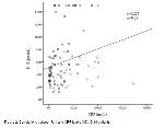
    In this study, 90 patients with COVID-19 infections were retrospectively evaluated. There was no significant difference in sex (p>0.05, X2=1.589) between the groups. Of the sample, 35 subjects were classified into the mild/moderate disease group while 55 patients were classified into the severe disease group. The mean age was significantly higher in severe COVID-19 patients than in both mild-moderate COVID-19 patients and control subjects. It was determined that 76 patients (84.4%) were discharged while 14 patients (15.6%) were transferred to ICU or died. The increased mortality and transfer to the ICU rates in severe COVID-19 patients compared to mild-moderate COVID-19 patients were shown in Table 1. UII levels were significantly increased in severe COVID-19 patients compared to mild-moderate COVID-19 patients and control subjects (Figure 1A). Additionally, mild-moderate COVID-19 patients had increased levels of UII than the control subjects (Figure 1A). CRP, fibrinogen, and ferritin levels were significantly higher in severe COVID-19 patients compared to the mild-moderate COVID-19 patients (Table 1). Furthermore, the UII levels were significantly higher in COVID-19 patients who were transferred to ICU or exitus compared to discharged COVID-19 patients (Figure 1B). Baseline characteristics of patients and laboratory parameters, which were grouped according to the severity of COVID-19, were demonstrated in Table 2. When we evaluated the correlation between UII and other laboratory parameters, significant positive correlations were observed between CRP and UII levels in COVID-19 patients (r=0.229, p<0.05) (Figure 2). When the cut-off value of UII was regarded as ≥5.26 pmol/L by ROC analysis for the predicting to the severity of COVID-19, UII had an area under the curve (AUC) in the receiver operating characteristic (ROC) curve of 0.688 (0.580-0.796; 95% Confidence Interval; p<0.01). The UII level of 5.26 was considered as the cut-off threshold between mild-moderate and severe COVID-19 patients. Accordingly, UII had a sensitivity of 80% and specificity of 59% (ROC curve) (Figure 3).


    Büyütmek İçin Tıklayın
    Table 1: The ratios of COVID-19 patients according to study end-points.


    Büyütmek İçin Tıklayın
    Figure 1: The UII levels according to groups and study end-points


    Büyütmek İçin Tıklayın
    Figure 2: Correlations between U-II and CRP levels COVID-19 patients


    Büyütmek İçin Tıklayın
    Figure 3: A) the cut-off level of UII in patients with mild-moderate and severe COVID-19, B) receiver operating characteristic (ROC) curve indicating sensitivity and specificity of UII in the prediction of severity of COVID-19


    Büyütmek İçin Tıklayın
    Table 2: Baseline characteristics of patients and laboratory parameters in patients with COVID-19 and controls

  • Başa Dön
  • Özet
  • Giriş
  • Materyal ve Metot
  • Bulgular
  • Tartışma
  • Kaynaklar
  • Tartışma
    Our study results showed that UII levels were significantly higher in both mild/moderate COVID-19 and severe COVID-19 patients compared to the control subjects. Moreover, there were more prominent increases in the severe COVID-19 patients and in patients who died or were transferred to ICU. In the correlation analysis, it was determined that the UII levels only correlated with the CRP levels. Additionally, it was determined that UII had a sensitivity of 80% and specificity of 59% in the differentiation of severe COVID-19 patients from mild/moderate COVID-19 patients.

    UII is known to be the most potent vasoconstrictor, and it has many physiological and pathophysiological activities, such as vasoconstrictor and vasodilator actions, cell proliferation, pro-fibrosis, and inflammatory effects. UII is involved in the process of inflammatory injury and plays a prominent role in the onset and development of inflammatory diseases. Although urotensin and its receptor are much more abundant in the central nervous system, it is expressed in the various tissues, such as the lung, kidney, spleen, and small intestine in addition to circulating in human plasma 6,14. Particularly, the highest expression of urotensin mRNA was observed in the mice lung 15, which suggested a role of UII/UII-related peptide in respiratory function. In another study, consistent with this hypothesis, it was demonstrated that UII in vitro induced a concentration-dependent contraction of airways and pulmonary blood vessels in monkeys 16. Furthermore, Kristof et al. described the cellular distribution of the urotensin II system in the normal lung 17. Accordingly, it was demonstrated that U-II played active roles in several lung diseases, such as pulmonary hypertension, lung adenocarcinoma, and sepsis-induced lung injury. Previous studies also demonstrated that chronic hypoxia induction resulted in enhanced contractile activity in the airways and enhanced pulmonary responses to UII in rats 16,18. Moreover, increased UII secretion with hypoxia was demonstrated in both in vitro and in vivo analyses 19,20. Same as chronic hypoxia, acute hypoxia results in increased UII secretion 21. Contrary to this response in rats, UII did not exhibit this response in pulmonary arteries isolated from humans 18,22. In another study, the cellular localization of UII and URP in patients with lymphangioleiomyomatosis was defined 17. On the other hand, synthetic UII stimulated A549 cell proliferation and accelerated the growth of A549 tumor xenografts in mice 23. Additionally, it was demonstrated that UII mRNA and UII immunoreactivity were also present in human lung adenocarcinoma cell line A549.These results suggest that UII may contribute to the pathogenesis of lung adenocarcinoma. UII promotes the release of IL‐6, TNF-α, and MMP‐9 (speculated to be due to the increased activation of NF‐κB) in the tumor microenvironment, and this may be one of the most important mechanisms by which UII promotes lung adenocarcinoma growth 24. In another study, the administration of UII resulted in increased expression of TNF-α, IL1 β, and IL6 mRNA in human endothelial cells 25. Additionally, it was reported that UII and its receptor contributed to the sepsis-induced lung damage of chronically diabetic mice, and this damage was prevented by the urotensin-II receptor antagonist 12. Many previous studies reported that UII antagonists reduced the inflammatory cytokine response 26-28. Dose-dependent reduction in the mRNA expressions of TNF-α, IL1β, IL6, and NFκB with UII antagonist in mice lung tissue and reduced serum TNF-α level were also reported in another study 12. For these reasons, it can be considered that UII modulates the inflammatory response.

    All the evidence collected until today indicates that increased inflammatory response plays an important role in COVID-19, and the severity of COVID-19 is increased by the inflammatory cytokine storm. Accordingly, cytokines and chemokines lead to the accumulation of immune cells and activate the immune responses 29. For these reasons, it is important to determine the markers to monitor the progression of the disease and ensure the early treatment of patients. An anti-inflammatory treatment approach may play an important role while controlling the inflammation in the progression of COVID-19 1. To date, several inflammatory factors, such as IL-6, TNF-α, CRP, and PCT, were determined as independent factors in the prediction of the severity of COVID-19 30-32. In this study, the results demonstrated increased levels of UII in COVID-19 patients, especially in severe cases, and it was observed that UII was correlated with CRP. To our knowledge, this is the first study that demonstrates the increased levels of UII in COVID-19 patients and the correlation between UII and CRP. CRP is a sensitive systemic marker, which can be used as an indicator of inflammation. This study is preliminary in terms of demonstrating that UII can be involved in the pathogenesis of COVID-19, and it may be a potential biomarker for monitoring the progression of the disease. However, the relationship between U-II and the inflammatory markers, such as IL-6 and TNF alpha, were not evaluated in this study.

    This study has some limitations. Firstly, our sample size was relatively small, and the study was conducted in a single healthcare center. Secondly, the lack of follow-up and post-treatment data may play significant roles while determining the significance of UII in this disease.

    In conclusion, increased levels of UII may be related to disease severity in COVID-19 patients. Initial evaluation of UII in these patients can help predict the adverse outcomes. In future studies, larger sample sizes and participation from several healthcare centers can determine the role of UII on COVID-19 pathogenesis and treatment approaches.

    Acknowledgments: This work has not received any financial support.

  • Başa Dön
  • Özet
  • Giriş
  • Materyal ve Metot
  • Bulgular
  • Tartışma
  • Kaynaklar
  • Kaynaklar

    1) Stebbing J, Phelan A, Griffin I, et al. COVID-19: Combining antiviral and anti-inflammatory treatments. Lancet Infect Dis 2020; 20: 400-402.

    2) Cheng K, Wei M, Shen H, et al. Clinical characteristics of 463 patients with common and severe type coronavirus disease (In Chinese). Shanghai Med J 2020; 1-15.

    3) Gao Y, Li T, Han M, et al. Diagnostic utility of clinical laboratory data determinations for patients with the severe COVID-19. J Med Virol 2020; 92: 791-796.

    4) Qin C, Zhou L, Hu Z, et al. Dysregulation of immune response in patients with COVID-19 in Wuhan, China. Clin Infect Dis 2020; 71: 762-768.

    5) Zeng F, Huang Y, Guo Y, et al. Association of inflammatory markers with the severity of COVID-19: A meta-analysis. Int J Infect Dis 2020; 96: 467-474.

    6) Ong KL, Lam KS, Cheung BM. Urotensin II: Its function in health and its role in disease. Cardiovasc Drugs Ther 2005; 19: 65-75.

    7) Totsune K, Takahashi K, Arihara Z, et al. Role of urotensin II in patients on dialysis. Lancet 2001; 358: 810-811.

    8) Totsune K, Takahashi K, Arihara Z, Sone M, Ito S, Murakami O. Increased plasma urotensin II levels in patients with diabetes mellitus. Clin Sci 2003; 104: 1-5.

    9) Russell FD, Meyers D, Galbraith AJ, et al. Elevated plasma levels of human urotensin-II immunoreactivity in congestive heart failure. Am J Physiol Heart Circ Physiol 2003; 285: 1576-1581.

    10) Cheung BM, Leung R, Man YB, Wong LY. Plasma concentration of urotensin II is raised in hypertension. J Hypertens 2004; 22: 1341-1344.

    11) Yang Y, Zhang J, Chen X, et al. UII/GPR14 is involved in NF-κB-mediated colonic inflammation in vivo and in vitro. Oncology Reports 2016; 36: 2800-2806.

    12) Ugan RA, Cadirci E, Halici Z, Toktay E, Cinar I. The role of urotensin-II and its receptor in sepsis-induced lung injury under diabetic conditions. Eur J Pharmacol 2018; 818: 457-469.

    13) NIH Coronavirus Disease 2019 (COVID-19) Treatment Guidelines. Downloaded from https://www.covid19treatmentguidelines.nih.gov/ (Last Updated: December 17, 2020).

    14) Vaudry H, Leprince J, Chatenet D, et al. International union of basic and clinical pharmacology. XCII. urotensin II, urotensin II-related peptide, and their receptor: from structure to function. Pharmacol Rev 2015; 67: 214-258.

    15) Dubessy C, Cartier D, Lectez B, et al. Characterization of urotensin II, distribution of urotensin II, urotensin II-related peptide and UT receptor mRNAs in mouse: Evidence of urotensin II at the neuromuscular junction. J Neurochem 2008; 107: 361-374.

    16) Hay DW, Luttmann MA, and Douglas SA. Human urotensin-II is a potent spasmogen of primate airway smooth muscle. Br J Pharmacol 2000; 131: 10-12.

    17) Kristof AS, You Z, Han Y, Giaid A. Protein expression of urotensin II, urotensin-related peptide and their receptor in the lungs of patients with lymphangioleiomyomatosis. Peptides 2010; 31: 1511-1516.

    18) MacLean MR, Alexander D, Stirrat A, et al. Contractile responses to human urotensin-II in rat and human pulmonary arteries: Effect of endothelial factors and chronic hypoxia in the rat. Br J Pharmacol 2000; 130: 201-204.

    19) Prosser HC, Forster ME, Richards AM, Pemberton CJ. Urotensin II and urotensin II-related peptide (URP) in cardiac ischemia-reperfusion injury. Peptides 2008; 29: 770-777.

    20) Zhang Y, Li J, Cao J, et al. Effect of chronic hypoxia on contents of urotensin II and its functional receptors in rat myocardium. Heart Vessels 2002; 16: 64 -68.

    21) Gao S, Oh YB, Shah A, et al. Urotensin II receptor antagonist attenuates monocrotaline-induced cardiac hypertrophy in rats. Am J Physiol Heart Circ Physiol 2010; 299: 1782-1789.

    22) Stirrat A, Gallagher M, Douglas SA, et al. Potent vasodilator responses to human urotensin-II in human pulmonary and abdominal resistance arteries. Am J Physiol Heart Circ Physiol 2001; 280: 925-928.

    23) Wu YQ, Song Z, Zhou CH, et al. Expression of urotensin II and its receptor in human lung adenocarcinoma A549 cells and the effect of urotensin II on lung adenocarcinoma growth in vitro and in vivo. Oncol Rep 2010; 24: 1179-1184.

    24) Zhou CH, Wan YY, Chu XH, et al. Urotensin II contributes to the formation of lung adenocarcinoma inflammatory microenvironment through the NF-kB pathway in tumor-bearing nude mice. Oncol Lett 2012; 4: 1259-1263.

    25) Park SL, Lee BK, Kim Y, Lee BH, Jung Y. Inhibitory Effect of an Urotensin II Receptor Antagonist on Proinflammatory Activation Induced by Urotensin II in Human Vascular Endothelial Cells. Biomol Ther (Seoul) 2013; 21: 277-83.

    26) Cadirci E, Halici Z, Yayla M, et al. Blocking of urotensin receptors as new target for treatment of carrageenan induced inflammation in rats. Peptides 2016; 82: 35-43.

    27) Liu LM, Liang DY, Ye CG, Tu WJ, Zhu T. The UII/UT system mediates upregulation of proinflammatory cytokines through p38 MAPK and NF-kappa B pathways in LPS-stimulated kupffer cells. PLoS One 2015; 10: 0121383.

    28) Onat AM, Turkbeyler IH, Pehlivan Y, et al. The efficiency of a urotensin II antagonist in an experimental lung fibrosis model. Inflammation 2012; 35 (3): 1138-1143.

    29) Xu Z, Shi L, Wang Y, et al. Pathological findings of COVID-19 associated with acute respiratory distress syndrome. Lancet Respir Med 2020; 8: 420-422.

    30) Liu F, Li L, Xu M, et al. Prognostic value of interleukin-6, C-reactive protein, and procalcitonin in patients with COVID-19. J Clin Virol 2020; 127: 104370.

    31) Ji P, Zhu J, Zhong Z, et al. Association of elevated inflammatory markers and severe COVID-19: A meta-analysis. Medicine (Baltimore) 2020; 99: 23315 32. Azkur AK, Akdis M, Azkur D, et al. Immune response to SARS-CoV-2 and mechanisms of immunopathological changes in COVID-19. Allergy 2020; 75: 1564-1581.

  • Başa Dön
  • Özet
  • Giriş
  • Materyal ve Metot
  • Bulgular
  • Tartışma
  • Kaynaklar
  • [ Başa Dön ] [ Özet ] [ PDF ] [ Benzer Makaleler ] [ Yazara E-Posta ] [ Editöre E-Posta ]
    [ Ana Sayfa | Editörler | Danışma Kurulu | Dergi Hakkında | İçindekiler | Arşiv | Yayın Arama | Yazarlara Bilgi | E-Posta ]