[ Ana Sayfa | Editörler | Danışma Kurulu | Dergi Hakkında | İçindekiler | Arşiv | Yayın Arama | Yazarlara Bilgi | E-Posta ]
Fırat Üniversitesi Sağlık Bilimleri Tıp Dergisi
2023, Cilt 37, Sayı 1, Sayfa(lar) 066-079
[ Özet ] [ PDF ] [ Benzer Makaleler ] [ Yazara E-Posta ] [ Editöre E-Posta ]
Sinapik Asit Sisplatin Kaynaklı Periferik Nöropatiyi İyileştirir: İn Vivo ve İn Vitro Bir Çalışma
Caner YILDIRIM1, Hatice YAMANER1, Mustafa ÖRKMEZ2, Sibel CANGİ3, Mehmet Akif BOZDAYI2, Demet TAŞDEMİR2, Sena CEVİK1
1Gaziantep University, Faculty of Medicine, Department of Physiology, Gaziantep, TÜRKİYE
2Gaziantep University, Faculty of Medicine, Department of Medical Biochemistry, Gaziantep, TÜRKİYE
3Gaziantep University, Faculty of Medicine, Department of Pathology, Gaziantep, TÜRKİYE
Anahtar Kelimeler: Sisplatin, nöropati, nosiseptif davranış deneyleri, sinapik asit, sitotoksisite, in vivo ve in vitro çalışma
Özet
Amaç: Platin bileşikleri, özellikle sisplatin, en etkili antitümör ajanlar arasındadır. Bununla birlikte, ilacın ciddi yan etkileri vardır. Sisplatinin sitotoksik etkileri nedeniyle kullanımı sınırlıdır. Kanser kemoterapisinde kısıtlayıcı faktörlerden biri nörotoksisitedir. Sinapik asitin antioksidan ve antienflamatuar bir etkinliği olduğundan, sisplatin nöropatisi üzerindeki koruyucu etkilerinin araştırılması amaçlandı.

Gereç ve Yöntemler: Spraque Dawley erkek sıçanlar rastgele beş gruba ayrıldı: grup I, normal kontrol; grup II, taşıyıcı; grup III, CIS; grup IV, CIS+SA 20 mg/kg; ve grup V, CIS + SA 40 mg/kg. Çalışmanın tamamlanmasının ardından hayvanlar üzerinde davranışsal nosiseptif çalışmalar ve sinir iletim hızı ölçümleri yapılmıştır. Hayvanlar daha sonra sakrifiye edilerek siyatik sinir dokusunda ve kanda oksidatif stres göstergeleri ve proinflamatuar sitokin seviyeleri ELISA yöntemi kullanılarak değerlendirildi. Siyatik sinir dokusunun histopatolojik boyaması yapıldı. Ek olarak, sinapik asiti sisplatinin antitümör aktivitesi üzerindeki olası etkisi sitotoksisite deneyleri ile değerlendirildi.

Bulgular: Sisplatine maruz kalan sıçanlarda, somatosensoriyel disfonksiyon, motor inkoordinasyon ve siyatik sinir motorik iletim hızında azalma görüldü. CMAP latens süresi artmıştı. Biyokimyasal olarak oksidatif stres belirteçleri ve proinflamatuar sitokin seviyeleri yükselmişti. Histopatolojik değerlendirmede akson dejenerasyonu, ödem ve fibrozis saptandı. Sinapik asit, oksidatif stresi ve proinflamatuar sitokin düzeylerini azaltmıştır. Sinapik asit ayrıca CMAP ve sinir lifi mimarisini de restore ettiği tespit edilmiştir.

Sonuç: Sinapik asit tedavisi, sisplatinin neden olduğu istenmeyen etkileri iyileştirmiştir. Sinapik asidin sisplatinin antitümör etkinliğini azaltmadan restoratif etkilere sahip olduğu değerlendirilmiştir.

  • Başa Dön
  • Özet
  • Giriş
  • Materyal ve Metot
  • Bulgular
  • Tartışma
  • Kaynaklar
  • Giriş
    During the 1960s, cisplatin was discovered to possess cytotoxic properties, making it one of the most effective therapeutic agents used to treat several solid tumors, such as small cell lung cancer, ovary cancer, testis cancer, head and neck cancer, and endometrial cancer 1. For over half a century, platinum agents, particularly cisplatin, have been the most effective antitumor agents. However, the drug has serious adverse effects. The use of cisplatin is limited due to its cytotoxic effects, such as neurotoxicity, nephrotoxicity, and hepatotoxicity 2. Peripheral neuropathy is among the main factors restricting progress during cancer treatment, manifesting as neuropathic pain in over 50% of patients receiving cisplatin during cancer chemotherapy 2-4. This condition is usually caused by the use of cisplatin, and it causes a reduction in dose or early suspension of chemotherapy, thereby adversely affecting the prognosis.

    Sinapic acid (SA) is a hydroxycinnamic acid derivative and polyphenol compound commonly found in various oilseed plants, cereals, vegetables, and fruits. In recent years, various in vivo/in vitro studies of SA and its main derivatives (sinapine, 4-vinylsingol, sinapoyl esters and syringaldehyde) have been carried out by many researchers, and as a result of these studies, it has been proven that they possess broad-spectrum pharmacological properties. These features are mainly anti-inflammatory, antioxidant, antimicrobial, anxiolytic, neuroprotective, cardioprotective, hepatoprotective, antihyperglycaemic, and anticancer properties 5-7. Sinapic acid has been shown to protect neurons against amyloid beta-induced Alzheimer's disease 8, 6-OHDA-induced Parkinson's disease 9, and kainic acid-induced neuronal damage 10. It was demonstrated to improve cognitive performance in mice suffering from scopolamine-induced amnesia 11. Sinapic acid has been demonstrated to have a nephroprotective effect in rats to cisplatin-induced nephrotoxicity by preventing renal dysfunction, oxidative stress, and an increase in free radicals 12. Sinapic acid has also been shown to have cytotoxic effects on prostate cancer cell lines (PC-3 and LNCaP) as well as human laryngeal carcinoma cell lines (HEp-2) 13,14. We hypothesized that sinapic acid could protect against cisplatin-induced neuropathy due to its strong antioxidant and anti-inflammatory properties. Due to the toxicity induced by chemotherapeutic drugs, an effective struggle against the tumor is impossible because the treatment is discontinued or the dose is lessened. The goal of this study is to reduce the neurotoxic effects of cisplatin and to alleviate the adverse effects.

  • Başa Dön
  • Özet
  • Giriş
  • Materyal ve Metot
  • Bulgular
  • Tartışma
  • Kaynaklar
  • Materyal ve Metot
    Research and Publication Ethics: This research was carried out in Gaziantep University Experimental Animal Research Center (GAUNDAM) in accordance with the ethical rules after it was approved by the Gaziantep University local ethics committee with decision number 2021/59 and protocol number 229

    Experimental Design: This research was carried out at the Gaziantep University Experimental Animal Research Center in accordance with the ethical rules. The Gaziantep University Experimental Animals Research Center provided Sprague Dawley male rats (8–10 weeks old, 200 ± 20 g weight, n = 35) for this experiment. We kept the animals in the animal room for a week to allow them to get used to their new surroundings. Throughout the experiment, the animals were given ad libitum access to pellet feed and drinking water. All animals were housed in cages with a 12-hour light/dark photoperiod in automated air-conditioned rooms with a constant temperature of 21±2°C.

    The animals were grouped as follows:

    Control group: The animals were not given any intervention for five weeks.

    Sham group: For five weeks (weekly at around 10:00 a.m. on Mondays), a physiological saline solution was given intraperitoneally (i.p.). For five weeks, a diluted DMSO solution was given intragastrically (daily at around 5:00 p.m.).

    CIS group: For five weeks (weekly at around 10:00 a.m. on Mondays), a single dose of cisplatin (3 mg/kg) was given i.p. 15.

    CIS+LSA group: For five weeks (daily at around 5:00 p.m.), SA (20 mg/kg) was given intragastrically [16, 17]. For five weeks (weekly at around 10:00 a.m. on Mondays), a single dose of cisplatin (3 mg/kg) was given i.p. (15).

    CIS+HSA group: For five weeks (daily at around 5:00 p.m.), SA (40 mg/kg) was given intragastrically 16,17. For five weeks (weekly at around 10:00 a.m. on Mondays), a single dose of cisplatin (3 mg/kg) was given i.p. 15.

    Drugs and Chemicals: All chemicals were purchased from Santa Cruz Biotechnology (Santa Cruz, CA, USA). SA was dissolved in dimethyl sulfoxide (DMSO) 12, and the DMSO was then diluted with physiological saline. The final volume of DMSO administered to the animals did not exceed 10%, and the animals were given this volume for five weeks. Cisplatin was dissolved in physiological saline. All solutions were freshly prepared before the experiment.

    In Vivo Experiments
    The rotarod motor activity test: The rotarod motor activity test is a frequently used technique for assessing motor coordination (Ugo Basile, Comerio, Italy). After acclimating the animals to the device and environment, a ramp protocol was used, beginning at 5 rpm and gradually increasing to 40 rpm for 300 seconds 18,19. The animals ran on the rotarod equipment 3 times, with a 5-minute break between each run. Group averages were then computed from the time spent by the animals on the rod.

    The Hot/Cold plate test: This test was used to evaluate thermal hypoalgesia or hyperalgesia. A plate encircled by plexiglass cylinders (Ugo Basile, Comerio, Italy, model 35100) was heated to 55°C. On the hot plate, the animal was left alone. The time interval between when the animal was put on the plate and when its feet were withdrawn was measured. Rats were not allowed to remain on the plate longer than 30 seconds (cut-off) to avoid tissue injury and/or excess suffering, despite the fact that their usual response time is between 5 and 20 seconds 20,21. The cold plate test was performed to determine the presence of cold allodynia and cold hyperalgesia (model 35100, Ugo Basile, Comerio, Italy). Cold allodynia was evaluated at a temperature of 10°C, whereas cold hyperalgesia was assessed at a temperature of 4°C 22,23. Rats were only left on the plate for 50 seconds to avoid tissue injury.

    The Tail-Flick test: The tail-flick test was used to determine the central effects of the pain threshold. For the tail-flick test, a photosensor was positioned beneath the area where the tail is inserted. The rat pulled its tail when it felt pain due to the thermal light (235 mW/cm2 and 50°C temperature) generated by the photosensor area at a constant distance from the 3–4 cm of the animal’s tail end in a special chamber (Ugo Basile 37360 Comerio, Italy). The duration between when the thermal light is applied and when the tail is pulled was then noted. To ensure that the animal would not suffer excessively, the duration was limited to 10 seconds 20.

    The von frey filament test: This technique is used to determine the mechanical pain threshold. The rats were contained in an apparatus coated in plexiglass and supported by a metal perforated wire base. To assess the withdrawal threshold, a mechanical stimulus (von Frey filaments [0.4–15 g, logarithmically incremental stiffness; Bioseb, Vitrolles, France]) was applied to the hind paw, mid-plantar surface of each rat. Three times in series, the animals’ hind paw withdrawal responses were measured, with a minimum of 10 seconds between each measurement. The mechanical pain threshold was determined as the average of these 3 measurements 24-26.

    The adhesive removal test: The adhesive removal test was used to assess sensorimotor function. Animals were expected to detect and remove rectangular labels (Kinesio bands) (10x10 mm) placed on the plantar surfaces of both forepaws 27. For this purpose, after the adhesives adhered to the plantar surface of the forehand, the rats’ time to notice and remove these adhesives was measured.

    Following each behavioral observation, the cylinder and plate surfaces were carefully cleaned with 70% ethyl alcohol to eliminate any scent clues left by the previous animal.

    Electrophysiological Experiments: The animals were anesthetized (with a combination of 10 mg/kg xylazine and 80 mg/kg ketamine) after the behavioral experiments were completed 28. The body temperatures of the animals were monitored utilizing a rectal probe, and the animals’ body temperatures were maintained at 36–37°C via the heating plate.

    Measurement of Nerve Conduction Velocity: Each animal’s right sciatic nerve was stimulated non-invasively with an animal stimulation electrode (AD instrument, product code: MLA0320) from 2 points 10 millimeters apart (proximal and distal) (Powerlab 4/25 t-276 models). The frequency of the electrical impulse was adjusted to 0.5 Hz and the duration to 0.1 ms. The negative electrode was then inserted at the greatest diameter of the gastrocnemius muscle, the positive electrode was inserted at the hindfoot 2-3 interosseus muscle, and the reference electrode on the opposite extremities. To determine the conduction velocity of the motoric branch of the sciatic nerve, the difference between the latency time of the compound muscle action potential (CMAP) induced by stimulation given from the proximal point and the latency time of the compound muscle action potential after stimulation given from the distal point was divided by the distance between the 2 points. In addition, the amplitude of the CMAP was measured from peak to peak. The following variables are required to measure nerve conduction velocity; range: 20uV, low pass; 2 kHz, high pass; 0.3 Hz, anti-alias; off, rate; 40 k/s, Powerlab; 4/25 t-276 models, software; LabChart v8.1.16). The MNCV was reported in meters per second 29.

    Biochemical Analyses: After the nerve conduction velocity studies were completed, all rats were anesthetized with a combination of ketamine hydrochloride (80 mg/kg, (Pfizer Ilac, Istanbul, Turkey) and xylazine hydrochloride (10 mg/kg, Rompun; Bayer Turk Ilac Ltd., Istanbul, Turkey). Then, blood was taken from the inferior vena cava with a 5 mL injector under anesthesia. Blood samples were maintained for around 20 minutes for coagulation at room temperature. The blood samples were centrifuged at 4000 rpm at +4°C for 10 minutes, and sera were separated. We separated serum samples into Eppendorf tubes, which were then kept at -80°C until analysis. And then rat's sciatic nerves harvested. After that, the tissues were treated with Phosphate Buffer Saline (PBS) (PH 7.4). The tissues were then weighed using a precision balance, and PBS was added 9 times their weight (fresh tissue (gr)/PBS (mL): 1/10). The tissues were then homogenized in PBS with a tissue homogenizer and centrifuged at 5000 RPM at +5 oC. The supernatant samples were collected and kept in labeled Eppendorf tubes at -80 °C until the study day.

    Determination TAC, TOC and index of oxidative stress (OSI): TAC and TOS levels were determined using a colorimetric technique developed by Erel 30,31. The measurement was carried out using a Beckman Coulter AU480 autoanalyzer with a commercially available TOC kit (Rel Assay Diagnostics, Gaziantep, Turkey), and the findings were calculated as μmol H2O2 Eq/L. The ratio of TOC to TAC was accepted as the Oxidative Stress Index (OSI). Calculation of the OSI; It was calculated as an arbitrary unit using the formula TOC (μmol H2O2 Eq/L)/(TAC (mmol Trolox Eq/L) x 10) 32.

    Determination of TNF-α, IL-6, IMA levels: The levels of tumor necrosis factor-alpha (TNF-α), interleukin-6 (IL-6), and ischemia-modified albumin (IMA) in the serum samples were determined with the enzyme-linked immunosorbent assay (ELISA) method using commercial kits (FineTest, Cat. Nos: ER1393 [TNF-α], ER0042 [IL-6], ER1108 [IMA], China). The color intensities were measured using an ELISA reader at 450 nm (Biotek ELx800, USA).

    Histopathological Evaluation: The right sciatic nerve was isolated, and 1 cm of nerve tissue was fixed with formaldehyde solution (10%) for 24 hours and then made ready for blocking in the tissue tracking device. Then, 5μm thick sections were taken from the materials obtained from paraffin blocks in a microtome device, deparaffinized, and examined under a light microscope by staining with Hemotoxylin-Eosin (H-E) (200x and 400x). In addition, sections with a thickness of 5μm for histochemical staining will be deparaffinized, then stained with a Masson-Trichrome stain. A pathologist who was blinded to the study evaluated stained slides under a light microscope (Olympus BX51, Tokyo, Japan) (200x and 400x).

    Influence of Sinapic Acid on the Cytotoxicity of Cisplatin In Tumor Cells
    Adenocarcinomic human alveolar basal epithelial cells cancer cell line (A549) and a human non-tumorigenic lung epithelial cell line (BEAS-2B):
    The effect of Sinapic acid on cisplatin’s antitumor activity was evaluated using an adenocarcinomic human alveolar basal epithelial cells cancer cell line (A549) and a human non-tumorigenic lung epithelial cell line (BEAS-2B). In our study, lung adenocarcinoma cell line A549 (American Type Culture Collection, LGC Standards GmbH, Wesel, Germany), pleura (BC-3), and normal bronchial epithelium (BEAS-2B) cell lines transformed with simianvirus40 (American Type Culture Collection, LGC Standards GmbH, Wesel, Germany) were used. A549 and BEAS-2B cell cultures were obtained as previously described in RPMI-1640 medium containing 10% FCS, 100 I.U/ml penicillin, and 100 μg/ml streptomycin. The cells utilized in the studies were cultured in a 75 cm2 culture vessel (Falcon; BD Biosciences, USA) equipped with an incubator set to 37°C, 5% CO2, and 95% humidity. 5 mL HBSS was used to wash the cells (Hank Solution Balanced Salt Solution).

    Assay of cell viability: The viability of the cells was evaluated by the MTT (3-[4,5-dimethylthiazol-2-yl]-2,5-diphenyl tetrazoliumbromide) method. Briefly, the culture medium was replaced with serum free medium containing 1 mg/ml MTT (Sigma) and incubated at 37ºC for 30 min. Then the MTT solution was poured and DMSO (Sigma) was placed on the cells. The change in color was read at 550 nm with a colorometric reader (spectrophotometer) 33,34.

    Cytotoxicity evaluation using MTT viability assay: Sinapic acid was dissolved in dimethylsulfoxide (DMSO, Sigma) and cisplatin was dissolved in physiological saline and prepared for cytotoxicity experiments at the concentrations determined in serum-free ('serum free, SF') medium. According to reference publications, 1, 3, 10, 30, 100 μM cisplatin doses will be used to determine the toxic dose of cisplatin 35. A549 and BEAS-2B cells were cultured in 96-well plates until they covered 70-80% of the surface, and then they were exposed to cisplatin at the determined doses. It was determined that cisplatin killed more than 50% of cells in post-exposure cancer cells (A549) but did not affect healthy cells (BEAS-2B). Similarly, the non-toxic dose of sinapic acid was determined by performing cell viability studies at different doses (1, 3, 10, 30, 100 μM) only on healthy cells (BEAS-2B). We investigated sinapic acid's effect on cisplatin's antitumoral activity to see whether it may be administered to prevent cisplatin-induced peripheral neuropathy. The determined cisplatin and sinapic acid doses were evaluated in cell lines (A549) and (BEAS-2B).

    Statistical Analysis: The statistical Package for the Social Sciences (SPSS) Version 23.0 (SPSS Inc., Chicago, USA) package program was used to perform statistical analysis in this study. The Shapiro-Wilk test was performed to assess the normality of data distribution. For the normally distributed data, one-way variance analyses (ANOVA) were utilized. Following that, the Tukey posthoc test was used to establish which group the difference stemmed from. The descriptive statistics for numerical variables were expressed as group mean±standard deviation. The significance level was set at p<0.05. Finally, the graphics were drawn using OriginLab (OriginPro 2018 (64-bit; SR1 b9.5.1.195).

  • Başa Dön
  • Özet
  • Giriş
  • Materyal ve Metot
  • Bulgular
  • Tartışma
  • Kaynaklar
  • Bulgular
    In Vivo Nociception Experiments
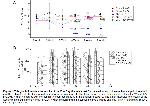
    Sinapic acid alleviated mechanical allodynia caused by cisplatin: The presence of mechanical allodynia was determined using the Von Frey filament test. Briefly, ten von Frey filaments of increasing diameters (0.4, 0.6, 1, 1.4, 2, 4, 6, 8, 10, and 15 g) were sequentially applied vertically, with twisting of the hairs, to the hind paw’s plantar surface for 5 seconds until positive responses were elicited; these included withdrawal, licking, and flinching of the hind paw. If a rat showed a positive response, the less stiff filament was used for the next trial, while the stiffer one was used on rats displaying no withdrawal or licking 2,38. At baseline, there were no significant differences in the response to mechanical stimulation with von Frey filaments between groups (p>0.05) (Fig. 1A). After the 4th dose of cisplatin was administered, withdrawal thresholds were significantly lower in the CIS group than in the control group (p< 0.001). At weeks 5th and 6th, the CIS group continued to demonstrate considerable reduction in paw withdrawal thresholds as compared to the control and sham groups (p<0.001). Starting at the 4th week, both high-dose and low-dose sinapic acid treatments (HSA and LSA, respectively) prevented the decrease in the withdrawal threshold level caused by cisplatin (p<.001). It was determined that cisplatin-induced mechanical allodynia was not observed in animals receiving sinapic acid (Fig. 1A).


    Büyütmek İçin Tıklayın
    Figure 1: This graph illustrates the results of the Von Frey filaments test for the evaluation of the mechanical pain threshold and the rotarod tests for the evaluation of motor coordination. A. This trace shows the Von frey filament test results. *** and ### p<0.001. * when compared with the control. # when compared with the cisplatin groups. B. The graph illustrates the results of the weekly rotarod experiment. * and # p<0.05. * when compared with the control and p<0.05 using one-way ANOVA followed by Tukey’s test as a post-hoc test

    The administration of cisplatin to rats resulted in impaired motor coordination: After the animals had become habituated to the rotarod, they were run on it for 300 seconds, beginning at 5 rpm and progressively rising to 40 rpm. The animals were run in three trials, with 5-minute breaks between them. Elapsed times at which animals fell from the rotarod were recorded. Averages of 3 experiments per animal were determined as a measure of motor performance. Tests were performed weekly, and we found that the motor coordination of animals exposed to cisplatin for 5 weeks was significantly impaired compared to the control group when assessed at the 5th and 6th weeks (p<0.05) (Fig. 1B). Although both HSA and LSA treatments improved cisplatin-impaired motor coordination, these effects were not statistically significant at any of the time points (p>0.05).

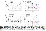
    Nociceptive behavior experiments: In the tail-flick test, tail withdrawal latency was found to be significantly longer in cisplatin-exposed rats compared to control and sham group rats in the 3th and 4th weeks (p<0.05), and this effect became more pronounced over the remainder of the experimental period (p<0.001) (Fig. 2A). By the 4th week, the HSA group showed significant reductions in the cisplatin-prolonged withdrawal thresholds (p<0.05); however, no statistically significant change was observed in the LSA group (p>0.05). Both sinapic acid doses significantly reduced the effect of cisplatin on withdrawal latency measured at last week (p<0.001) (Fig. 2A).


    Büyütmek İçin Tıklayın
    Figure 2: The graph depicts the results of hot/cold plate, and tail-flick. A. This trace shows the Tail Flick test results. B. In the hot plate test, there was a difference between the groups from the 3rd week. C. In terms of the cold plate 10°C test and D. In the cold plate 4°C test, there was a substantial difference between the groups. * and # p<0.05. *** and ### p<0.001. * and # p<0.05 * when compared with the control. # when compared with the cisplatin groups and p<0.05 using one-way ANOVA followed by Tukey’s test as a post-hoc test

    Paw withdrawal latency time in the hot plate test was shown to be significantly longer in cisplatin-treated rats than in control and sham group rats after the 3rd week (p<0.001) (Fig. 2B) and for the remainder of the experiment (p<0.001). Starting in the 3rd week, LSA administration suppressed the effect of cisplatin treatment on the paw withdrawal latency (p<0.001). Although HSA treatment also decreased the paw withdrawal latency time prolonged by cisplatin, no statistically significant change was observed (p>0.05) (Fig. 2B).

    The temperature of the cold plate was kept at 4°C for cold hyperalgesia or 10°C for cold allodynia (23). At baseline, there was no significant difference in the response to the cold stimulus at 10°C and 4°C between groups. It was determined that the rats' withdrawal latency times were prolonged in response to cold stimuli in the 10°C and 4°C cold plate test by cisplatin administration. The effect of cisplatin was statistically significant in both cold plate tests after the third week (p<0.001), (Fig 2C and 2D). At low and high doses, sinapic acid significantly reduced the prolonged response latency induced by cisplatin treatment at 4°C cold stimuli (p<0.001) (Fig 2D). At 10°C cold stimulus, sinapic acid significantly reduced the prolonged response latency induced by cisplatin treatment at both low and high doses in the 6th week only (p<0.001) (Fig 2C).

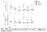
    Sinapic acid treatment alleviated the impairment of sensory function caused by cisplatin: We investigated the effects of cisplatin on sensory function using a modified adhesive removal test 27,39. We recorded the measure of the latency until the animal shaking its paw, licking it, and touching his front two paws together to see how long it took for the animal to become notified of the adhesive tape on its paw. We also recorded the time required to remove the adhesive tape. According to findings from this experiment, animals receiving cisplatin took significantly longer to notice and remove the tape from their forelegs from the third week than those receiving a control group (p<0.001) (Fig 3A,3B). Sinapic acid treatment, at low and high doses, varies degree significantly reduced the time required to detect and remove the tape from the animals' feet when compared to cisplatin treatment (p<0.001) (Fig 3A,3B). As a result of this experiment, sinapic acid treatment alleviated the impairment of sensory function caused by cisplatin.


    Büyütmek İçin Tıklayın
    Figure 3: The graph illustrates the average results of the weekly adhesive tape detect time and removal time. A. The group averages of the time to notice the adhesive tape B. Adhesive tape removal time. * and # p<0.05. *** and ### p<0.001. * and # p<0.05 * when compared with the control. # when compared with the cisplatin groups and p<0.05 using one-way ANOVA followed by Tukey’s test as a post-hoc test

    Electrophysiological parameters in the evaluation of peripheral neuropathy induce by cisplatin: The motor conduction velocity (MNCV) and compound muscle action potential (CMAP) of the sciatic nerve were assessed electrophysiologically. After the CMAP recordings were obtained, the latency time, amplitude, and MNCV values were calculated offline. It was then determined whether electrophysiological parameters differed between groups. As a result of this evaluation, in comparison to the control group, the CIS group had prolonged latency times, smaller CMAP amplitude, and the slowest mean in terms of MNCV (p<0.001) (Fig 4). Sinapic acid at both high and low doses dramatically decreased the prolonged latency time by cisplatin (p<0.05) (Fig 4A). It also significantly increased MNCV of sciatic nerve values that were affected by cisplatin, which allowed them to return to approximately their previous levels. This effect, was found statistically significant (p<0.05) (Fig 4B). The effect of sinapic acid on CMAP amplitude was not significant (p>0.05) (Fig 4C). Fig 4D depicts a representative illustration of CMAP recordings obtained from animals.


    Büyütmek İçin Tıklayın
    Figure 4: The graph illustrates the electrophysiological parameters and representative CMAP data. A. the mean latency of the experimental groups is shown. B. the mean conduction velocity of the motor branch of the sciatic nerve. C. The graphic depict that the amplitude of the CMAP. D. The graph displaying the CMAP traces that are the most representative of the mean values

    Results of Biochemical Parameters
    Sinapic acid alleviated the oxidative damage caused by cisplatin administration: In all groups, both in sciatic nerve tissue and in serum, antioxidant and oxidative parameters were measured to evaluate how cisplatin and sinapic acid affect oxidant and antioxidant capacity. Firstly, in sciatic nerve tissue, To determine the protective and ameliorating effects of LSA and HSA against cisplatin-induced oxidative stress, the CIS group was compared with the CIS plus LSA group and CIS plus HSA group. In addition, the CIS plus LSA group was compared with the CIS plus HSA group to assess the superiority of the two supplements against each other. When CIS group was compared to each of the control and sham groups, TOS, OSI, and TAC levels were found to be significantly different (for TOC and OSI; (p<0.001) and for TAC level; (p<0.05)). When compared to CIS group, TOS, OSI, and TAC levels were found to be decreased in CIS plus LSA group (for TOC and TAC p<0.05 and for OSI index level (p<0.001). Additionally, all of the oxidative parameters were found decreased in CIS plus HSA group compared to CIS group (for TOC and TAC; (p<0.05) and for OSI index; (p<0.001)). When the CIS plus LSA group was compared to the CIS plus HSA group, no significant differences in oxidative stress markers were observed (p>0.05) (Table 1). Additionally, we evaluated serum oxidative stress and antioxidant capacity. It was determined that the oxidative stress and antioxidant capacity of serum were found to be similar to those of sciatic nerve tissue. In compared to the control and sham groups, cisplatin significantly increased oxidative stres (for TOC and OSI index; (p<0.001)), but sinapic acid treatment decreased considerably effect of cisplatin, at both doses (p<0.05) (Table 2).


    Büyütmek İçin Tıklayın
    Table 1: The table illustrating the total oxidant and antioxidant capacity in sciatic nerve tissue, as well as the levels of proinflammatory cytokines in the experimental groups


    Büyütmek İçin Tıklayın
    Table 2: The table illustrating the total oxidant and antioxidant capacity of serum in the experimental groups

    Sinapic acid inhibits cisplatin's effect on proinflammatory cytokines: Table 1 shows that TNF-α levels in the CIS group were significantly increased compared with the control and sham groups (p<0.001). It was determined that administration of sinapic acid at doses CIS plus 20 mg/kg (LSA), CIS plus 40 mg/kg (HSA), significantly declined the TNF-α levels compared with the CIS group (p<0.001, p<0.001 respectively). As demonstrated in Table 1, the CIS group's IL6 level was significantly higher than the control group's (p<0.001). Sinapic acid at a dose of CIS plus 20 mg/kg (LSA) and CIS plus 40 mg/kg (HSA) substantially decreased IL6 levels when compared to the CIS group (p<0.001, p<0.001 respectively). Additionally, we determined the levels of ischemia modified albumin in sciatic nerve tissue. When CIS group was compared to each of the control and sham groups, IMA levels were found to be significantly different (p<0.001). When compared to CIS group, IMA levels were found to be decreased in CIS plus LSA group and CIS plus HSA group (p<0.001, p<0.001, respectively) (Table 1).

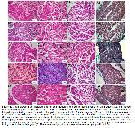
    The results of histopathological analysis: Figure 5 shows representative images of the sciatic nerve slices taken from the rats in each of the five groups. There were no observations of demyelination area, neuronal degeneration, or axon vacuolization in the sciatic nerve evaluations of the control (A1-A3) and sham groups (B1-B3). Axons that were parallel, regular, uniform, and thick were seen in these groups. CIS group axons, on the other hand, were not regular and parallel, indicating that there was neuronal degeneration, axonal fragmentationand vacuolization in the group (C1-C3). As demonstrated by the asterisk (C2), there was edema between nerve fibers, and hyperplasic schwann cells were detected in several locations of the nerve fibers as well (shown with black arrowhead in C2). It was determined that when CIS group was compared to the control group, certain myelinated nerve fibers showed significant reductions in axon diameter (C3). With the administration of sinapic acid, these pathological findings diminished (D1-D3 and E1-E3). CIS plus low dose sinapic acid and CIS plus high dose sinapic acid group's outlook was similar to that of the control group. The rate of neuronal degeneration, vacuolization, axonal fragmentation, and the number of hyperplasic Schwann cells was reduced (FIG 5). Masson Trichrome staining was also used to determine the extent of fibrosis in sciatic nerve tissue. While no fibrotic regions were identified between nerve fibers in animal of control and sham groups (A4,B4), intense fibrotic areas were observed between nerve fibers in rats of CIS groups (shown with white arrow in C4). When compared to the CIS group, it was determined that the sinapic acid treatment significantly reduced the density of fibrotic areas (D4,E4) (Fig 5).


    Büyütmek İçin Tıklayın
    Figure 5: Graph showing the histopathological examination of the sciatic nerve tissues of the groups, H&E and masson trichrome staining. A. Control, B. Sham, C. CIS, D.CIS+LSA, and E. CIS+HSA. In both the control and sham groups, no histopathological findings were observed on histopathological examination. Parallel, regular, uniform, and thick axons were seen in these groups (A and B).However, in the cisplatin group (C), significant edema between the fibers (C2, asteriks), axonal degeneration (C3, black arrow), and Schwann cell hyperplasia (C2, black arrow) were seen. These histopathological findings are among the findings of neuropathy. Additionally, cisplatin-induced significant fibrosis was observed by Masson's trichrome staining (C4, white arrow). In the control and sham groups, no fibrotic areas were found. Sinapic acid treatment (D and E) considerably decreased the histopathological findings seen in the cisplatin group (C). There were hardly any fibrotic areas in the treatment groups (D4 and E4)

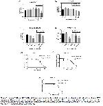
    Cisplatin's cytotoxicity was unaffected by sinapic acid: We showed that sinapic acid has a protective role against the adverse effects of cisplatin-induced peripheral neuropathy at both doses, according to behavioral, electrophysiological, biochemical and electrophysiological experimental results. We do not know yet how co-administration of sinapic acid and cisplatin will affect cisplatin's antitumoral efficacy. To see whether sinapic acid may be utilized as a co-drug to prevent cisplatin-induced peripheral neuropathy, we needed to look at its possible influence on cisplatin's antitumoral effect. In order to investigate this, we conducted an in vitro cell culture experiment with any of the cancer cell lines for which cisplatin is commonly used. According to our findings, sinapic acid showed cytotoxic activity on both BEAS-2B and A549 cell lines at a concentration of 100 μm (P<0.05), (p<0.001), respectively (FIG 6-A1,A2). In addition, although it showed a cytotoxic effect on A549 cell line at 30 μm concentration (p<0.001), no cytotoxicity was observed on BEAS-2B cell line at the same concentration (FIG 6-A1,A2). In sinapic acid-cisplatin co-administration, it was determined that sinapic acid did not cause any change in the antitumoral activity of cisplatin in both BEAS-2B and A549 cell lines (FIG 6-B1,B2).


    Büyütmek İçin Tıklayın
    Figure 6: Human A549 and BEAS-2B cell lines were treated with cisplatin alone or in combination with sinapic acid in an MTT experiment. Sinapic acid is abbreviated as SA, while cisplatin is abbreviated as CIS. NC=Negative Control. The bars represent the means and standard deviations. *Statistically significant difference compared with the negative control (p<0.05), as measured by one-way ANOVA followed by Tukey's post hoc test

  • Başa Dön
  • Özet
  • Giriş
  • Materyal ve Metot
  • Bulgular
  • Tartışma
  • Kaynaklar
  • Tartışma
    This is the first study to investigate the protective effects of sinapic acid on cisplatin toxicity in vivo using electrophysiological, behavioral, biochemical, and histopathological methods, as well as the effect of sinapic acid cisplatin combination on cisplatin's antitumoral efficacy using in vitro methods. According to the conclusions of our investigation, sinapic acid reduced the adverse effects of cisplatin without reducing its antitumoral efficiency.

    Evaluation of Nociceptive Behaviour Experiments and Electrophysiological Measurements: Behavioral, electrophysiological, and histopathological evaluations were performed to confirm that peripheral neuropathy developed in cisplatin-treated rats. Thermal hypoalgesia, mechanical allodynia, and motor incoordination were observed as a consequence of a cisplatin injection (3 mg/kg i.p.) given once a week for five weeks as evidenced by increased paw and tail withdrawal latency times in hot plate, in both cold plate experiments and in tail-flick test, respectively. These results are consistent with our previous reports 29. As a result of a review of the literature, it was determined that cisplatin-induced neuropathy can induce thermal hyperalgesia in some studies 40-43, while thermal hypoalgesia was also reported in some studies 44-46. The reason for this situation is unclear. It was observed that both low and high doses of sinapic acid considerably alleviated cisplatin's this adverse effects. The latency time in the hot-cold plate tests and tail flick experiments was found to be reduced in the CIS plus LSA and CIS plus HSA groups compared to the CIS group. Also, cisplatin administration significantly impaired motor coordination in rats. These findings consistent with previous studies on the cisplatin-induced peripheral neuropathy model 29,40,41,47,48. Although treatment with sinapic acid improved motor skills, the effect was not statistically important. The cisplatin-treated animals required significantly longer to detect the adhesive band on their paws than the control and sham groups. The time it took for these animals to remove the adhesive tape was also quite prolonged. This indicates a somatosensory dysfunction induced by cisplatin. It was determined that the time required for cisplatin-treated rats to detect and remove the adhesive tape after sinapic acid treatment was decreased. In cisplatin-treated animals noticed and removed the adhesive tape late, indicating that they have sensory and motor impairment 27. Sinapic acid treatment alleviated these impairments to an extent and ameliorates cisplatin-related adverse effects.

    Our electrophysiological data indicate that cisplatin considerably decreases the motor conduction velocity of the sciatic nerve when compared to the control and sham groups. Similarly, it was shown that cisplatin considerably extended the latency period of CMAP in rats. These results corroborated previous literature reports 49-51. It is determined that sinapic acid treatment dramatically increased the motoric conduction velocity of the sciatic nerve, which had been damaged by cisplatin, and the prolonged latency time was greatly diminished.

    Evaluation of Oxidative Stress and Proinflammatory Cytokines:Cisplatin-induced neuropathy is thought to occur as a result of oxidative stress, inflammation, mitochondrial dysfunction, DNA damage, and apoptosis of nervous system cells 52. Total oxidative capacity (TOC) determination are the most commonly used oxidative stress indicators. Consistent with the literature, our results suggest increased oxidative stress in cisplatin-treated rats, as shown by we observed a significant decrease in TAC levels accompanied by an increase in TOC in the sciatic nerve 53,54. In both serum and sciatic nerve tissue, we observed a reduction in TOC and an increase in TAC levels after the administration of sinapic acid. Previous studies have indicated that sinapic acid has an antioxidant effect and may provide protection against several diseases. Behavioral and electrophysiological studies have shown that antioxidant compounds such as resveratrol, mesna, alpha lipoic acid, coenzyme Q10 that prevent the formation of free radicals provide protection against cisplatin-induced peripheral neuropathy 49,55.

    We observed that a significant increase in proinflammatory cytokines, such as IL6 and TNF-alpha, in the sciatic nerve tissue of rats treated with cisplatin. In previous studies, it was reported that the administration of cisplatin caused an increase in TNF- alpha, IL6 levels and showed a significant proinflammatory response 56. In our study, the level of proinflammatory cytokines elevated by cisplatin was significantly reduced with sinapic acid treatment. It has been shown in previous studies that sinapic acid has an anti-inflammatory effect 57,58,59. TNF-α and IL6 have been previously reported to be associated with the development of neuropathic pain 60-63.

    We determined that cisplatin administration significantly increased the IMA level in sciatic nerve tissue as compared to the control and sham groups. Cisplatin's antitumoral effect has been suggested to be related to its effect on the tumor's vascular system, potentially causing degeneration of the vasa nervorum 64. Cisplatin may have induced to sciatic nerve ischemia by degeneration of the vasa nervorum in peripheral nerve fibers. As a result, the level of IMA in the sciatic nerve tissue may be increased. Sinapic acid's ability to decrease the level of IMA in sciatic nerve tissue may have have contributed to its therapeutic effect.

    Sinapic Acid Reduced the Side Effects of Cisplatin Without Compromising Cisplatin's Antitumor Effects: Sinapic acid has been previously reported to have anticancer and antiproliferative properties 65, various cancers such as prostate 13, pancreatic 66, and colon cancer 67. Despite the fact that sinapic acid has significant therapeutic biological activity, the absence of significant side effects increases the attraction of this compound. However, there is minimal evidence on sinapic acid's toxicity 68. Because of its antioxidant and anti-inflammatory properties, it was found in our study that it improved the symptoms of cisplatin-induced peripheral neuropathy. The failure to establish a strategy to avoid the adverse effects of a cancer medicine such as cisplatin, which has remarkable results in the fight against cancer and the restriction of its usage as a result of its side effects diminish the efficacy of the drug. When cisplatin is co-administered with sinapic acid, we found that its side effects are decreased without compromising its antitumoral efficacy. This is may be significant development in terms of the future.

    In conclusion, as shown electrophysiologically, biochemically, histopathologically, and behaviorally, cisplatin-induced peripheral neuropathy may be greatly mitigated by the co-administration of sinapic acid. Cisplatin-treated peripheral neuropathy's rats treated with SA showed alleviated neuropathy findings and also SA mitigated impaired electrophysiological parameters measured from the sciatic nerve, behaviourally findings such as mechanical allodynia, thermal hypoalgesia, and somatosensory dysfunction. The SA treatment reduced oxidative stress, inflammation, and fibrosis of the sciatic nerve in rats with neuropathy. In this connection, we conclude that SA's neuroprotective properties are owing to its antioxidant and anti-inflammatory characteristics. Moreover, in vitro studies demonstrate that cisplatin's antitumor efficacy is not diminished when co-administered with sinapic acid. In this study, we demonstrate for the first time that SA has a protective effect against the destructive effects of cisplatin-induced peripheral neuropathy. The findings suggest SA may have great therapeutic potential in the reduction of peripheral neuropathy symptoms induced by cisplatin. This research has limitations due to the absence of a control group of animals given only synapic acid. There was no group that showed the effects of sinapic acid alone in healthy control animals, hence it was impossible to demonstrate the effects of sinapic acid on healthy animals.

    Author Contributions: CY: planning the project, performing in vivo experiments and electrophysiological recordings, sacrification of animals and sample collection. CY, HY and SC: performed the analysis of the electrophysiological data and drafted the manuscript. SC; histopathological stain and evaluation. MO, MAB; biochemical analyzes. DT; conducted the in vitro studies. All authors read and approved the final version of the manuscript.

    Conflict of Interests: The authors declare that they have no conflict of interest.

    Funding: This work was supported by the Research Project Management Unit University of Gaziantep [grant number: TF.YLT.21.32] for funding this study. We gratefully the Research Project Management Unit University of Gaziantep.

  • Başa Dön
  • Özet
  • Giriş
  • Materyal ve Metot
  • Bulgular
  • Tartışma
  • Kaynaklar
  • Kaynaklar

    1) Dasari S, PB Tchounwou. Cisplatin in cancer therapy: molecular mechanisms of action. Eur J Pharmacol 2014; 740: 364-378.

    2) Friesland A, Z Weng, M Duenas, et al. Amelioration of cisplatin-induced experimental peripheral neuropathy by a small molecule targeting p75 NTR. Neurotoxicology 2014; 45: 81-90.

    3) Gutierrez-Gutierrez G, M Sereno, A Miralles, E Casado-Saenz, E Gutierrez-Rivas. Chemotherapy-induced peripheral neuropathy: clinical features, diagnosis, prevention and treatment strategies. Clin Transl Oncol 2010; 12: 81-91.

    4) Strumberg D, S Brugge, MW Korn, et al. Evaluation of long-term toxicity in patients after cisplatin-based chemotherapy for non-seminomatous testicular cancer. Ann Oncol 2002; 13: 229-236.

    5) Chen CY. Sinapic Acid and Its Derivatives as Medicine in Oxidative Stress-Induced Diseases and Aging. Oxid. Med. Cell. Longev 2016; 2016:1-10.

    6) Engels C, A Schieber, MG Ganzle. Sinapic acid derivatives in defatted Oriental mustard (Brassica juncea L.) seed meal extracts using UHPLC-DAD-ESI-MSn and identification of compounds with antibacterial activity. Eur Food Res Technol 2012; 234: 535-542.

    7) Yun KJ, DJ Koh, SH Kim, et al. Anti-inflammatory Effects of Sinapic Acid through the Suppression of Inducible Nitric Oxide Synthase, Cyclooxygase-2, and Proinflammatory Cytokines Expressions via Nuclear Factor-kappa B Inactivation. J. Agric. Food Chem 2008; 56: 10265-10272.

    8) Lee HE, DH Kim, SJ Park, et al. Neuroprotective effect of sinapic acid in a mouse model of amyloid beta(1-42) protein-induced Alzheimer's disease. Pharmacol Biochem Behav 2012; 103: 260-266.

    9) Zare K, A Eidi, M Roghani, AH Rohani, The neuroprotective potential of sinapic acid in the 6-hydroxydopamine-induced hemi-parkinsonian rat. Metab Brain Dis 2015; 30: 205-213.

    10) Kim DH. BH Yoon, WY Jung, et al. Sinapic acid attenuates kainic acid-induced hippocampal neuronal damage in mice. Neuropharmacology 2010; 59: 20-30.

    11) Karakida F, Y Ikeya, M Tsunakawa, et al. Cerebral protective and cognition-improving effects of sinapic acid in rodents. Biol Pharm Bull 2007; 30: 514-519.

    12) Singh HP, TG Singh, R Singh. Sinapic acid attenuates cisplatin-induced nephrotoxicity through peroxisome proliferator-activated receptor gamma agonism in rats. J Pharm Bioallied Sci 2020; 12: 146-154.

    13) Eroglu C, E Avci, H Vural, E Kurar. Anticancer mechanism of Sinapic acid in PC-3 and LNCaP human prostate cancer cell lines. Gene 2018; 671: 127-134.

    14) Janakiraman K, S Kathiresan, AVA Mariadoss. Influence of sinapic acid on induction of apoptosis in human laryngeal carcinoma Cell Line. 2015.

    15) Herradon E, C Gonzalez, JA Uranga, et al. Characterization of Cardiovascular Alterations Induced by Different Chronic Cisplatin Treatments. Front. pharmacol 2017; 8: 1-15.

    16) Alaofi AL. Sinapic Acid Ameliorates the progression of streptozotocin (STZ)-Induced Diabetic nephropathy in RatsviaNRF2/HO-1 mediated pathways. Front Pharmacol 2020; 11: 1-11.

    17) Raish M, A Ahmad, YA Bin Jardan, et al. Sinapic acid ameliorates cardiac dysfunction and cardiomyopathy by modulating NF-kappaB and Nrf2/HO-1 signaling pathways in streptozocin induced diabetic rats. Biomed Pharmacother 2021; 145: 1-13.

    18) Chen L, W Chen, X Qian, Y Fang, N Zhu. Liquiritigenin alleviates mechanical and cold hyperalgesia in a rat neuropathic pain model. Sci Rep 2014; 4: 1-4.

    19) Griffiths LA, NA Duggett, AL Pitcher, SJL Flatters. Evoked and Ongoing Pain-Like Behaviours in a Rat Model of Paclitaxel-Induced Peripheral Neuropathy. Pain Res Manag 2018; 2018: 1-11.

    20) Singh P, K Kongara, D Harding, et al. Comparison of electroencephalographic changes in response to acute electrical and thermal stimuli with the tail flick and hot plate test in rats administered with opiorphin. BMC Neurol 2018; 18: 1-10.

    21) Yamamoto T, N Nozaki-Taguchi, T Chiba. Analgesic effect of intrathecally administered orexin-A in the rat formalin test and in the rat hot plate test. Br J Pharmacol 2002; 137: 170-176. 22. Kim SO, HJ Kim. Berberine ameliorates cold and mechanical allodynia in a rat model of diabetic neuropathy. J Med Food 2013; 16: 511-517.

    23) Sakurai M, N Egashira, T Kawashiri, et al. Oxaliplatin-induced neuropathy in the rat: Involvement of oxalate in cold hyperalgesia but not mechanical allodynia. Pain 2009; 147: 165-174. 24. Alsalem M, M Haddad, SA Aldossary, et al. Effects of dual peroxisome proliferator-activated receptors alpha and gamma activation in two rat models of neuropathic pain. PPAR Res 2019; 2019: 1-9.

    25) Han FY, BD Wyse, MT Smith. Optimization and pharmacological characterization of a refined cisplatin-induced rat model of peripheral neuropathic pain. Behav Pharmacol 2014; 25: 732-740.

    26) Lin H, BH Heo, MH Yoon. A New Rat Model of Cisplatin-induced Neuropathic Pain. Korean J Pain 2015; 28: 236-243.

    27) Mao-Ying QL, A Kavelaars, K Krukowski, et al, The anti-diabetic drug metformin protects against chemotherapy-induced peripheral neuropathy in a mouse model. PLoS One 2014; 9: 1-8.

    28) Kar F, C Hacioglu, H Senturk, DB Donmez, G Kanbak. The Role of oxidative stress, renal ınflammation, and apoptosis in post ıschemic reperfusion ınjury of kidney tissue: The protective effect of dose-dependent boric acid administration. Biol Trace Elem Res 2020; 195: 150-158.

    29) Yildirim C, S Cevik, H Yamaner, et al. Boric acid improves the behavioral, electrophysiological and histological parameters of cisplatin-induced peripheral neuropathy in rats. J Trace Elem Med Biol 2022; 70: 1-10.

    30) Erel O. A novel automated direct measurement method for total antioxidant capacity using a new generation, more stable ABTS radical cation. Clin. Biochem 2004; 37: 277-285.

    31) Erel O. A new automated colorimetric method for measuring total oxidant status. Clin. Biochem 2005; 38: 1103-1111.

    32) Harma M, M Harma, A Kocyigit, O Erel. Increased DNA damage in patients with complete hydatidiform mole. Mutat Res 2005; 583: 49-54.

    33) Shigematsu N, T Kawashiri, D Kobayashi, et al. Neuroprotective effect of alogliptin on oxaliplatin-induced peripheral neuropathy in vivo and in vitro. Sci Rep 2020; 10: 1-8.

    34) Grela E, Ząbek A, Grabowiecka A. Interferences in the Optimization of the MTT Assay for Viability Estimation of Proteus mirabilis. Avicenna J Med Biotechnol. 2015; 7:159-67.

    35) Zhu J, VA Carozzi, N Reed, et al. Ethoxyquin provides neuroprotection against cisplatin-induced neurotoxicity. Sci Rep 2016; 6: 1-10.

    36) Wang H, CJ Heijnen, N Eijkelkamp, et al. GRK2 in sensory neurons regulates epinephrine-induced signalling and duration of mechanical hyperalgesia. Pain 2011; 152: 1649-1658.

    37) Bouet V, M Boulouard, J Toutain, et al. The adhesive removal test: a sensitive method to assess sensorimotor deficits in mice. Nat Protoc 2009; 4: 1560-1564.

    38) Liu CH, MM Ma, CL Wen, RU Zaman, OJ Olatunji. Antiallodynic and anti-hyperalgesia effects of Tiliacora triandra against cisplatin-induced peripheral neuropathy. All Life 2021; 14: 441-449. 39. Shabani M, MH Larizadeh, S Parsania, V Hajali, A Shojaei. Evaluation of destructive effects of exposure to cisplatin during developmental stage: no profound evidence for sex differences in impaired motor and memory performance. Int J Neurosci 2012; 122: 439-448.

    40) Ta LE, AJ Bieber, SM Carlton, et al. Transient Receptor Potential Vanilloid 1 is essential for cisplatin-induced heat hyperalgesia in mice. Mol. Pain 2010; 6: 1-15.

    41) Ta LE, PA Low, AJ Windebank. Mice with cisplatin and oxaliplatin-induced painful neuropathy develop distinct early responses to thermal stimuli. Mol. Pain 2009; 5: 1-11.

    42) Seto Y, F Okazaki, K Horikawa, et al. Influence of dosing times on cisplatin-induced peripheral neuropathy in rats. BMC Cancer 2016; 16: 1-10.

    43) Shahid M, F Subhan, N Ahmad, RDE Sewell. The flavonoid 6-methoxyflavone allays cisplatin-induced neuropathic allodynia and hypoalgesia. Biomed Pharmacother 2017; 95: 1725-1733.

    44) Wahdan SA, DA Elsherbiny, SS Azab, E El-Demerdash. Piceatannol ameliorates behavioural, biochemical and histological aspects in cisplatin-induced peripheral neuropathy in rats. Basic Clin Pharmacol Toxicol 2021; 129: 486-495.

    45) Abdelsameea AA, SL Kabil. Mitigation of cisplatin-induced peripheral neuropathy by canagliflozin in rats. Naunyn Schmiedebergs Arch Pharmacol 2018; 391: 945-952.

    46) Authier N, J Fialip, A Eschalier, F Coudore. Assessment of allodynia and hyperalgesia after cisplatin administration to rats. Neurosci Lett 2000; 291: 73-76.

    47) Bhadri N, T Sanji, H Madakasira Guggilla, R Razdan. Amelioration of behavioural, biochemical, and neurophysiological deficits by combination of monosodium glutamate with resveratrol/alpha-lipoic acid/coenzyme Q10 in rat model of cisplatin-induced peripheral neuropathy. Sci World J 2013; 2013: 1-8.

    48) Boehmerle W, P Huehnchen, S Peruzzaro, M Balkaya, M Endres. Electrophysiological, behavioral and histological characterization of paclitaxel, cisplatin, vincristine and bortezomib-induced neuropathy in C57Bl/6 mice. Sci Rep 2014; 4: 63-70.

    49) Sharawy N, L Rashed, MF Youakim. Evaluation of multi-neuroprotective effects of erythropoietin using cisplatin induced peripheral neurotoxicity model. Exp Toxicol Pathol 2015; 67: 315-322.

    50) Brouwers EE, AD Huitema, W Boogerd, JH Beijnen, JH Schellens. Persistent neuropathy after treatment with cisplatin and oxaliplatin. Acta Oncol 2009; 48: 832-841.

    51) Demkow U, B Biatas-Chromiec, A Stelmaszczyk-Emmel, et al. The cardiac markers and oxidative stress parameters in advanced non-small cell lung cancer patients receiving cisplatin-based chemotherapy. EJIFCC 2011; 22: 6-15.

    52) Topal I, A Ozbek Bilgin, F Keskin Cimen, et al. The effect of rutin on cisplatin-induced oxidative cardiac damage in rats. Anatol J Cardiol 2018; 20: 136-142.

    53) Saadati H, S Noroozzadeh, H Esmaeili, et al. The neuroprotective effect of mesna on cisplatin-ınduced neurotoxicity: Behavioral, electrophysiological, and molecular studies. Neurotox Res 2021; 39: 826-840.

    54) Kumar P, K Sulakhiya, CC Barua, N Mundhe. TNF-alpha, IL-6 and IL-10 expressions, responsible for disparity in action of curcumin against cisplatin-induced nephrotoxicity in rats. Mol Cell Biochem 2017; 431: 113-122.

    55) Ansari MA, M Raish, A Ahmad, et al. Sinapic acid mitigates gentamicin-induced nephrotoxicity and associated oxidative/nitrosative stress, apoptosis, and inflammation in rats. Life Sci 2016; 165: 1-8.

    56) Ansari MA, M Raish, A Ahmad, et al. Sinapic acid ameliorate cadmium-induced nephrotoxicity: In vivo possible involvement of oxidative stress, apoptosis, and inflammation via NF-kappaB downregulation. Environ Toxicol Pharmacol 2017; 51: 100-107.

    57) Bin Jardan YA, MA Ansari, M Raish, et al. Sinapic acid ameliorates oxidative stress, ınflammation, and apoptosis in acute doxorubicin-ınduced cardiotoxicity via the NF-kappaB-Mediated pathway. Biomed Res Int 2020; 2020: 1-10.

    58) Arruda JL, RW Colburn, AJ Rickman, MD Rutkowski, JA DeLeo. Increase of interleukin-6 mRNA in the spinal cord following peripheral nerve injury in the rat: potential role of IL-6 in neuropathic pain. Brain Res Mol Brain Res 1998; 62: 228-235.

    59) Leung L, CM Cahill. TNF-alpha and neuropathic pain-a review. J Neuroinflammation 2010; 7: 1-11. 60. Myers RR, VI Shubayev. The ology of neuropathy: An integrative review of the role of neuroinflammation and TNF-alpha axonal transport in neuropathic pain. J Peripher Nerv Syst 2011; 16: 277-286.

    61) Wagner R, RR Myers. Endoneurial injection of TNF-alpha produces neuropathic pain behaviors. Neuroreport 1996; 7: 2897-2901.

    62) Kirchmair R, DH Walter, M Ii, et al. Antiangiogenesis mediates cisplatin-induced peripheral neuropathy: Attenuation or reversal by local vascular endothelial growth factor gene therapy without augmenting tumor growth. Circulation 2005; 111: 2662-2670.

    63) Badr DA, ME Amer, WM Abd-Elhay et al. Histopathological and genetic changes proved the anti-cancer potential of free and nano-capsulated sinapic acid. Appl Biol Chem 2019; 62:1-10.

    64) Huang Z, H Chen, P Tan, et al. Sinapic acid inhibits pancreatic cancer proliferation, migration, and invasion via downregulation of the AKT/Gsk-3beta signal pathway. Drug Dev Res 2021; 83: 721-734.

    65) Balaji C, J Muthukumaran, N Nalini. Chemopreventive effect of sinapic acid on 1,2-dimethylhydrazine-induced experimental rat colon carcinogenesis. Hum Exp Toxicol 2014; 33: 1253-1268.

    66) Pandi A, VM Kalappan, Pharmacological and therapeutic applications of sinapic acid-an updated review. Mol Biol Rep 2021; 48: 3733-3745.

  • Başa Dön
  • Özet
  • Giriş
  • Materyal ve Metot
  • Bulgular
  • Tartışma
  • Kaynaklar
  • [ Başa Dön ] [ Özet ] [ PDF ] [ Benzer Makaleler ] [ Yazara E-Posta ] [ Editöre E-Posta ]
    [ Ana Sayfa | Editörler | Danışma Kurulu | Dergi Hakkında | İçindekiler | Arşiv | Yayın Arama | Yazarlara Bilgi | E-Posta ]