[ Ana Sayfa | Editörler | Danışma Kurulu | Dergi Hakkında | İçindekiler | Arşiv | Yayın Arama | Yazarlara Bilgi | E-Posta ]
Fırat Üniversitesi Sağlık Bilimleri Tıp Dergisi
2004, Cilt 18, Sayı 3, Sayfa(lar) 157-162
[ Özet ] [ PDF ] [ Benzer Makaleler ] [ Editöre E-Posta ]
IMPORTANCE OF CHROMOSOME 8 GAIN AND c-myc GENE AMPLIFICATION IN HIGH GRADE PROSTATE CANCER
Ebru ETEM1, Halit ELYAS1, Hüseyin YÜCE1, Nusret AKPOLAT2, Mustafa Kemal ATIKELER3
1Department of Medical Biology and Genetics, Faculty of Medicine, Fırat University, Elazığ - TÜRKİYE
2Department of Pathology, Faculty of Medicine, Fırat University, Elazığ - TÜRKİYE
3Department of Urology, Faculty of Medicine, Fırat University, Elazığ - TÜRKİYE
Keywords: Prostate cancer, gene amplification, FISH, c-myc, PIN.
Summary
Recently, cytogenetic and molecular biological studies have identified the band 8q24, where the located c-myc gene is commonly amplified in prostate cancer, especially in advanced and recurrent ones. Our objectives in this study were to define the role of c-myc and the gain of chromosome 8 in prostatic carcinogenesis and to evaluate the genetic relationship between prostatic intraepithelial neoplasia (PIN) and carcinomas. We examined a total of 30 specimens, including 9 adenocarcinoma, 6 PIN and 15 benign prostatic hyperplasia (BPH) by Fluorescence in situ Hybridization (FISH). The gain of chromosome 8 identified in 88.8% of adenocarcinomas was associated in metastatic prostate cancer. The most frequent anomaly in PIN and carcinoma was a gain of chromosome 8, and the presence of this anomaly strongly correlated with a high Gleason Score. We believe that the basic mechanism in overexpression of c-myc gene may not be amplification. Our results indicate that the basic mechanism of c-myc gene overexpression may be simple gain of chromosome 8 or gain of ‘’8q’’. Our results agree with findings of other authors that PIN is probably a procurser of carcinoma.
  • Top
  • Summary
  • Introduction
  • Methods
  • Results
  • Disscussion
  • Conclusion
  • References
  • Introduction
    Prostate cancer is the most frequent malignancies and the second leading cause of cancer deaths among males in the Western World. The clinical course of the disease is higly complex and genetic factors underlying tumorigenesis are poorly understood 1. An understanding of the genetic events that accompanied with the progression of the most likely precursor lesion, PIN, to prostatic adenocarcinoma and the subsequent development of metastases may be useful for prevention, early detection and treatment 2-4.

    Recent cytogenetic and molecular biological studies have established that band 8q24 is commonly amplified in prostate cancer, especially in advanced and recurrent prostate cancer. c-myc activation usually occurs at later stages of the carcinoma in human and is usually a poor prognostic marker 4. The c-myc gene is mapped to this region, and the gene family appears to play an important role in the regulation of cellular proliferation and differantiation. Aberrrant expression of these genes contributes to the pathogenesis of numerous human neoplasms and has been implicated in the apoptotic process 5,6. A significant association was found between elevated htert (human telomerase reverse transcriptase) expression and c-myc overexpression. It is likely that the ability of c-myc protein to stimulate expression of htert and thereby enhance telomerase activity represents an important step in prostate tumorigenesis 7,8

    FISH analysis of interphase cells with centromere specific and region-specific probes is useful for the detection of numerical chromosomal abnormalities and genetic alterations in solid tumors such as prostatic carcinomas that are often difficult to analyze by conventional cytogenetic analysis 2,9. The most common alterations in prostate cancer are loss of 8p and gain of 8q, which have been detected in more than 80% and 90% of the cases, respectively. The gain of 8q is often associated with the loss of 8p especially in hormone-refractory carcinomas and in distant metastases 10-12.

    Our aims in this study were to define the role of c-myc and the gain of chromosome 8 in prostatic carcinogenesis and evaluate the genetic relationship between PIN and adenocarcinomas.

  • Top
  • Summary
  • Introduction
  • Methods
  • Results
  • Disscussion
  • Conclusion
  • References
  • Methods
    Patient Selection

    Prostate cancer, PIN and BPH cases were collected for our study at Firat Medical Center between March 2001 and August 2002. Prostate adenocarcinomas and PIN were histopathologically diagnosed as high-grade.

    Sample Preparation and Histopathological Evaluation

    We analyzed a total of 30 cases, including 9 prostate adenocarcinoma, 15 benign prostatic hyperplasia and 6 prostatic intraepithelial neoplasia. Two patients with prostate cancer had metastases (One case had bone metastases and the other one had pelvic lymph node metastases), two patients had recurrent and this patients had received hormone therapy. All remaining cases included 26 newly diagnosed untreated patients. To determine the criteria for FISH anomalies, 15 samples of benign prostatic hyperplasia tissue obtained by retropubic prostectomy were also analyzed. The tumors selected had high Gleason Scores 6-10. Tumor grade was classified according to the Gleason Score and Cell Cytology. May Grunwold-Giemsa (MGG) and Papanicolaou Gynaekologie (PAP) was made for cell cytology. Cell cytology and Gleason Score were evaluated by expert pathologist.

    Fluorescence in situ Hybridization (FISH)

    Slides were prepared so as to use touch preparation protocol. Specimens were touched lighty on precleaned slides, which can be performed in a short time. After air-drying at room temperature, these slides were fixed with fixing solution (3:1 methanol:acetic acid) and stored at -20ºC until use. Target slides were denatured in 2XSSC /70% formamide, pH 7, at 67ºC for 6 min and dehydrated in graded ethanol. Dual-labeling hybridization was performed using 10ml of the hybridization mixture containing fluorescein direct labeled chromosome 8 alpha-satellite probe and rhodamine direct-labelled c-myc probe. Probes were denatured at 76ºC for 10 min and applied to the target slides. Hybridization was performed overnight at 37ºC in moist chamber. Posthybridization washes were performed with %50 formamide/ 2XSSC three times for 10 min, 2XSSC for 5 min, and 2XSSC/Nonidet P-40 for 5 min at 42ºC. Counterstaining was specifically prepared by mixing 2µl of PI to 8µl of DAPI and then used. The number of FISH signals was counted with a Nicon microscope equipped with a three color filter. At least 100 nuclei was evaluated. FISH signals were counted according to the criteria described previously 13. Gain of Chromosome 8: ≥20% nuclei with three or more signals for centromeric 8. C-myc gene amplification: more c-myc signals than sentromeric 8 signals or c-myc/centromeric 8 ratio ≥ 1.10.

    Statistical Analysis

    Statistical analysis was carried out using the SPSS programme (Statistical Packages of Social Sciences, SPSS for Windows, Version 9.0, Inc, Chicago, IC, USA). Fischer’s exact test was used to determine the association between copy number aberrations and tumor recurrence as well as metastases and Gleason Score. The p value of less than 0.05 was considered significant.

  • Top
  • Summary
  • Introduction
  • Methods
  • Results
  • Disscussion
  • Conclusion
  • References
  • Results
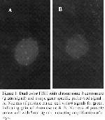
    Total of 30 cases were successfully analyzed. We found an 88.8% gain of chromosome 8 centromere and 100% c-myc extra copy number of adenocarcinomas exhibiting 3 or more positive signals for chromosome 8 centromere or c-myc in 20% or more of the cells. The gain of chromosome 8 and c-myc gene extra copy number are summarized in Table 1. Figure 1 shows typical FISH results for chromosome 8 and c-myc gene.


    Click Here to Zoom
    Table 1: Classification of patients with prostate adenocarcinoma on the basis of FISH findings.


    Click Here to Zoom
    Figure 1: Dual-color FISH with chromosome 8 centromere (green signal) and c-myc gene specific probe (red signal). A: Nucleus of prostate cancer cell with 6 signals for green, indicating gain of chromosome 8. B: Nucleus of prostate cancer cell with 5 red signals, indicating amplification of c-myc.

    According to the extra copies of c-myc, specimens could be divided into three groups; i. those with simple gain of a whole chromosome 8 (no increase in c-myc copy number relative to the chromosome 8 centromere), which was identified in 88% and 26.6%, in adenocarcinoma and PIN, respectively. ii. those with an intermediate increase in c-myc copy number relative to the chromosome 8 centromere, which was found in adenocarcinomas and PIN in 22.2% and 16.6%, respectively. iii. those with substantial amplification of c-myc (large increases in c-myc copy number relative to the chromosome 8 centromere), which was found in 11.1% of prostate adenocarcinoma, but not in PIN and BPH. Case 6, 8, and 14 are not included in the amplification evaluation to the study because of the existence of extra copy number of c-myc in centromeric 8 signals.

    There was no correlation between gain of chromosome 8 or c-myc extra copy number and recurrence or metastases (p=0.12, p= 0.95 and p=0.14, p=0.95), respectively. However, the correlation was found between gain of basic chromosome 8 centromere and metastases with linear correlation test (p=0.038). Average signals numbers seen in most cells for chromosome 8 centromere and c-myc in cases with adenocarcinoma are illustrated in Table 2.


    Click Here to Zoom
    Table 2: A statistically significant difference was found between PIN and carcinomas for signals obtained from c-myc and chromosome 8 in most of cells (p<0.05).

  • Top
  • Summary
  • Introduction
  • Methods
  • Results
  • Disscussion
  • Conclusion
  • References
  • Discussion
    Recent studies have indicated that several chromosomes (7, 8, 10 and Y) play important roles in tumorigenesis and tumor progression of prostate cancer 1,14. Numerical chromosomal anomalies were found in 67%, 68%, and 96% of foci of PIN, carcinoma, and metastases, respectively. Chromosome 8 alterations, including loss of 8p21-22 and gain of 8q24, are commonly observed in prostate carcinoma. Sato et al. reported that alterations of c-myc were associated with both systemic progression and patient deaths 15. We found extra copies of c-myc in chromosome 8 centromere in 16% of PIN (case14), 22% of cancer (case 6 and 8).

    A variety of factors may contribute to gain of c-myc. Simple gain of the whole chromosome 8 can account for many cases with extra copies of c-myc. Chromatid separation in proliferative cells will result in an apparent increase in the number of region-specific probe signals. Some studies have also reported loss of 8p concurrent with gain of the long arm of chromosome 8 (8q) sequences in advanced prostatic cancer. This combination of events occurring on the same chromosome–loss of 8p seguences and gain of 8q sequences-suggests formation of i(8q) chromosomes in advanced prostate tumors 16. Alers et al. (1997) reported that overrepresentation of 8q sequences, most likely by isochromosome 8q formation, is involved in metastatic spread to the bone 17.

    Brown et al reported that anomalies of chromosomes 8 and/or 7, present in 14 of the 16 cases (88%) aneusomic by FISH and high-grade tumors, were more likely to be aneuploid on FISH (18). The present study showed similar findings with an 88.8% gain of chromosome 8 in adenocarcinomas.

    Oncogene amplification is one mechanism that leads to stepwise progression in solid tumors. Moreover, oncogene amplification may be a useful indicator of progressional prognosis in various human cancers 19. c-myc amplification is not common in prostate cancer specimens, although FISH has been demonstrated to be a sensitive technique for detecting changes in gene copy number. Miyoshi et al. reported that c-myc gene amplification was detected in 8%, 19%, and 46% of PIN, carcinoma and metastases of prostate 20. Bubendorf et al. reported no cases of high level myc amplification in primary tumors 21. Alers et al. previously found c-myc amplification in 8% of primary prostate tumors and c-myc gene amplification which correlates with high levels of myc protein expression 22. Mark et al. reported that an increased copy number in c-myc oncogene copy number was not a prominent finding in their cohort of prostate cancer patients 13. In our study, c-myc gene amplification was found in 11.1% of prostate cancer cases. Interestingly, c-myc gene amplification has been shown to occur in a case with primary prostate cancers without metastases and untreated prostate cancers in the present study. Our results support the findings of others that c-myc gene amplification or copy number increase is not common in prostate cancer specimens.

    Jenkins et al. found extra copies of c-myc in 50% of PIN foci, 44% of cancer, and 92% of lymph node metastases and these were usually observed simultaneously with gain of chromosome 8 centromere 4. Our study demonstrated that extra copies of c-myc were found in 100% of adenocarcinoma and 33.3% PIN.

    Bastacky et al. have been show 36% in group B ( high-grade PIN, HGPIN/no PC) and 69% in group A (HGPIN/PC) for chromosome 8/c-myc. Using a cutoff of 4, between Group A and Group B were found statistically different 23. The present study found statistically significant difference between PIN and carcinomas for signals obtained from c-myc and chromosome 8 in most cells (p<0.05). This finding is important because it helps to differentiate between adenocarcinoma and PIN.

    This report suggests that amplification and overexpression of c-myc alone with another gene(s) mapped to 8q, may play a key role in the progression and evaluation of prostatic carcinoma. Overall frequencies of extra c-myc copy anomalies and numeric chromosomal anomalies in PIN and carcinoma were similar, suggesting that they share a similar underlying pathogenesis. Thus, these findings suggest that PIN is a precursor of carcinoma. We conclude that an increase in c-myc oncogene copy number was not a prominent finding in our cohort of prostate cancer patients. We believe that the basic mechanism in overexpression of c-myc gene may not be amplification. Our results indicate that the basic mechanism of c-myc gene overexpression may be gain of simple chromosome 8 or gain of ‘’8q’’. Our results concerning c-myc amplification and gain of chromosome 8 in prostate cancer in Turkish patients are consistent with the results observed in prostate cancer in Western countries and suggest that these genetic and chromosomal changes may be associated with the development and progression of prostate cancers.

  • Top
  • Summary
  • Introduction
  • Methods
  • Results
  • Discussion
  • Conclusion
  • References
  • Conclusion
    Hyperploidy of chromosomes 8 are a common finding in high grade prostate cancer and predictive of follow-up prostate cancer. This gain is associated with distant tissue metastases. FISH is a powerful method to definition in chromosomal gain or loss in cancer tissue. But, cellular proliferation studies are necessary when using interphase cytogenetics to ascertain gene amplification. We advice that FISH studies with probes specific for 8p, 8q, and chromosome 8 centromere are used.

    Acknowledgement

    We thank Dr. O. Akyol for his fruitful collabration in reviewing the manuscript.

  • Top
  • Summary
  • Introduction
  • Methods
  • Results
  • Discussion
  • Conclusion
  • References
  • References

    1) Verma RS, Manikal M, Conte RA, Godec CJ. Chromosomal basis of adenocarcinoma of the prostate. Cancer Invest 1999; 17: 441-447.

    2) Alers CA, Krijtenburg PJ, Vissers KJ, Bosman FT, van der Kwast TH, van Dekken H. Interphase cytogenetics of prostatic adenocarcinoma and precursor lesions: analysis of 25 radical proctectomies and 17 adjacent prostatic intraepithelial neoplasia. Genes Chromosomes Cancer 1995; 59: 241-250.

    3) Bostwick DG, Brawer MK. Prostatic intraepithelial neoplasia and early invasion in prostate cancer. Cancer 1997; 59: 788-794.

    4) Jenkins RB, Qian J, Lieber MM, Bostwick DG. Detection of c-myc amplification and chromosomal anomalies in metastatic prostatic carcinoma by fluorescence in situ hybridization. Cancer Res 1997; 57: 524-531.

    5) Sommer A, Bousset K, Kremmers E, Austen M, Luscher B. Identification and characterization of specific DNA-binding complexes containing members of the Myc/Max/Mad network of transcriptional regulators. J Biol Chem 1998; 273: 6632-6642.

    6) Luscher B, Larssan LG. The basic region/helix-loop-helix/leucine zipper domain of Myc protooncoproteins: Function and regulation. Oncogene 1999; 18: 2955-2966.

    7) Latil A, Vidaud D, Valeri A, Fournier G, Vidaud M, Lidereau R, Cussenot O, Biache I. Htert expression correlates with myc overexpression in human prostate cancer. Int J Cancer 2000; 89: 172-176.

    8) Cerni C. Telomeres, telomerase and MYC. An update. Mutat Res 2000; 462: 31-47.

    9) Qian J, Bostwick DG, Takahashi S, Borell TJ, Herath JF, Lieber MM, Jenkins RB.. Chromosomal anomalies in prostatic intraepithelial neoplasia and carcinoma detected by fluorescence in situ hybridization. Cancer Res 1995; 55: 5408-5414.

    10) Visakorpi T, Kallioniemi AH, Syvanen AC, Hyytinen ER, Karhu R, Tammela T, Isola JJ, Kallioniemi OP. Genetic changes in primary and recurrent prostate cancer by comparative genomic hybridization. Cancer Res 1995; 55: 342-347.

    11) Cher ML, Bova GS, Moore DH, Small EJ, Carroll PR, Pin SS, Epstein JI, Isaacs WB, Jensen RH. Genetic alterations in untreated metastases and androgen-independent prostate cancer detected by comparative genomic hybridization. Cancer Res 1996; 56: 3091-3102.

    12) Vocke CD, Pozzatti RO, Bostwick DG, Florence CD, Jennings SB, Strup SE, Duray PH, Liotta LA, Emmert-Buck MR, Linehan WM. Analysis of 99 microdissected prostate carcinomas reveals a high frequency of allelic loss on chromosome 8p12-21. Cancer Res 1996; 56: 2411-2416.

    13) Mark HF, Samy M, Santoro K, Mark S, Feldman D. Fluorescent in situ hybridization study of c-myc oncogene copy number in prostate cancer. Exp Mol Pathol 2000; 68: 65-69.

    14) Karashima T, Taguchi T, Yoshikawa C, Kamada M, Kasahara K, Yuri K, Shuin T. Numerical chromosomal changes in metastatic prostate cancer following anti-androgen therapy: flourescence ın situ hybrization analysis of Japanese cases. Cancer Genet Cytogenet 2000; 120: 148-154,

    15) Sato K, Qian J, Slezak JM, Lieber MM, Bostwick DG, Bergstralh EJ, Jenkins RB. Clinical significance of alterations of chromosome 8 in high-grade, advanced, nonmetastatic prostate cancer. J Natl Cancer Inst 1999; 91: 1574-1580.

    16) Macoska JA, Beheshti B, Rhim JS, Hukku B, Lehr J, Pienta KJ, Squire JA. Genetic characterization of immortalized human prostate epithelial cell cultures: Evidence for structural rearrangements of chromosome 8 and i(8q) chromosome formation in primary tumor-derived cells. Cancer Genet Cytogenet 2000; 120: 50-57.

    17) Alers JC, Krijtenburg PJ, Rosenberg C, Hop WC, Verkerk AM, Schroder FH, van der Kwast TH, Bosman FT, van Dekken H. Interphase cytogenetics of prostatic tumor progression: specific chromosomal abnormalities are involved in metastasis to the bone. Lab Invest 1997; 77: 437-438.

    18) Brown JA, Alcaraz A, Takahashi S, Persons DL, Lieber MM, Jenkins RB. Chromosomal aneusomies detected by fluorescent in situ hybridization analysis in clinically localized prostate carcinoma. J Urol 1994; 152:1157-1162.

    19) Schwab M, Amler LC. Amplification of cellular oncogenes: a predictor of clinical outcome in human cancer. Genes Chromosomes Cancer 1990; 1: 181-193.

    20) Miyoshi Y, Uemura H, Fujinami K, Mikata K, Harada M, Kitamura H, Koizumi Y, Kubota Y. Fluorescence in situ hybridization evaluation of c-myc and androgen receptor gene amplification and chromosomal anomalies in prostate cancer in japanese patients. Prostate 2000; 43: 225-232.

    21) Bubendorf L, Konomen J, Koivisto P, Schraml P, Moch H, Gasser TC, Willi N, Mihatsch MJ, Sauter G, Kallioniemi OP. Survey of gene amplifications during prostate cancer progression by high throughput fluorescence in situ hybridization on tissue microarrays. Cancer Res 1999; 59: 803-896.

    22) Alers JC, Rochat J, Krijtenburg PJ, Hop WC, Kranse R, Rosenberg C, Tanke HJ, Schroder FH, van Dekken H. Identification of genetic markers for prostatic cancer progression. Lab Invest 2000; 80: 931-942

    23) Bastacky S, Cıeply K, Sherer C, Dhır R, Epsteın JI, Use of Interphase fluorescence In Situ Hybridization in Prostate Needle Biopsy Specimens With Isolated High-Grade Prostatic Intraepithelial Neoplasia as a Predictor of Prostate Adenocarcinoma on Follow-Up Biopsy. Hum Pathol 2004; 35: 281-289

  • Top
  • Summary
  • Introduction
  • Methods
  • Results
  • Discussion
  • Conclusion
  • References
  • [ Başa Dön ] [ Özet ] [ PDF ] [ Benzer Makaleler ] [ Editöre E-Posta ]
    [ Ana Sayfa | Editörler | Danışma Kurulu | Dergi Hakkında | İçindekiler | Arşiv | Yayın Arama | Yazarlara Bilgi | E-Posta ]