 |
 |
| [ Ana Sayfa | Editörler | Danışma Kurulu | Dergi Hakkında | İçindekiler | Arşiv | Yayın Arama | Yazarlara Bilgi | E-Posta ] | |
| Fırat Üniversitesi Sağlık Bilimleri Tıp Dergisi | |||||
| 2006, Cilt 20, Sayı 6, Sayfa(lar) 445-446 | |||||
| [ Özet ] [ PDF ] [ Benzer Makaleler ] [ Yazara E-Posta ] [ Editöre E-Posta ] | |||||
| Non-traumatic Cecal Perforation in a Female Patient : Report of a Case | |||||
| Ziya ÇETİNKAYA1, Mustafa GİRGİN1, Ibrahim H.ÖZERCAN2, Refik AYTEN1 | |||||
| 1Fırat University Faculty of Medicine Department of General Surgery Elazığ-TURKEY 2Fırat University Faculty of Medicine Department of Pathology Elazığ-TURKEY |
|||||
| Keywords: Cecal perforation | |||||
| Summary | |||||
A case of closed cecal perforation in a 38-years-old patient was reported . In the case presented,
no history of trauma, and no other etiological factors reported in literature was present. After
debridement of cecum wall of 1-2 cm. diameter so as to contain the perforation hole, it was
suturized primaryly and appendectomy performed. In the histopathologic study; acute necrotizan
inflammation in perforated area was reported. The patient externed from the hospital with full recovery without any complication and this case was one of the rare cecum perforation cases, who, unlike other cases, displayed none of the few probable etiological factors reported in literature. |
|||||
| Introduction | |||||
Cecal perforation is an uncommen condition that is clinically difficult to diagnose and
differentiate from acute apandicitis. Physical examination failed to differentiate these two
disease entities. Ultrasonography (US) and Computerized Tomography (CT) were
reported to be useful in the early diagnosis of cecal diverticulitis. It is, however, difficult
to determine which patient should require further image study.1 In some cases of
colonic pseudoobstruction, (Ogilvie’s syndrome) cecum distention results in cecum
perforation. According to Laplace’s law, more severe progressive distention is observed
in the cecum. This implies that tensile strength of the colonic wall will be exceeded in
the cecum that has the greatest diameter 2. We herein present a case of cecal perforation, which was incidentally encountered and in the management of this rare condition, primary repair of the cecum with appendectomy was performed. |
|||||
| Case Presentation | |||||
A 38-years-old female patient was referred to the hospital for an abdominal pain
which had started in epigastric region twelve hours ago. The pain was progressive in
nature and localized in the right abdomen by time. The patient stated that she first had
a stab-like temporary abdominal pain a week ago which was regressed spontaneously.
The patient mentioned that she sometimes had swelling on her abdomen and usually
suffered from constipation. She had had an operation of bilateral subtotal thyroidectomy
7 years ago and she has been on 100 μg thyroksin daily. Physical examination of the
patient revealed that, her general condition was good and there were a tenderness,
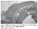
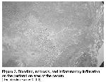
local defence, and rebound tenderness in right lower abdomen. The level of blood leucocyte was 16700/ mm3 and there was not any abnormal value in biochemical parameters. AP chest radiograph, abdominal radiograph and abdominal ultrasound were normal. On the basis of history and physical examination, she was operated with the preoperative diagnosis of acute appendicitis. The circulation of ceacum was well and there was local inflammation. There was not any periceacal absess. Also there was no inflammation in pelvic region. Ileum was observed to be normal up to 100 cm length. There was no Meckel diverticulum. After depridment of ceacum wall of 1-2 cm diameter so as to contain the perforation hole, it was suturized primaryly and appendectomy was performed. The operation was completed after placing a drain into paracolic gutter. There was no complication occured in the postoperative controls. Postoperative thyroid function analysis was normal. In postoperative interrogation of patient, it was revealed that there was no history of trauma. In the histopathological study; appendix was exhibiting lymphoid hyperplasia (Figure 1), acute inflammation infiltration was seen in the omentum. As a result, an acute necrotizan inflammation in perforated area were determined (Figure 2).
|
|||||
| Discussion | |||||
Non-traumatic cecum perforation is a very rare
condition. Non-traumatic clinical conditions developing
cecum perforation reported in literature are pseudoobstructions
of colon 2,3, mechanical intestinal
obstruction in colon 4,5, cecal diverticulitis 1, and
salmonella infection 6. In the case presented, there was no history of trauma, and no other etiological factors reported in literature was determined. It is difficult to estimate the presence of cecum perforation prior to surgery in cases with cecum wall tension with no pathology. More than 70% of the patients with cecal diverticulitis are operated with the diagnosis of preoperative acute appendicitis. Ultrasonography and Computer Tomography can be useful in the early diagnosis of cecal diverticulitis conditions. However, it is very difficult to decide for which patients further examinations are necessary. The anatomical distribution of the diverticula in colon vary in different countries. Diverticula mostly appear in the left colon in developed Western countries and in USA. In Asian countries, on the other hand, most of the diverticula are located in the right colon, especially in cecum and the ascending colon. Right colon diverticulosis occur at relatively younger ages and prominently more frequent in men. Most of the diverticula are pseudodiverticula. Solitary diverticula, on the other hand, occur congenitally and comprise all the layers of the intestinal wall. Although our case’s condition resembled cecal diverticulitis, histopathological tests did not reveal cecal diverticulitis and perforation developing from the diverticula. Surgical therapy approach in cecum perforation varies depending on the underlying etiology. Primary repair, cecostomy, and ileocecal resection with ileostomy are among the therapy options for cecal perforation. There was no pathology to cause cecum perforation at the basis of our case. The region of perforation in the cecum was closed with appendices epiploica, the inflamation was restricted, and there was no fecal contamination in the surrounding tissues. Our preference of surgical approach in this patient was primary repair following biopsy from the side of the perforation, and debritement. No complications arose in our patient after the operation. In conclusion, this case was one of the rare cecum perforation cases, who, unlike other cases, displayed none of the few probable etiological factors reported in literature. |
|||||
| References | |||||
1) Fang J-F, Chen R-J, Lin B-C, Hsu Y-B, Kao J-L, Chen M-F.
Agressive resection is indicated for cecal diverticulitis. Am J
Surg 2003; 185:135-140.
2) Noory N, Abbaszadeh E. Cecal dilatation and perforation
after cesarean section. Int J Gynecol Obstet 2003;81:47-48.
3) Meier C, Di Lazzaro M, Decurtins M. Ogilvie syndrome with
cecal perforation. A rare complication after isolated thoracic
trauma. Case report and current literature review. Swiss
Surg 2000;6(4):184-91.
4) Butterworth SA, Webber EM. Meconium Thorax: A case of
Bochdalek hernia and cecal perforation in a neonate with
Job’s syndrome. J Pediatr Surg 2002;37(4):673-674.
|
|||||
| [ Başa Dön ] [ Özet ] [ PDF ] [ Benzer Makaleler ] [ Yazara E-Posta ] [ Editöre E-Posta ] | |||||
 |
| [ Ana Sayfa | Editörler | Danışma Kurulu | Dergi Hakkında | İçindekiler | Arşiv | Yayın Arama | Yazarlara Bilgi | E-Posta ] |

