
Deneysel Hiperkolesterolemide Melatoninin Oksidatif Stres Parametreleri, Dimetilarginin Dimetilaminohidrolaz ve Histopatolojik Değişiklikler Üzerine Etkileri
Volume 36, Issue 1
March 2022 · Pages 074-079
Deneysel Hiperkolesterolemide Melatoninin Oksidatif Stres Parametreleri, Dimetilarginin Dimetilaminohidrolaz ve Histopatolojik Değişiklikler Üzerine Etkileri
Effects of Melatonin on the Oxidative Stress Parameters, Dimethylarginine on Dimethylaminohydrolase and Histopathological Changes in Experimental Hypercholesterolemia
Gülnur ASLAN1, Dilşad SEZGİN1, Ali Osman ÇERİBAŞI2, Dilek ARSLAN ATEŞŞAHİN3, Kezban CAN ŞAHNA4, Kazım ŞAHİN5, Engin ŞAHNA1
2Firat University Faculty of Veterinary, Department of Pathology Elazig, TÜRKİYE
3Firat University Faculty of Science, Department of Biology, Elazig, TÜRKİYE
4Firat University Faculty of Veterinary, Department of Virology Elazig, TÜRKİYE
5Firat University Faculty of Veterinary, Department of Animal Nutrition, Elazig, TÜRKİYE
Amaç: Bu çalışmada, melatoninin deneysel hiperkolesterolemide karaciğer dokusunda oksidatif stres parametreleri, nitrik oksit metabolizmasında rol oynayan dimetilarginin dimetilaminohidrolazın (DDAH) ekspresyon düzeyleri ve histopatolojik değişiklikler üzerine etkileri araştırıldı.
Gereç ve yöntem: Sıçanlar 4 gruba ayrıldı: a) Kontrol, b) Hiperkolesterolemi (HCT), c) Kolesterol diyetiyle eşzamanlı olarak uygulanan melatonin (HCT-MEL, profilaktik), d) Kolesterol diyeti ile beslenen ve sadece son 2 hafta içinde uygulanan melatonin (HCT-2MEL, terapötik olarak) grupları.
Bulgular: Hiperkolesterolemi ile MDA düzeylerinde artış gözlenmesine rağmen, DDAH protein ekspresyonu ve GSH düzeylerinin azaldığı görüldü. MDA düzeyleri, GSH aktivitesi ve DDAH ekspresyonu melatonin uygulamalarıyla normalize edildi. Karaciğer dokusunda hiperkolesterolemi ile sinüzoidal konjesyon, bulanık şişkinlik, periportal hücre infiltrasyonu, periasiner yağlaması ve apoptoz gibi önemli değişiklikler gözlendi. HCT-MEL grubunda periportal ve midzonal bölgedeki yağın şiddeti, HCT grubuna göre azaldı. İki tedavi grubu karşılaştırıldığında histopatolojik olarak profilaktik melatonin terapötik kullanımdan daha etkiliydi.
Sonuç: Melatonin, diyetle indüklenen hiperkolesterolemide antioksidan etkiye sahip hepatoprotektif ve/veya terapötik bir ajan ve karaciğer patojenezinde histopatolojik değişiklikleri önlemede nitrik oksit metabolizması regülatörü olabilir.
Objectives: In this study, it was aimed to investigate the effects of melatonin on oxidative stress parameters, expression levels of dimethylarginine dimethylaminohydrolase (DDAH) which involved in nitric oxide metabolism, and histopathological changes in the liver tissue in experimental hypercholesterolemia.
Material and Methods: Rats were divided into 4 groups: a) Control, b) Hypercholesterolemia (HCT), c) Melatonin administrated concurrently with cholesterol diet (HCT-MEL, prophylactically), d) Melatonin administrated only during the last 2 weeks of cholesterol diet feeding (HCT-2MEL, therapeutically) groups.
Results: Although an increase was observed in the MDA levels, the DDAH protein expression and GSH levels were found to decrease with hypercholesterolemia. MDA levels, GSH activity and DDAH expression were normalized with melatonin administrations. Significant changes such as sinusoidal congestion, cloudy bloating, periportal cell infiltration, periacinary lubrication, and apoptosis were observed in the liver tissue by hypercholesterolemia. In the HCT-MEL group, the severity of the fat in the periportal and midzonal region was lowere compared to the HCT group. Histopathologically when the two treatment groups are compared, prophylactic melatonin was more effective than its therapeutic use.
Conclusion: Melatonin may be a hepatoprotective and/or therapeutic agent with antioxidant effect, and regulator of nitric oxide metabolism in prevention of histopathological changes in liver pathology in diet-induced hypercholesterolemia.
Introduction
The liver has an important role in the regulation of plasma cholesterol levels. Maintain a healthy function of the liver is necessary to overcome hypercholesterolemia and its related pathologies such as the development of atherosclerosis and cardiovascular diseases [1].Lipids which accumulated with hypercholesterolemia leads to increased production of oxidative stress that play a role in the atherogenic process [2]. Oxidative stress which is the consequence of cellular lipid overload contributes to hepatic inflammatory injury and fibrogenesis [3,4].
Endothelial cell injury is a critical mechanism for the onset and progression of atherosclerosis. Hypercholesterolemia, an important risk factor for atherogenesis, has been reported to increase the production of reactive oxygen species (ROS) and leads to vascular dysfunction [5].
Nitric oxide (NO), an important homeostatic agent to control vascular tone and blood flow, is produced in liver sinusoidal endothelial cells with endothelial nitric oxide synthase (eNOS). eNOS is activated by stimuli such as blood flow shear stress [6]. However, vascular dysfunction induced by hypercholesterolemia has been declared to be associated with endothelial injury, mostly through NO-dependent processes [7]. Reduced endothelial NO synthesis leads to endothelial dysfunction resulting from disruption of endothelium-dependent vasodilatation [5].
Melatonin is derived from serotonin and is produced in the pineal gland [8]. Melatonin scavenges free radicals, activates antioxidant defense enzymes, normalizes lipid and blood pressure profile, inhibits inflammation [9] and increases NO bioavailability [10]. It exhibits atheroprotective effects in different atherosclerotic pathological signaling processes; endothelial-induced adhesion inhibits the formation of molecules, reduces fatty acid leakage into the endothelial layer [11].
In this study, effects of melatonin on the changes in dimethylarginine dimethylaminohydrolase (DDAH) which is an enzyme taking the role of nitric oxide metabolism, and oxidative stress parameters (MDA, GSH) which take part in the development of liver damage and atherosclerosis in hypercholesterolemic rats were investigated.
Material and Method
Animals and Experimental Design: 28 adult male Sprague-Dawley rats (300±50 g) were housed under standard laboratory conditions. A commercial pellet diet and fresh drinking water were given ad libitum. The hypercholesterolemic diet was prepared by adding cholesterol 2%+cholic acid 0.5% to the standard diet. Cholesterol and colic acid amounts were determined according to Butun et al [15]. Prepared feeds were stored at +4 °C during use.Rats were divided into 4 groups (n:7). (i) Control group, rats fed with standard diet; (ii) HCT group, rats fed with the hypercholesterolemic diet for 8 weeks; (iii) HCT-MEL group; rats fed with hypercholesterolemic diet and melatonin was administrated daily by i.p. injection at 5 mg/kg concurrently with cholesterol; (iv) HCT-2MEL group; rats fed with hypercholesterolemic diet and melatonin was administrated by i.p. the injection only lasts 2 weeks (10 mg/kg). The dose of melatonin used was based upon an earlier study presenting a positive result of 10 mg of melatonin per kilogram on rats [12].
Biochemical Analyses: At the end of 8 weeks, the rats were decapitated under urethane anesthesia. Liver tissue was taken by laparotomy. Blood was centrifuged at 3,000 x g for 10 minutes to obtain serum and was used in the analysis of liver function tests. Serum and liver tissue were stored at -80 °C for further analysis. Some of them were placed in 10% formaldehyde and separated for histopathological analysis.
Liver Function Tests: Alanine aminotransferase (ALT), aspartate transaminase (AST), gamma-glutamyl transferase (GGT) levels were measured in the autoanalyzer (Siemens Healthcare Diagnostics Tarrytown, USA) using commercial kits (Siemens Healthcare Diagnostics Tarrytown, USA).
Measurement of Oxidative Stress Markers: The MDA concentration was measured by a modified method of Ohkawa et al. [13] spectrometrically at 532 nm. The method is based on reaction with thiobarbituric acid and 1,1,3,3 tetraethoxypropane was used as a standard. Results were expressed as nmol mL homogenate-1. Tissue GSH concentrations were determined by the method of Ellman [14] and dithiolnitrobenzoic acid was reduced by sulfuryl compounds to form a disulfide compound. This yellow complex was measured at 412 nm and expressed as µmol mg protein-1.
Western Blot Protocol: DDAH protein expression levels were analyzed by this technique [15]. All the primary and secondary antibodies were purchased from Abcam (Abcam, Cambridge, UK). Densitometric analyses of the bands were studied with Image J program (National Institute of Health, Bethesda, USA).
Histopathological Method: For histopathological investigations, tissue samples taken from the liver were detected in 10% buffered neutral formalin solution. Paraffin blocks were prepared from the tissues. The blocks were cut in 5 µm thickness and stained according to the hematoxylin-eosin (HE) method and examined in the light microscope [16]. Changes in liver tissue were scored as no (0), mild (+), moderate (++) and severe (+++) lesions. Scoring results were evaluated statistically.
Chemicals: The cholesterol used in the production of hypercholesterolemic feed was obtained from Ambresco (Solon, USA) while colic acid was supplied by Alfa Aesar (Karlsruhe, Germany). Melatonin was purchased by Sigma Aldrich Inc. (St. Louis, MO. A.B.D).
Statistical Analyses: The power analysis applied before the study showed that it would be enough to include 7 rats per group to evaluate the available parameters (power=0.80, α=0.05, β=0.1). Data were expressed as arithmetic means±S.D. All statistical analyses were studied with SPSS 21.0 pack program. Normality of the distribution within the groups was evaluated with the Shapiro–Wilk test. Raw data for each experiment satisfied the assumptions of normality and homogeneity of variance. While the one-way ANOVA test was used for statistical comparisons, the differences between the groups were determined by the Post Hoc Tukey test. The significance level was accepted as p<0.05.
Results
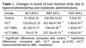
Results of Biochemical Analysis: Serum AST and GGT levels were increased in the HCT group when compared with the control group and decreased in both melatonin groups when compared to the HCT group significantly (p=0.004, p=0.001). Increasing serum ALT level with HCT when compared with the control decreased with melatonin but these changes were not statistically significant (p=0.423, Table 1).
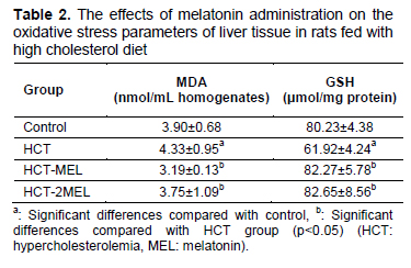
MDA and GSH levels were determined as oxidative stress parameters. Liver tissue MDA levels were increased in the HCT group when compared with the control group and decreased in both melatonin groups when compared with the HCT group (p=0.003, p=0.004). When we analyzed the changes in GSH levels between the groups, we found that it decreased in the HCT group when compared with the control group. However, GSH levels increased significantly in the HCT-MEL group and HCT-2MEL group when compared with the HCT group (p=0.007, Table 2).
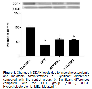
DDAH level decreased by 59.16% in the HCT group when compared with the control group and also increased by 42.68% in the HCT-MEL group (p=0.002) and 40.08% in HCT-2MEL group (p=0.012) when compared with the HCT group. All these changes were statistically significant (Figure 1).
Histopathological Results: When microscopic results in liver tissue were analyzed, the severity of lesions determined in the liver in all groups and it was summarized in table 3. In the control group (Figure 2A) and HCT-MEL group (Figure 2D), livers were found to have a normal histological appearance. In the control group (Figure 3A) and HCT-MEL group (Figure 3D), ın the conserved cytoplasm, it was observed that the hepatocytes with prominent nuclei and nuclei formed the cordial structures. The most significant histological changes were detected in the HCT group. In the HCT group, macro-vesicular lubrication was more pronounced in the periportal and midzonal regions (Figure 3/B). There were mononuclear cell infiltrations in some periportal areas.
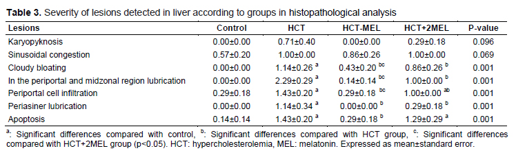
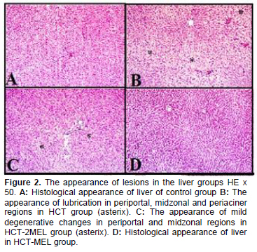
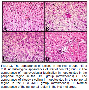
Cloudy swelling was observed in the hepatocytes in the periosper region together with mild fatty tissue. Karyopyknosis formed in the nuclei of some hepatocytes observed in oiling (Figure 3B). In the HCT-2MEL group, the severity of the fat in the periportal and midzonal region decreased compared to the HCT group, (Figure 2C) blurred puffiness in hepatocytes in these regions was a more prominent microscopic change (Fig
Serum transaminase levels are considered a sensitive marker in liver injury. Amin et al. [17] showed that liver transaminases (ALT, AST and GGT) increased in hypercholesterolemic rats at 3 and 6 weeks of age compared to controls. In hepatic steatosis in non-alcoholic fatty liver disease, ALT, AST and oxidative stress parameters have been shown to decrease serum levels in melatonin doses of 10 mg/kg [18]. On thioacetamide-induced liver fibrosis, melatonin administered for 4 weeks was reported to have a positive effect on ALT and AST [19]. In our study, increased ALT, AST and GGT levels in the hypercholesterolemia group were decreased in melatonin groups but changes in ALT levels did not reach statistical significance. Similarly, in another study, ALT values of fibrosis stage 1 and 4 were not significantly increased compared to stage 0 [20].
Oxidative stress plays a critical role in the inflammatory process in the liver by increasing the migration and activation of inflammatory cytokines and mediators. In an experimental study, a high cholesterol diet has been shown to trigger oxidative stress in plasma, liver and aortic tissue [21]. Some animal studies have shown that melatonin inhibits hypocholesterolemia and lipid peroxidation [22]. GSH and MDA effectively reduce the level of lipid peroxidation, therefore it is considered as an indicator of oxidative stress in the liver [23]. MDA, the final product of lipid peroxidation, has been shown to activate inflammation [24]. It has been observed that melatonin treatment increased the antioxidant enzymes SOD, catalase and glutathione peroxidase activity and decreased GSH levels in the liver [25]. In a study, it was shown that melatonin administration for seven days did not change liver MDA levels in a mouse model of carbon tetrachloride-induced liver damage [26]. But in another study, liver MDA levels were reduced when treatment with melatonin administrated for 84 days in a rat model of carbon tetrachloride injury [27].
NO, which is synthesized from L-arginine by NOS in endothelial cells, plays a pivotal role in maintainance of vascular structure and function, and it is generally described as an ‘endogenous anti-atherosclerotic molecul’. It was reported that ADMA, a major endogenous inhibitor of NOS, could reduce NO production and decrease acetylcholine-induced vasodilator responses [28]. It was studied that the elevation of circulating ADMA level is involved in endothelial dysfunction in some cardiovascular abnormalities [29]. Decrease in activity of DDAH, a major hydrolase of ADMA, causes accumulation of ADMA under cardiovascular abnormalities [30]. DDAH is known to be highly susceptible to oxidation. In our study, melatonin treatment significantly increased the DDAH level compared to the hypercholesterolemia group. Since the ADMA level will decrease due to the increase in DDAH, melatonin may increase the level of DDAH and protect the tissue from endothelial damage.
DDAH activity is accepted as the main determinant of endogenous ADMA concentration [31]. In an experimental study, the hypercholesterolemic diet has been shown to increase ADMA levels and decrease DDAH activity and arginine/ ADMA ratio [32]. In a rat study, it has been shown that melatonin treatment increased DDAH activity thus, reduce the level of ADMA and thereby inhibit kidney damage caused by bile duct ligation [33]. Similarly, atherogenic lipid profile and serum ADMA levels were increased in rats exposed to high fructose diet, and oral melatonin treatment was reported to have positive effects on these parameters [34]. In a clinical study, high ADMA level has been shown to reduce the drop in blood pressure due to increased endogenous melatonin release at night [35].
The accumulated evidence shows that the parameters counted as inflammation and oxidative stress indicator in liver tissue and plasma are associated to clinical and histological findings. Observations regarding the most significant histopathological changes in the HCT groups support the finding that cholesterol may damage the liver [36]. In our study, significant changes such as sinusoidal congestion, cloudy bloating, periportal cell infiltration, periasiner lubrication and apoptosis were observed in the liver tissue by hypercholesterolemia. Melatonin prevented all these changes. In a recent study, Hong and colleagues [27] showed protective antifibrotic effects of melatonin on hepatic fibrosis induced by carbon tetrachloride in experimental rats. It has been reported that liver damage observed in ApoE -/- hypercholesterolemic mouse model was significantly reduced by oral support of melatonin [21]. Melatonin has a free radical scavenger (receptor-independent) effect directly against reactive oxygen and nitrogen types, while indirectly (receptor-dependent) upregulates antioxidant enzymes and downregulates pro-oxidant enzymes [35]. Besides that, it protects cellular function by showing antiapoptotic and anti-inflammatory effects with local receptor-mediated functions [37].
As a result, melatonin may influence NO bioavailability by modulating DDAH levels. The therapeutic administration of melatonin may also have effective consequences for liver protection. Melatonin may be a hepatoprotective and/or therapeutic agent as an antioxidant and regulator of NO metabolism to prevent histopathological changes in liver pathogenesis in diet-induced hypercholesterolemia.
Discussion
In our study, while liver function tests and an oxidative stress product MDA increased significantly; an antioxidant enzyme GSH and DDAH which plays a role in NO metabolism decreased significantly with high cholesterol diet. Sinusoidal congestion, cloudy bloating, periportal cell infiltration, periasiner lubrication and apoptosis were observed in the liver tissue by hypercholesterolemia in the histopathological analysis. Both melatonin treatments protected liver tissue by normalizing these parameters. However, the effect of prophylactic (HCT-MEL) melatonin was more successful than therapeutic (HCT-2MEL) use in the histopathological analysis.References
- Protective Effect of Melatonin on Oxidant and Antioxidant Systems in Ethanol-Induced Liver Damage
- ETANOL İLE OLUŞTURULMUŞ KARACİĞER HASARINDA MELATONİNİN OKSİDAN VE ANTİOKSİDAN SİSTEMLER ÜZERİNDEKİ KORUYUCU ETKİSİ
- CCL4’ün SEBEP OLDUĞU KARACİĞER HASARI ÜZERİNE NİGELLA SATİVA TOHUMUNUN KORUYUCU ETKİLERİ
- SOY İZOFLAVONLARIN KARBON TETRAKLORÜRE (CCL4) BAĞLI KARACİĞER HASARI VE PLAZMA PARAOKSONAZ İLE ARİLESTERAZ AKTİVİTE DÜZEYLERİNE OLAN ETKİLERİ
- Tıkanma Sarılıklı Ratlarda Egb-761 in Etkileri