
Posterior Superior Alveolar Arterin KIBT ile Değerlendirilmesi: Retrospektif Bir Çalışma
Volume 38, Issue 1
March 2024 · Pages 032-036
Posterior Superior Alveolar Arterin KIBT ile Değerlendirilmesi: Retrospektif Bir Çalışma
Evaluation of the Posterior Superior Alveolar Artery Using CBCT: A Retrospective Study
Rıdvan GÜLER1, Nedim GÜNEŞ1
Amaç: Bu çalışmanın amacı, konik ışınlı bilgisayarlı tomografi (KIBT) ile posterior superior alveolar arterin (PSAA) prevalansını, çapını, yerini ve alveolar kret tepesine olan oblik mesafe ile ilişkisini belirlemektir.
Gereç ve Yöntem: Çalışamızda 300 hastadan çeşitli nedenlerle alınan Konik Işınlı Bilgisayarlı Tomografi verileri retrospektif olarak incelenmiştir. Kesitsel görüntülerde PSAA’in prevelansı, lokalizasyonu (iç, orta, dış), arter çapları ve alveoler kret tepesine olan oblik mesafeleri belirlenmiştir. Elde edilen tüm veriler istatistiksel analizler ile değerlendirildi.
Bulgular: PSAA tüm maksiller sinüslerin %63'ünde görüldü. Arter her iki cinsiyette en sık intraosseöz (%65) olarak tespit edildi. PSAA’in lateral sinüs duvarının dış korteksine yakın yerleşimi ise en düşüktür. PSAA’in maxiller sinüs lateral duvarındaki konumu incelendiğinde, istatistiksel olarak kadınlar ve erkekler arasında PSAA konumu açısından anlamlı bir fark olmadığı saptandı (p=0.516). Ortalama PSAA çapı 1.42±0.50 mm bulundu. Sağ ve sol sinüsler için bu değerler sırasıyla 1.42±0.50 ve 1.33±0.50 mm olarak tespit edildi. 2 mm’in üzerinde olduğu vakalara nadir rastlanmıştır. Arterin alveol kretinin alt sınırına olan uzaklığı 18.20±4.80 mm olup bu değer sağ sinüs için 18.10±4.80 mm, sol sinüs için 18.20±4.80 mm idi. Sağ-sol karşılaştırmalarında arter çapı (p=0.811) ve alveoler krete olan uzaklık mesafe ölçümleri açısından anlamlı bir farklılık bulunamamıştır (p=0.840).
Sonuç: KIBT kesitlerinde PSAA gözlenmesi sıktır. Bundan dolayı maksiller sinus bölgesinde cerrahi girişimler esnasında komplikasyonların önlenmesi için intraossöz anastomozlarının ve maksiller sinüs morfolojisinin, preoperatif olarak KIBT ile değerlendirilmesi büyük önem taşımaktadır.
Objective: The aim of this study was to determine the prevalence, diameter, and location of the posterior superior alveolar artery (PSAA) and its relationship with the oblique distance to the apex of the alveolar crest using cone-beam computed tomography (CBCT).
Materials and Methods: This retrospective study analyzed Cone-Beam Computed Tomography data that was taken from 300 patients for various reasons. The prevalence, localization (inner, middle, outer), arterial diameters, and oblique distances to the alveolar crest apex of the PSAA were determined from cross-sectional images. All the data were evaluated using statistical analysis.
Results: The PSAA was seen in 63% of all maxillary sinuses with the artery most frequently detected as intraosseous (65%) in both genders. The location of the PSAA close to the outer cortex of the lateral sinus wall was the least common. There was no statistically significant difference in the PSAA position between men and women (p=0.516). The mean PSAA diameter was 1.42±0.50 mm, with values of 1.42±0.50 mm and 1.33±0.50 mm for the right and left sinuses, respectively. Cases of a diameter greater than 2 mm were rare. The distance of the artery to the lower border of the alveolar crest was 18.20±4.80 mm. These values of 18.10±4.80 mm and 18.20±4.80 mm for the right and left sinuses. In right-left comparisons, no significant difference was found in terms of arterial diameter (p=0.811) and distance measurements to the alveolar crest (p=0.840).
Conclusion: PSAA is common in CBCT sections. Therefore, preoperative evaluation of intraosseous anastomoses and maxillary sinus morphology with CBCT is of great importance in order to prevent complications during surgical interventions in the maxillary sinus region.
Introduction
The posterior superior alveolar artery (PSAA) and the infraorbital artery (IOA) are branches of the maxillary artery, supplying the lateral wall and sinus membrane of the maxillary sinus. The blood supply of the maxillary sinus and Schneiderian membrane originates from the maxillary artery [1,2]. Both arteries give off extraosseous and intraosseous branches that then anastomose around the maxillary sinus. The course of the intraosseous branch of the PSAA in the buccal wall of the sinus may be straight or U-shaped [3]. Knowledge of the vascular structure of the maxillary sinus, the position, and height of the PSAA in the alveolar bone is essential to avoid major bleeding complications during surgical procedures [4]. Pneumatization due to tooth loss can increase the volume of the maxillary sinus, causing changes in the position of the main vessels compared to other anatomical structures, which is crucial for patients requiring posterior dental implants [5,6].The maximum diameters of the PSAA and IOA can reach 2 mm and 2.7 mm, respectively. The risk of bleeding severity increases depending on the size of the artery diameter. Due to the potential bleeding risk, these vessels should be taken into account during surgical procedures. Therefore, the anatomy of the region should be carefully examined before surgical applications [1,4].
Cone-beam computed tomography (CBCT) is a valuable imaging technique for dentomaxillofacial imaging due to its lower radiation dose compared to standard medical computed tomography scans. kV, mA, exposure time, and increase in the field of view increase the radiation dose regardless of the type of imaging technique used [7]. The radiation dose obtained from a medical computed tomography compared to a standard dental extraction scan with CBCT has been reported to be 1.5 to 12.3 times higher than CBCT scans [8,9]. For this reason, CBCT is very useful in examining the anatomical structures associated with sinus surgery, to increase the success rate of surgical procedures and to reduce complications [10].
The aim of this study was to evaluate the prevalence, diameter, localization, and relationship of the PSAA with the oblique distance, which is the alveolar crest, using CBCT.
Material and Method
Research and Publication Ethics: This study was conducted in accordance with the Principles of the Declaration of Helsinki. Ethical approval of the study was obtained from Dicle University Faculty of Dentistry Dean's Clinical Research Ethics Committee (Date: 29/06/2022, Protocol code: 2022-36).This retrospective study included 300 patients who visited Dicle University Faculty of Dentistry, Department of Oral and Maxillofacial Surgery, for various reasons, such as paranasal sinus infections, orthodontic planning, pre-dental implant evaluation, impacted wisdom tooth surgery, and planning before Le-Fort I osteotomy. CBCT data were retrospectively scanned and used for the analysis. Patients outside the age range of 18-80 years, patients with tumor-cyst formation in the examined area and operated, patients in whom evaluation was not possible due to image scattering caused by patient or device-related errors during radiological imaging, patients with metallic filling or crown-bridge prosthesis were not included in the study.
CBCT images obtained using an i-Cat (Imaging Sciences International Hatfield PA, USA) device with irradiation parameters of 20.27 mA, 120 kVp and 14.7 sec were evaluated by 2 researchers on axial, sagittal and coronal sections 0.2 mm cross section with the i-Cat-Vision (Imaging Sciences International Hatfield PA, USA) program CBCT images of a total of 300 patients, 135 (45%) male and 165 (55%) female, aged between 18 and 80 (mean age 42) who met the inclusion and exclusion criteria were used in the study.
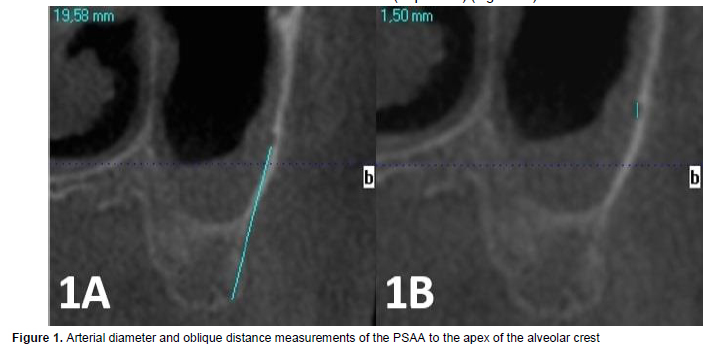
The incidence of PSAA on the lateral wall of the maxillary sinus, artery diameters (Figure 1b), localization (inner, middle, outer) and oblique distance to the alveolar crest apex, if present, were measured (Figure 1a).
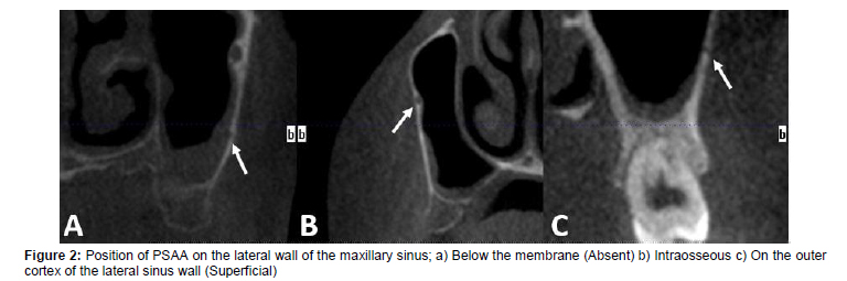
Based on this data, arterial localizations were divided into 3 classes:
1. Below the membrane (Absent) (Figure 2a),
2. Intraosseous (Figure 2b),
3. On the outer cortex of the lateral sinus wall (Superficial) (Figure 2c).
Statistical Analysis: In this study, the suitability of continuous variables to normality distribution was evaluated with the Kolmogorov-Smirnov test, and their homogeneity was evaluated with the Levene Test. Additionally, the power of the test was calculated statistically using power analysis. While examining the changes in parameters for different groups, t-test was used in independent groups and Chi-Square tests were used to evaluate the frequencies of variables. By applying 95% confidence interval in statistical analysis tests; Descriptive statistical analyzes were performed using R version 3.2.3 (2015-12-10), Copyright © 2015 The R Foundation for Statistical Computing free software computer package program. In statistical evaluations, p<0.05 was considered significant.
Results
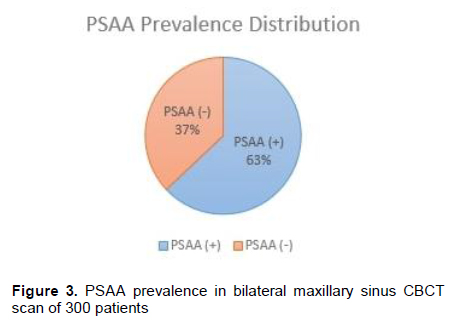
In the study, a total of 600 maxillary sinuses were evaluated in bilateral maxillary sinus CBCT scans of 300 patients. PSAA was detected in 378 (63%) of the 300 bilateral maxillary sinus CBCT images (Figure 3).
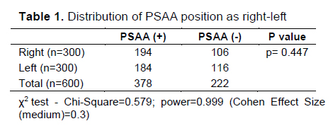
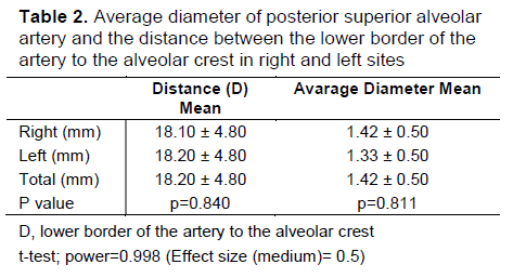
Of these, 194 (51.3%) were located in the right maxillary sinus, while 184 (48.7%) were located in the left maxillary sinus (Table 1). In right-left comparisons, no significant difference was found in terms of distribution of PSAA (p=0.447). The mean PSAA diameter was recorded as 1.42 ± 0.50 mm. These values were recorded as 1.42 ± 0.50 and 1.33 ± 0.50 mm for the right and left sinuses, respectively. The artery diameter was 1 mm in 38%; 1–2 mm in 54% and 2 mm in 8%. While the rate of arterial diameter between 1-2 mm was found to be the highest in both men and women, the cases with arterial diameters above 2 mm were found less frequently. The distance of the artery to the lower border of the alveolar crest was recorded as 18.20±4.80 mm, which was 18.10±4.80 mm for the right sinus and 18.20±4.80 mm for the left sinus. In right-left comparisons, no significant difference was found in terms of arterial diameter (p=0.811) and distance measurements from the alveolar crest (p=0.840). (Table 2).
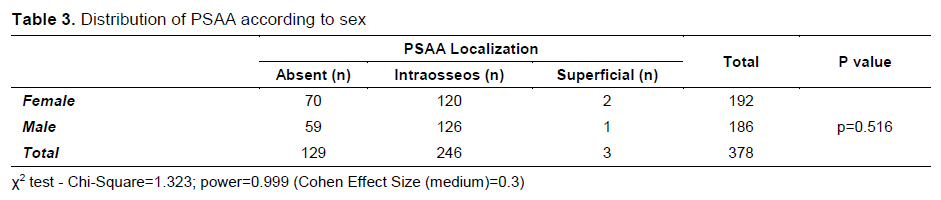
When the position of PSAA on the lateral wall of the maxillary sinus was examined, it was found that there was no statistically significant difference between men and women in terms of PSAA position (p=0.516). It was determined that the PSAA is located mostly intraosseously (middle) in both women and men, while this rate is 62.5% in women, it is 67.7% in men. In women, the location of the PSAA close to the outer cortex of the lateral sinus wall was found to be 6.8%. On the other hand, the rate of placement close to the outer cortex of the lateral sinus wall was found to be 5.3% in men (Table 3).
In a study conducted by Tassoker in 2022, the incidence of maxillary PSAA was reported as 86.5% on the right and 84.5% on the left in 200 CBCT scans [14]. Aksoy et al. reported PSAA in 366 (61%) of 300 bilateral maxillary sinus CBCT images. In addition, 191 (52.2%) of them were located in the right maxillary sinus, while 175 (47.8%) were in the left maxillary sinüs [11]. In a 2011 study by Güncü et al. [3], it was reported that the prevalence of arteries were 64.5% and mostly intraosseous (68.2%). Ilgüy et al. [17] reported the prevalence of PSAA in the maxillary sinus as 89.3%, Tehranchi et al. [15] as 87%, Kim et al. [18] as 52%, and Chitszai et al. [16] as 71%. In 2007 Mardinger et al. [19] found the incidence of maxillary intraosseous ducts to be 55% in 208 maxillary sinus CT images. In the present study, the PSAA prevalence was found to be 63% in 300 bilateral maxillary sinus CBCT scans. Of these, 194 (51.3%) were located in the right maxillary sinus, while 184 (48.7%) were located in the left maxillary sinus. According to the results of the studies in the literature, it can be thought that PSAA is present in all samples and the prevalence differences are possible due to the use of different techniques and devices, the small diameter of the canal and the lack of accurate observation.
Güncü et al. [3] reported the mean PSAA diameter as 1.3 mm, Kim et al. [18] as 1.52 mm, Tehranchi et al. [15] as 1.29 mm, Atul et al. [20] as 0.63 mm, Danesh-Sani et al. [21] as 1.17mm, Zhitian et al. [22] as 0.96 mm, and Chitsazi et al. [16] as 1.37 mm. Aksoy et al. [11] reported that the mean PSAA diameter was between 1-2 mm in their studies. İlgüy et al. reported that the PSAA diameter was less than 1 mm in their study, contrary to the literature. In the present study, the mean diameter of the PSAA canal was found to be 1.42 mm, and the values vary between 0.20 mm and 3.50 mm. These values for the right and left sinuses were 1.42±0.50 and 1.33±0.50 mm, respectively. The present study shows parallelism with the literature. In addition, in the present study, no significant difference was found in terms of arterial diameter and distance to alveolar crest in right-left comparisons.
Tassoker et al. [14] reported the incidence of the artery in the intraosseous position as 50% on the right and 51.5% on the left. İlgüy et al. [17] found that the artery was in the intraosseous position in 71.1%. Güncü et al. [3] also found that the PSAA was localized interosseous in 68.2% in their study, Tehranchi et al. [15] at a rate of 47%, Chitsazi et al. [16] 73.2%, Danesh-Sani et al. [21] similarly, found 69.6%. In this study, the frequency of intraosseous localization of the PSAA was found to be 65%. As in the present study, the PSAA is more likely to be seen in the intraosseous location. This is important in terms of complications in surgical procedures to be performed in the region. In addition, when the position of the PSAA on the lateral wall of the maxillary sinus was examined in the study, it was found that there was no statistically significant difference in the PSAA position between women and men.
Oblique distance between the PSAA lower border and crest was found to be 16.88±3.46 mm (16.79±3.79 mm in females, 17.00±2.94 mm in males) by Ilgüy et al. [17], 18±4.90 mm by Guncu et al. [3], 16 ±3.50 mm by Elian et al. [23], 16.70±3.96 mm (15.94±4.06 mm in females and 17.50±3.69 mm in males) by Tehranchi et al. [15], 16.90 mm by Mardinger et al. [19], and 18.90 mm by Kim et al. [18]. In the present study, the distance of the artery to the lower border of the alveolar crest was 18.20±4.80 mm, which was 18.10±4.80 mm for the right sinus and 18.20±4.80 mm for the left sinus. In right-left comparisons, no significant difference was found in terms of distance measurements of the artery to the alveolar crest. It can be thought that these differences in distance measurements between studies may be due to anatomical variation in the positions of the arteries.
In conclusion, CBCT scanning is a valuable imaging modality for assessing the presence of arteries in maxillary sinus-related surgeries. According to the results of this study, CBCT cross sectional analysis showed the prevalence of PSAA to be high (63%). The average PSAA diameter was found to be 1.4 mm. Due to the high prevalence, it is of great importance to evaluate intraosseous anastomoses and maxillary sinus morphology with CBCT preoperatively in order to prevent complications during surgical interventions in the maxillary sinus region. Although there are differences in each patient, the findings from this study suggest that the upper border of the lateral window should be limited to 18 mm from the apex of the alveolar crest in surgical procedures to avoid any potential vascular damage.
Discussion
The blood supply of the maxillary sinus is important in terms of implant applications and sinus augmentation procedures to be applied in the region [11]. In cases of long-term edentulism, both the resorption of the alveolar bone and the downward sagging of the maxillary sinus in the edentulous region will lead to the inability to find the necessary bone for implant application. In this case, maxillary sinus augmentation is applied to obtain vertical bone height [12,13]. As a result of a better understanding of biological healing and the development of bone graft materials used for maxillary sinus augmentation, endosseous implant placement in the atrophic posterior maxilla has become a common procedure in modern times [3]. In the present study, the presence and location of the PSAA was able to be observed with CBCT scans. The artery was observed in 63% of the sinuses and was mostly intraosseous (65%).Figures
References
- Removal of a Dental Implant from the Maxillary Sinus by Caldwell–Luc Surgery: A Case
- Removal of a Third Molar and Dentigerous Cyst from the Orbital Floor in Maxillary Sinus: A Case Report
- Multidisciplinary Approach to Antral Retention Cysts That Need Treatment- Case Series
- A Large Residual Cyst Involving the Maxillary Sinus: Case Report