Asemptomatik Bir Hastada İntrahepatik Portal Ven Anevrizması
Volume 26, Issue 3
December 2012 · Pages 131-133
Asemptomatik Bir Hastada İntrahepatik Portal Ven Anevrizması
Intrahepatic Portal Vein Aneurysm in an Asymptomatic Patient
Berhan GENÇ1, Aynur SOLAK1, Serkan GÜR1, Murat YALÇIN2
1Şifa Üniversitesi, Tıp Fakültesi, Radyoloji Anabilim Dalı, İzmir, TÜRKİYE 2Şifa Üniversitesi, Tıp Fakültesi, İç Hastalıkları Anabilim Dalı, İzmir, TÜRKİYE
Portal ven anevrizmaları sakküler ya da füziform olarak karşımıza çıkan nadir klinik patolojilerdir.
Çoğu anevrizmalar extrahepatik portal ven dalında lokalize olmakla birlikte nadiren intra hepatik
segmenttede görülebilir. Portal ven anevrizması nadir fakat önemli bir klinik durumdur. Biz burada
asemptomatik, portal hipertansiyon bulgusu olmayan intrahepatik portal ven anevrizmalı bir olgu
rapor ediyoruz.
Aneurysms of the portal venous system, localized fusiform or saccular dilatations are rare clinical
abnormalities. Most aneurysms are located in the extrahepatic segment and found rarely in
intrahepatic branches. Portal vein aneurysm is a very rare but an important condition. In this case
report, we report a case with portal vein aneurysm that was asymptomatic and had no sings
suggestive of portal hypertension.
Introduction
Portal vein aneurysm is a focal dilatation of the portal venous system. It can be in a
fusiform or saccular configuration. It is a rare vascular anomaly being increasingly
reported with more frequent use of radiologic investigations for the diagnosis and
screening of abdominal disorders[1,2]. Portal vein aneurysms are extremely rare.[3].
Two forms of this abnormality, namely, congenital and acquired, have been described.
Acquired aneurysms are more frequent and commonly associated with hepatic cirrhosis
and portal hypertension[4].
In this case report, we report a patient with intrahepatic portal vein aneurysm that
was asymptomatic and had no signs suggestive of portal hypertension.
Case Presentation
A 58-year old male was admitted to department of internal medicine for a prolonged
abdominal pain and dyspepsia with no sings suggestive of portal hypertension. There
was no history of jaundice, haematemesis, melanea, abdominal inflammation, or
trauma. No abdominal mass was palpated. Liver function study tests were normal.
Gastrointestinal system was initially investigated by abdominal ultrasonography that
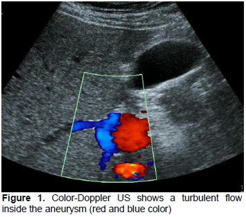
incidentally showed a portal vein aneurysm. Color-Doppler US (Figure 1) showed a
turbulent flow inside the aneurysm (red and blue color). The intrahepatic portal vein
aneurysm that was 30 mm in diameter was located at the connection point of the main
and the left portal veins. The intrahepatic branches of the portal vein and the mesenteric
vein were normal in caliber. The liver parenchyma was homogenous, and its size and
contours were normal. Etiologic factors like portal hypertension, trauma, surgery,
intervention to portal system, and hypercoagulability were not present in our patient.
Figure 1: Color-Doppler US shows a turbulent flow
inside the aneurysm (red and blue color)
There were no alterations in liver function, with normal bilirubin, albumin, and
protamin time. The transaminases and canalicular enzymes were also normal, showing
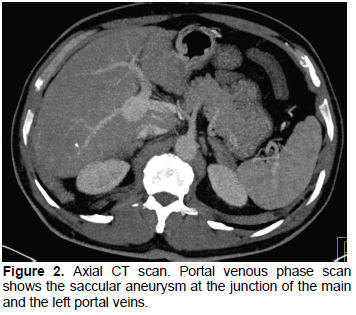
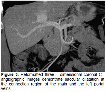
no hepatic cholestasis or hepatic injury. For further anatomical diagnosis, CT
angiography was performed. There was an aneurysm with a diameter of 30x30 mm at
the proximal site of the left portal vein (Figure 2, 3).
Figure 2: Axial CT scan. Portal venous phase scan
shows the saccular aneurysm at the junction of the main
and the left portal veins.
Figure 3: Reformatted three – dimensional coronal CT
angiographic images demontrate saccular dilatation at
the connection region of the main and the left portal
veins.
Discussion
Intrahepatic portal vein aneurysm is still a rare
vascular abnormality although new imaging modalities
have been developed[5]. Portal vein aneurysm can be
defined as a localized dilatation of the portal vein beyond
normal limits[6] and can be divided into extrahepatic and
intrahepatic types. Main portal vein and its intrahepatic
branches are the most commonly involved vessels
although any portion of the portal venous system might
be affected[7]. Although long debated, the pathogenesis
of portal venous system aneurysms has remained
controversial[5,8]. A portal venous system aneurysm
may be congenital or acquired as a result of weakening
of the vascular wall.
Chronic liver disease, portal hypertension,
pancreatitis trauma, and the effect of surgery have been
reported as acquired causes of portal venous system
aneurysm[2,5,9]. In our case, the aneurysm may be of
a congenital origin since there was no identifiable cause.
Majority of cases are clinically asymptomatic. In few
patients, abdominal pain due to compression of adjacent
structures, jaundice by compression of the biliary ducts,
or gastrointestinal bleeding due to rupture of biliary
system were reported[10]. Portal vein aneurysms are
frequently identified with US or Doppler US and
confirmed with dynamic or multiphasic CT. Treatment of
a portal vein aneurysm depends basically on the
patient's initial clinical symptoms and the presence of
associated portal hypertension[1]. Most portal venous
system aneurysms require no treatment and follow-up is
sufficient[5].
In conclusion, portal vein aneurysm is a vascular
pathology also seen in asymptomatic patients without
portal hypertension. Treatment is unnecessary in
asymptomatic cases but radiological follow-up is
warranted for complications of the aneurysm.
References
Brock PA, Jordan PH,Bart MH, Rose AG. Portal vein
aneurysm:a rare but important vascular condition. Surgery
1997; 121: 105-108.
Fulcher A, Turner M. Aneurysm of the portal vein and
superior mesenteric vein. Abdom Imaging 1997; 22: 287-292.
Feliciano PD, Cullen JJ, Corson JD. The management of
extrahepatic portal vein aneurysm: observe or treat? HPB
Surgery 1996; 10: 113-116.
Mucenic M, Rocka Md Mde S, Laudanna AA, Cancado EL.
Treatment by splenectomy of a portal vein aneurysm in
hepatosplenic schistosomiasis. Rev Inst Med Trop S Paulo
2002; 44; 261-264.
Lopez-Machado E, Mallorquin-Jimenez F, Medina-Benetiz
A, Ruiz-Carazo E, Cubero-Garcia M. Aneurysms of the
portal venous system: ultrasonography and CT findings.
Eur J Radiol 1998; 26: 210-214.
Yang DM, Yoon MH, Kim HS, et al. CT findigs of portal
vein aneurysm caused by gastric adenocarsinoma invading
the portal vein. Br J Radiol 2001; 74: 654-656.
Ozbek SS, Killi MR, Pourbagher MA et al. Portal venous
system aneurysm: report of five cases. J Ultrasound Med
1999; 18: 417-422.
Ohnami Y, Ishida H, Konno K, et al. Portal vein aneurysm:
report six cases and reviewof the literature. Abdom
Imaging 1997; 22: 281-286.
Atasoy KC, Fitoz S, Akyar G, Aytaç S, Erden I. Aneurysm
of the portal venous system; gray scale and color Doppler
ultrasonografic findings with CT and MRI corelation. Clin
Imaging 1998; 22: 414-417.
Mhanna T, Bernard P, Pilleul F, Partensky C. Portal
venous aneurysm: Report of two cases.
Hepatogastroentrology 2004; 51: 1162-1164.