
Diyabetik Ayak Ülserli Hastalarda Serum GDF-15, Seçilmiş Matriks Metalloproteinazlar ve TIMP-2 Düzeyleri: Kesitsel Karşılaştırmalı Bir Çalışma
Cilt 40, Sayı 3
Kasım 2026 · Sayfalar 215-222
Diyabetik Ayak Ülserli Hastalarda Serum GDF-15, Seçilmiş Matriks Metalloproteinazlar ve TIMP-2 Düzeyleri: Kesitsel Karşılaştırmalı Bir Çalışma
Serum GDF-15, Selected Matrix Metalloproteinases, and TIMP-2 in Patients with Diabetic Foot Ulcers: A Cross-Sectional Comparative Study
Abdullah ALTUĞ1, Mehmet Ferit GÜRSU2, Tuğçe KAYMAZ3
2Fırat University, Faculty of Medicine Department of of Medical Biochemistry, Elazığ, TÜRKİYE
3Hacı Bektaş Veli University, Faculty of Medicine Department of Molecular Biology and Genetics, Nevşehir, TÜRKİYE
Amaç: Bu çalışmanın amacı, diyabetik ayak ülseri (DAÜ) bulunan hastalarda serum büyüme farklılaşma faktörü-15 (GDF-15), matriks metalloproteinaz (MMP)-1, MMP-2, MMP-8, MMP-9 ve metalloproteinaz doku inhibitörü-2 (TIMP-2) düzeylerini değerlendirmek ve bu biyobelirteçlerin DAÜ varlığı ile seçilmiş klinik özelliklerle ilişkisini incelemektir.
Gereç ve Yöntem: Bu kesitsel karşılaştırmalı çalışmaya toplam 90 katılımcı dâhil edildi: 30 DAÜ hastası, 30 aktif ayak ülseri olmayan diyabet hastası ve 30 sağlıklı kontrol. Serum GDF-15, MMP-1, MMP-2, MMP-8, MMP-9 ve TIMP-2 düzeyleri ELISA yöntemiyle ölçüldü. Gruplar arası karşılaştırmalar uygun istatistiksel testlerle yapıldı; tanısal performans ROC analizi ile değerlendirildi.
Bulgular: Serum GDF-15 düzeyleri DAÜ grubunda anlamlı olarak daha yüksekti (1316.3±349.6 pg/mL; p=0.001). Buna karşılık, MMP-1 ve MMP-9 düzeyleri DAÜ grubunda anlamlı olarak daha düşüktü (her ikisi için p<0.001). MMP-2 ve MMP-8 düzeyleri, aktif ayak ülseri olmayan diyabet hastalarında diğer gruplara göre anlamlı olarak daha yüksekti (sırasıyla p=0.009 ve p<0.001). En yüksek tanısal performans MMP-1 (AUC=0.970; duyarlılık %96.7; özgüllük %93.3) ve MMP-9 için (AUC=0.950; duyarlılık %80.0; özgüllük %100.0) gözlendi; GDF-15 için AUC değeri 0.764 idi.
Sonuç: Serum GDF-15 düzeylerindeki artış ile MMP-1 ve MMP-9 düzeylerindeki azalma, DAÜ ile ilişkili ayırt edici bir biyobelirteç paterni oluşturabilir. Özellikle MMP-1 ve MMP-9, DAÜ varlığını ayırt etmede yüksek tanısal performans gösteren yardımcı aday biyobelirteçler olarak öne çıkmıştır. Aktif ayak ülseri olmayan diyabet hastalarında MMP-2 ve MMP-8’in farklı düzenlenmesi, diyabetik doku biyolojisinde farklı matriks yeniden düzenlenme yanıtlarını yansıtabilir.
Objective: This study aimed to evaluate serum growth differentiation factor-15 (GDF-15), matrix metalloproteinase (MMP)-1, MMP-2, MMP-8, MMP-9, and tissue inhibitor of metalloproteinase-2 (TIMP-2) levels in patients with diabetic foot ulcers (DFU) and to investigate their association with DFU presence and selected clinical features.
Materials and Methods: This cross-sectional comparative study included 90 participants: 30 patients with DFU, 30 patients with diabetes without active foot ulcers, and 30 healthy controls. Serum biomarker levels were measured by ELISA. Intergroup comparisons were performed using appropriate statistical tests, and diagnostic performance was evaluated by receiver operating characteristic (ROC) analysis.
Results: Serum GDF-15 levels were significantly higher in the DFU group (1316.3±349.6 pg/mL; p=0.001). In contrast, MMP-1 and MMP-9 levels were significantly lower in the DFU group (p<0.001 for both). MMP-2 and MMP-8 levels were significantly higher in patients with diabetes without active foot ulcers than in the other groups (p=0.009 and p<0.001, respectively). The highest diagnostic performance was observed for MMP-1 (AUC=0.970; sensitivity 96.7%; specificity 93.3%) and MMP-9 (AUC=0.950; sensitivity 80.0%; specificity 100.0%); the AUC for GDF-15 was 0.764.
Conclusion: Increased serum GDF-15 levels together with decreased MMP-1 and MMP-9 levels may constitute a distinctive biomarker pattern associated with DFU. MMP-1 and MMP-9 emerged as candidate adjunctive biomarkers for distinguishing DFU presence. The distinct regulation of MMP-2 and MMP-8 in patients with diabetes without active foot ulcers may reflect different matrix-remodeling responses in diabetic tissue biology.
Giriş
Diabetic foot ulcer (DFU) is one of the most serious complications of diabetes mellitus, associated with a marked increase in the risk of infection, hospitalization, amputation, and mortality. In current practice, DFU is recognized as a complex condition involving the convergence of neuropathy, ischemia, infection, and impaired wound healing. Clinical course tends to be more severe and treatment more challenging in cases accompanied by infection and peripheral artery disease [1,2].Although ulcer depth, infection status, perfusion, and tissue loss are key determinants in the clinical assessment of DFU, these parameters do not always fully reflect the underlying biological heterogeneity of the disease. In recent years, the question of whether DFU can be better characterized through circulating biomarkers has gained importance [2,3].
Growth differentiation factor-15 (GDF-15) is a stress-responsive cytokine that increases during cellular stress, mitochondrial dysfunction, hypoxia, and inflammatory stimuli. GDF-15 has been shown to be elevated in cardiometabolic and vascular diseases and has emerged as a marker reflecting systemic stress burden. Given the coexistence of tissue damage, chronic inflammation, infection, and ischemia in DFU, GDF-15 represents a meaningful candidate biomarker for investigation in this patient population [4,5].
Matrix metalloproteinases (MMPs) and their inhibitors, which are involved in extracellular matrix remodeling, play a central role in the regulation of wound healing. In diabetic wound healing, prolonged inflammation, impaired cellular migration, and loss of matrix degradation-synthesis balance render the MMP axis particularly important [6,7].
This study aimed to investigate serum GDF-15, MMP-1, MMP-2, MMP-8, MMP-9, and TIMP-2 levels in patients with diabetic foot ulcers and to evaluate the association of these biomarkers with DFU presence and selected clinical features. A secondary aim was to assess the potential diagnostic value of these biomarkers in distinguishing DFU by comparing with patients with type 2 diabetes without active foot ulcers and healthy controls.
Gereç ve Yöntem
Research and Publication Ethics: This study was designed as a single-center, cross-sectional comparative study conducted at a tertiary university hospital. Ethical approval was obtained from the local ethics committee (March 9, 2023; decision no: 2023/04-29). The study was supported by the Fırat University Scientific Research Projects Coordination Unit (FÜBAP; project no: TF.23.44). Written informed consent was obtained from all participants.Participants and Study Groups: A total of 90 participants were included in the study: (i) patients with DFU (DFU group, n=30), (ii) patients with diabetes without active foot ulcers (DM group, n=30), and (iii) healthy volunteers without diabetes (control group, n=30). Participants were enrolled according to predefined inclusion and exclusion criteria. The DFU group consisted of patients with diabetes who were hospitalized in the diabetic foot unit due to clinically significant diabetic foot ulcers requiring inpatient management. Patients were eligible for the DFU group if they had at least a Wagner grade 2 ulcer accompanied by DFU-related infection, poor glycemic regulation requiring hospitalization, and/or a wound requiring surgical debridement. DFU diagnosis was established by clinical evaluation. Ulcer severity in the DFU group was graded according to the Wagner classification [8]. Clinical variables, including osteomyelitis, peripheral artery disease, and surgical intervention history, were obtained from medical records.
The DM control group consisted of patients with diabetes who were evaluated during outpatient assessment and had no active foot ulcer, history of DFU, active infection, or known chronic inflammatory disease at the time of enrollment. The healthy control group consisted of volunteers without diabetes, active infection, chronic inflammatory disease, or known major comorbidities.
Exclusion Criteria: Active malignancy, advanced systemic disease, active systemic infection other than DFU-related infection, chronic inflammatory disease, immunosuppressive therapy, pregnancy, history of surgery within the last 6 months, and active psychiatric illness were accepted as exclusion criteria.
Blood Samples and Biomarker Measurements: Venous blood samples were collected at the time of hospitalization in the DFU group and during outpatient assessment in the other groups. Sera were stored at −80°C until analysis. Serum GDF-15, MMP-1, MMP-2, MMP-8, MMP-9, and TIMP-2 concentrations were measured using commercially available sandwich ELISA kits (Reed Biotech Ltd., Wuhan, China; 96T) according to the manufacturer’s instructions. Optical density was read using an EPOCH 2 microplate reader at 450 nm. The manufacturer-reported analytical sensitivities were 14.06 pg/mL for GDF-15, 4.52 pg/mL for MMP-1, 0.47 ng/mL for MMP-2, 1.61 pg/mL for MMP-8, 0.05 ng/mL for MMP-9, and 0.1 ng/mL for TIMP-2. The manufacturer-reported intra- and inter-assay coefficients of variation were below 8% for all assays. Routine biochemical parameters were measured using Siemens Atellica CH 930; complete blood count was measured using Sysmex XN-1000; HbA1c was measured by HPLC; and CRP, ESR, and PCT were measured by hospital routine methods.
Clinical Variables: Diabetes duration, inpatient treatment characteristics, relevant comorbid conditions, osteomyelitis status, surgical intervention, Wagner stage, and vascular assessment findings were obtained from medical records in the DFU group. During hospitalization, all patients in the DFU group received antibiotic therapy according to infectious disease consultation and basal-bolus insulin therapy for glycemic regulation. Major comorbid conditions, such as hypertension, ischemic heart disease, chronic kidney disease, anemia, chronic obstructive pulmonary disease, heart failure, and peripheral artery disease, were reviewed as background clinical variables. Peripheral artery disease was assessed by Doppler ultrasonography and/or CT angiography. Peripheral perfusion status was evaluated using the ankle-brachial index (ABI); ABI values between 0.90 and 1.30 were classified as preserved perfusion, while values <0.90 were classified as impaired perfusion [9].
Statistical Analysis: Statistical analyses were performed using SPSS 22.0. Categorical variables were expressed as numbers/percentages, and continuous variables as mean±SD or median (range), as appropriate. Normal distribution was assessed by the Kolmogorov–Smirnov test. Pearson chi-square, one-way ANOVA, and Student’s t-test were used for intergroup comparisons; Pearson correlation analysis was used for correlations between variables. Diagnostic performance was evaluated by ROC analysis. A p-value <0.05 was considered statistically significant.
Bulgular
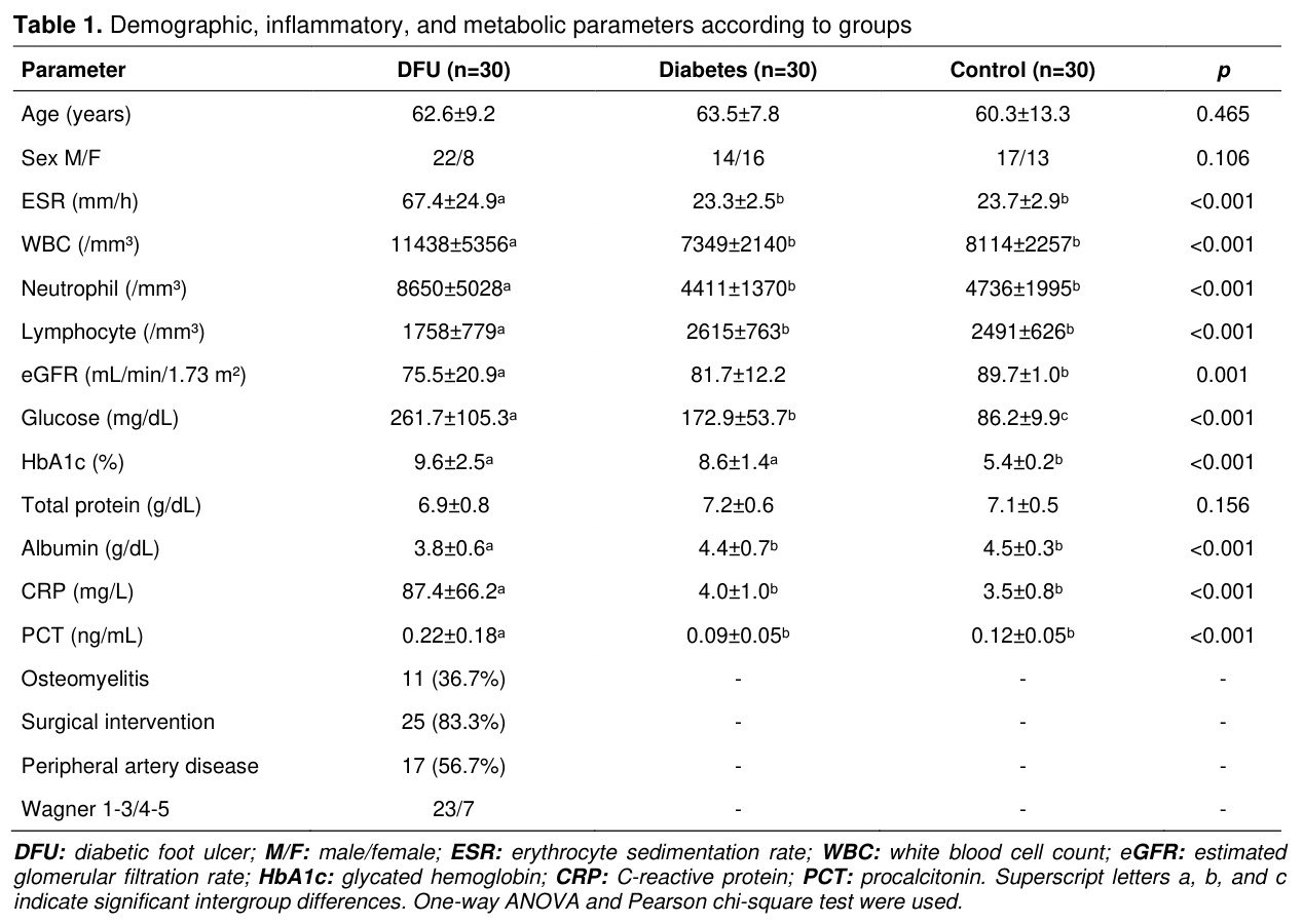
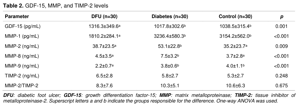
A total of 90 participants were included. No significant differences were found between groups regarding age and sex (p=0.465; p=0.106). In the DFU group, the median duration of diabetes was 20 years (range: 2–35 years; mean±SD: 17.4±8.7 years). ESR, WBC, neutrophil, CRP, and PCT levels were significantly higher in the DFU group (all p<0.001). Lymphocyte and albumin levels were lower in the DFU group (p<0.001). eGFR was lower in the DFU group compared to controls (p=0.001) (Table 1).GDF-15 was elevated in the DFU group (1316.3±349.6 pg/mL; p=0.001), while MMP-1 and MMP-9 were decreased (p<0.001). MMP-2 and MMP-8 levels were significantly higher in patients with diabetes without active foot ulcers than in the other groups (p=0.009 and p<0.001, respectively). No significant differences were observed in TIMP-2 or the MMP-2/TIMP-2 ratio (Table 2).
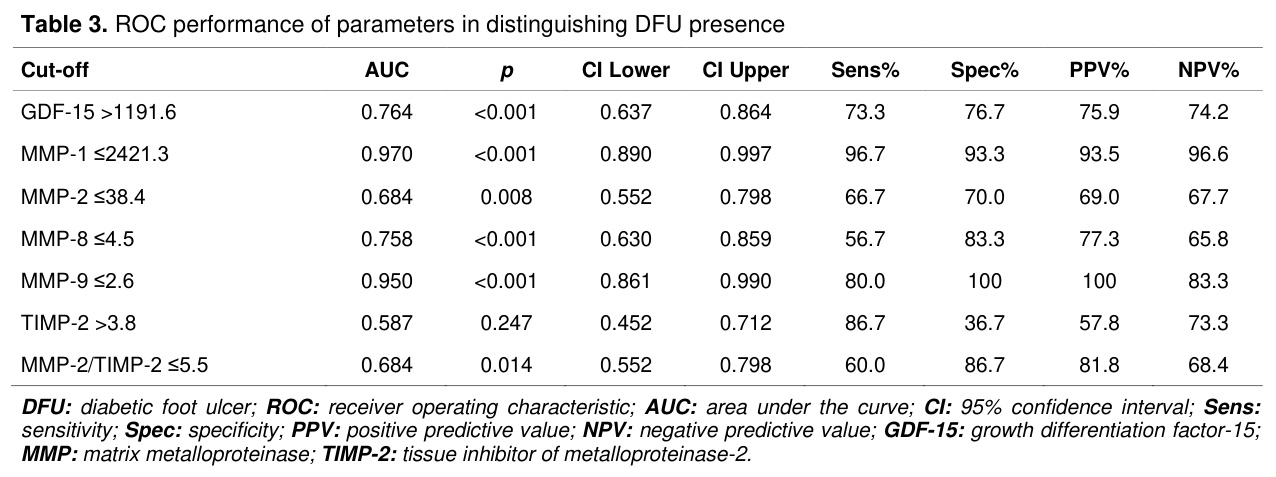
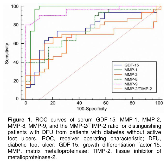
ROC analysis revealed that MMP-1 (AUC=0.970; sensitivity 96.7%; specificity 93.3%) and MMP-9 (AUC=0.950; sensitivity 80.0%; specificity 100.0%) had the highest diagnostic performance for distinguishing DFU (Table 3). The AUC for GDF-15 was 0.764 and for TIMP-2 was 0.587. The corresponding ROC curves are shown in Figure 1.
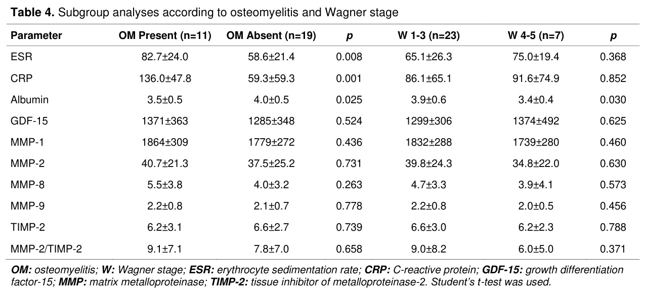
In subgroup analyses, ESR and CRP were higher (p=0.008; p=0.001) and albumin was lower (p=0.025) in the presence of osteomyelitis. Albumin was lower in the Wagner 4–5 group (p=0.030). No significant differences were found in GDF-15, MMPs, and TIMP-2 levels between subgroups (Table 4).
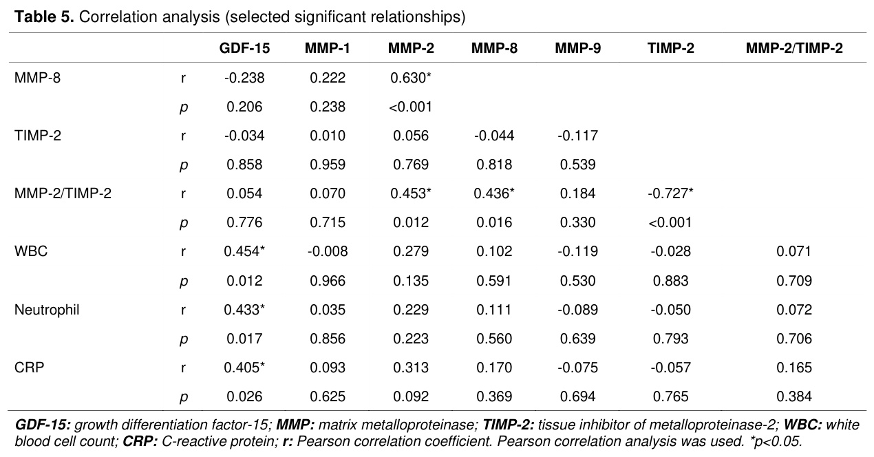
Correlation analysis revealed positive correlations between GDF-15 and WBC (r=0.454; p=0.012), neutrophil count (r=0.433; p=0.017), and CRP (r=0.405; p=0.026). A strong positive correlation was found between MMP-2 and MMP-8 (r=0.630; p<0.001). A negative correlation was observed between TIMP-2 and the MMP-2/TIMP-2 ratio (r=-0.727; p<0.001) (Table 5).
Tartışma
This study, by examining circulating GDF-15, selected MMPs, and TIMP-2 profiles at the systemic level in patients with DFU, suggests that diabetic foot ulcer is not merely a local wound process but carries a systemic disease component accompanied by coordinated dynamic changes in stress signaling, persistent inflammation, and ECM remodeling. The main findings are as follows: (i) significant elevation of serum GDF-15 levels and positive correlation with inflammatory markers in the DFU group, (ii) marked decrease in serum MMP-1 and MMP-9, (iii) elevation of MMP-2 and MMP-8 only in patients with diabetes without active foot ulcers, and (iv) high diagnostic performance of MMP-1 and MMP-9. These findings suggest that DFU biology may involve a stage-dependent biological transition extending from adaptive matrix remodeling to persistent inflammatory burden and relative regenerative exhaustion. However, this interpretation is hypothetical and cannot be definitively confirmed with the current cross-sectional data.GDF-15 is a stress-responsive cytokine belonging to the TGF-β superfamily, induced during cellular stress, mitochondrial dysfunction, hypoxia, and inflammatory activation [10-12]. Experimental and clinical studies have demonstrated that GDF-15 rises as a robust response to oxidative stress and tissue damage and represents a comprehensive indicator of systemic stress signaling [13,14]. The positive correlations observed between GDF-15 and WBC, neutrophil count, and CRP in the DFU group in the present study support this relationship. These findings are consistent with the data reported by Sendur et al. regarding the positive correlation between DFU severity and serum GDF-15 levels [5] and with the finding by Shikama et al. that elevated circulating GDF-15 is associated with adverse outcomes in lower extremity artery disease [15], while the strong association of GDF-15 with cardiometabolic mortality has been confirmed in large cohort studies [16,17]. GDF-15 has been shown to modulate integrin activation, thereby regulating leukocyte migration and immune cell responses [18]. Considering this mechanism, elevated GDF-15 levels may be associated with persistent inflammatory responses, and this process may indirectly affect protease dynamics. Indeed, the coexistence of GDF-15 elevation, neutrophil increase, and MMP-1/MMP-9 decrease observed in the present findings is biologically consistent with this hypothetical framework. On the other hand, the marked lymphopenia concurrently observed in the DFU group suggests that the modulatory effect of GDF-15 on immune cell populations may not be limited to the neutrophil axis.
The inflammatory burden of the DFU group should be considered an important determinant when interpreting the circulating biomarker profile. In the present study, ESR, WBC, neutrophil count, CRP, and PCT levels were significantly increased, whereas lymphocyte and albumin levels were decreased in patients with DFU, indicating a systemic inflammatory and catabolic state rather than an isolated local wound condition. This background may have contributed particularly to the elevation of GDF-15, which is known to respond to inflammatory, hypoxic, oxidative, and tissue injury-related stress signals [4,13,17]. Therefore, increased serum GDF-15 in DFU should be interpreted not as a DFU-specific signal alone, but as a marker integrating infection-related inflammation, metabolic stress, tissue damage, and vascular burden. Inflammatory activation may also influence matrix metalloproteinase-related remodeling responses, given their close relationship with extracellular matrix turnover and diabetic wound healing [6,7]. However, the absence of significant correlations between serum MMP-1/MMP-9 levels and conventional inflammatory markers suggests that the reduction in these proteases may not simply represent an acute-phase inflammatory response, but rather a distinct alteration in systemic matrix-remodeling biology.
One of the most striking findings of the present study is the significant decrease in circulating MMP-1 and MMP-9 levels in the DFU group, despite the frequently reported increased MMP activity in local wound fluid in the chronic wound literature [19,20]. This paradoxical finding can be explained by the compartmentalized nature of the biological response: the local proteolytic environment and systemic circulation levels may be independently regulated. Muller et al. reported that the MMP-1/TIMP-1 ratio in wound fluid may be valuable in predicting wound healing in diabetic foot ulcers [21]. However, since these findings pertain to the local wound fluid compartment, they should not be directly equated with serum levels. Therefore, the increased MMP activity reported in the local wound environment and the low serum MMP-1 and MMP-9 levels observed in the present study may reflect biological responses belonging to different compartments. Whether the decrease in serum MMP-1 and MMP-9 levels reflects reduced production or increased local consumption cannot be definitively distinguished with the current data. Another noteworthy point is that this decrease encompasses two functionally distinct proteases: MMP-1 is the primary degrader of type I–III fibrillar collagen, while MMP-9 is neutrophil- and macrophage-derived and exhibits activity directed at basement membrane components [22]. Specifically, MMP-1 has been identified as a critical collagenase for the initiation of keratinocyte migration and progression of re-epithelialization in the type I dermal collagen matrix [23]. Similarly, MMP-9 has been shown to proteolytically degrade pro-healing proteins such as dermatopontin in chronic wounds [24]. The simultaneous decrease of these two proteases with different cellular origins and substrate profiles may be compatible with the possibility of diminished adaptive remodeling capacity in severe DFU. An important additional finding supporting this interpretation is the absence of significant correlation between serum MMP-1 and MMP-9 levels and inflammatory markers. Immunometabolic reprogramming (metaflammation) may alter inflammatory and metabolic signaling pathways in diabetes, potentially creating a biological background that could affect circulating protease profiles [25]. Additionally, diabetes-related endothelial dysfunction may also contribute to the circulating protease profile [26]. Oxidative inflammation has been reported to impair tissue repair and wound healing in diabetes, potentially contributing to chronic wound biology [27].
On the other hand, the finding that MMP-2 and MMP-8 levels were highest in patients with diabetes without active foot ulcers, along with the strong positive correlation between these two markers, suggests that both proteases may reflect a distinct matrix-remodeling response in diabetic individuals without foot ulcers. MMP-2 (gelatinase A) has been demonstrated to contribute to angiogenic processes through proteolytic processing of extracellular matrix components [28]. However, selective MMP-2 inhibition has been reported not to significantly affect cutaneous wound healing [29], and the in vivo contribution of this enzyme to wound repair remains unclear. MMP-8 (collagenase-2) contributes to inflammation resolution through controlled collagen degradation and proteolytic inactivation of chemokines [30,31], and experimental mouse models have demonstrated that MMP-8 deficiency markedly impairs wound healing [31]. Indeed, MMP-8 has been reported to be the predominant collagenase in healing wounds and to contribute to structural integrity in wound repair [32]. Considering these functional profiles, the elevated MMP-2 and MMP-8 levels in patients with diabetes without active foot ulcers may represent an adaptive response aimed at maintaining tissue homeostasis under metabolic stress, or alternatively may reflect an early pathological process predisposing to ulcer development. Prospective cohort studies are needed to make this distinction.
The high AUC values obtained for MMP-1 and MMP-9 in ROC analysis support that these biomarkers strongly reflect the proteolytic remodeling disturbance associated with DFU. From a clinical perspective, however, these ROC findings should be interpreted cautiously. DFU is primarily diagnosed by clinical examination, wound inspection, and assessment of infection and perfusion status; therefore, serum MMP-1 and MMP-9 measurements should not be considered substitutes for standard clinical evaluation. Rather, their high AUC values suggest that these biomarkers may serve as adjunctive indicators of systemic matrix-remodeling alterations associated with DFU. While the high diagnostic performance of both MMP-1 and MMP-9 is noteworthy, particularly the high sensitivity of MMP-1 and the high specificity of MMP-9, it should be considered that limited sample sizes have the potential to inflate such extreme values. The lack of difference in serum TIMP-2 levels between groups suggests that matrix dysregulation cannot be fully explained by classic MMP-TIMP imbalance. TIMP-2 is known to serve both inhibitory and activating roles through the MT1-MMP/TIMP-2/MMP-2 ternary complex [22], and this dual biology limits the standalone diagnostic performance of TIMP-2. The moderate diagnostic performance of GDF-15 is consistent with this marker reflecting general metabolic and vascular stress burden rather than being specific to DFU. Although the present findings are exploratory and require external validation, these biomarkers may contribute in the future to multi-biomarker models for biological phenotyping, risk stratification, and monitoring of systemic disease burden in patients with diabetic foot ulcers.
In subgroup analyses, the significant increase in ESR and CRP along with the decrease in albumin in the presence of osteomyelitis is consistent with infection-related systemic inflammation and catabolic response; indeed, the diagnostic value of ESR and CRP in diabetic foot osteomyelitis has been confirmed in systematic reviews [33]. The hypoalbuminemia observed in advanced Wagner stages is a well-established clinical finding reflecting the combined effect of chronic inflammation and malnutrition [2]. Importantly, GDF-15, MMPs, and TIMP-2 levels did not show significant differences across osteomyelitis, PAD, surgical intervention, and Wagner stage subgroups within the DFU group. This finding suggests that these biomarkers have discriminative ability for identifying DFU presence but may be limited in grading disease severity.
The study has several limitations. The lower eGFR values observed in the DFU group do not entirely exclude the possible contribution of renal function in the interpretation of GDF-15 levels. Serum concentrations rather than enzymatic activity were evaluated; however, serum-based measurements offer a practical advantage in terms of clinical translatability. Importantly, circulating serum biomarker levels may not fully represent the local wound microenvironment. Local wound fluid contains proteases, inflammatory mediators, tissue degradation products, and cell-derived factors that may be regulated differently from systemic circulation. Therefore, the present findings should be interpreted as systemic biomarker patterns rather than direct measures of local wound proteolytic activity or local wound biology. The single-center design and limited sample size may have restricted the generalizability of the results.
Sonuç
In conclusion, this study suggests that increased serum GDF-15 levels together with decreased MMP-1 and MMP-9 levels may constitute a distinctive biomarker pattern in patients with DFU. In particular, MMP-1 and MMP-9 emerged as candidate adjunctive biomarkers with high diagnostic performance for distinguishing DFU presence. The complementary reflection of inflammatory and systemic stress response by GDF-15 and the distinct regulation of MMP-2 and MMP-8 in patients with diabetes without active foot ulcers suggest that DFU biology is not limited to the local wound process but may be associated with multidimensional systemic changes. However, given the cross-sectional design and limited sample size of the study, these findings need to be validated in larger, prospective, and independent cohorts. Future studies should prioritize the simultaneous evaluation of serum and local wound fluid biomarkers and the testing of multi-biomarker panels within diagnostic models.References
- Schaper NC, van Netten JJ, Apelqvist J, et al. Practical guidelines on the prevention and management of diabetes-related foot disease (IWGDF 2023 update). Diabetes Metab Res Rev 2024;40(3):e3657.
- Senneville E, Albalawi Z, van Asten SA, et al. IWGDF/IDSA guidelines on the diagnosis and treatment of diabetes-related foot infections (IWGDF/IDSA 2023). Diabetes Metab Res Rev 2024;40(3):e3687.
- Deng H, Li B, Shen Q, et al. Mechanisms of diabetic foot ulceration: A review. J Diabetes 2023;15(4):299-312.
- Lockhart SM, Saudek V, O’Rahilly S. GDF15: A Hormone Conveying Somatic Distress to the Brain. Endocr Rev 2020;41(4):bnaa007.
- Sendur SN, Firlatan B, Baykal G, Lay I, Erbas T. Serum growth differentiation factor-15 levels are associated with the severity of diabetic foot ulcer. Hormones (Athens) 2022;21(4):719-728.
- Chen J, Qin S, Liu S, et al. Targeting matrix metalloproteases in diabetic wound healing. Front Immunol 2023; 14: 1089001.
- Zhang WQ, Tang W, Hu SQ, et al. Effect of matrix metalloproteinases on the healing of diabetic foot ulcer: A systematic review. J Tissue Viability 2023;32(1):51-58.
- Wagner FW Jr. The dysvascular foot: a system for diagnosis and treatment. Foot Ankle 1981;2(2):64-122.
- Fitridge R, Chuter V, Mills J, et al. Editor’s Choice - The Intersocietal IWGDF, ESVS, SVS guidelines on peripheral artery disease in people with diabetes mellitus and a foot ulcer. Eur J Vasc Endovasc Surg 2023;66(4):454-483.
- Bootcov MR, Bauskin AR, Valenzuela SM, et al. MIC-1, a novel macrophage inhibitory cytokine, is a divergent member of the TGF-beta superfamily. Proc Natl Acad Sci USA 1997;94(21):11514-11519.
- Tsai VWW, Husaini Y, Sainsbury A, Brown DA, Breit SN. The MIC-1/GDF15-GFRAL Pathway in Energy Homeostasis: Implications for Obesity, Cachexia, and Other Associated Diseases. Cell Metab 2018;28(5):764-775.e5.
- Emmerson PJ, Wang F, Du Y, et al. The metabolic effects of GDF15 are mediated by the orphan receptor GFRAL. Nat Med 2017;23(10):1215-1219.
- Baek SJ, Eling T. Growth differentiation factor 15 (GDF15): A survival protein with therapeutic potential in metabolic diseases. Pharmacol Ther 2019;198:46-58.
- Wollert KC, Kempf T. Growth differentiation factor 15 in heart failure: an update. Curr Heart Fail Rep 2012;9(4):337-345.
- Shikama T, Otaki Y, Watanabe T, et al. Growth differentiation factor-15 and clinical outcomes in lower extremity artery disease. J Atheroscler Thromb 2024;31(6):964-978.
- Daniels LB, Clopton P, Laughlin GA, Maisel AS, Barrett-Connor E. Growth-differentiation factor-15 is a robust, independent predictor of 11-year mortality risk in community-dwelling older adults: The rancho bernardo study. Circulation 2011;123(19):2101-2110.
- Wallentin L, Hijazi Z, Andersson U, et al. Growth differentiation factor 15, a marker of oxidative stress and ınflammation, for risk assessment in patients with atrial fibrillation: Insights from the apixaban for reduction in stroke and other thromboembolic events in atrial fibrillation (ARISTOTLE) trial. Circulation 2014;130(21):1847-1858.
- Wischhusen J, Melero I, Fridman WH. Growth/differentiation factor-15 (GDF-15): From biomarker to novel targetable immune checkpoint. Front Immunol 2020;11:951.
- Lobmann R, Ambrosch A, Schultz G, et al. Expression of matrix-metalloproteinases and their inhibitors in the wounds of diabetic and non-diabetic patients. Diabetologia 2002;45(7):1011-1016.
- Krishnaswamy VR, Mintz D, Sagi I. Matrix metalloproteinases: The sculptors of chronic cutaneous wounds. Biochim Biophys Acta Mol Cell Res 2017;1864(11 Pt B):2220-2227.
- Muller M, Trocme C, Lardy B, et al. Matrix metalloproteinases and diabetic foot ulcers: The ratio of MMP-1 to TIMP-1 is a predictor of wound healing. Diabet Med 2008;25(4):419-426.
- Visse R, Nagase H. Matrix metalloproteinases and tissue inhibitors of metalloproteinases: Structure, function, and biochemistry. Circ Res 2003;92(8):827-839.
- Pilcher BK, Dumin JA, Sudbeck BD, et al. The activity of collagenase-1 is required for keratinocyte migration on a type I collagen matrix. J Cell Biol 1997;137(6):1445-1457.
- Krishnaswamy VR, Manikandan M, Munirajan AK, Vijayaraghavan D, Korrapati PS. Expression and integrity of dermatopontin in chronic cutaneous wounds: A crucial factor in impaired wound healing. Cell Tissue Res 2014;358(3):833-841.
- Hotamisligil GS. Inflammation, metaflammation and immunometabolic disorders. Nature 2017;542(7640):177-185.
- Beckman JA, Creager MA, Libby P. Diabetes and atherosclerosis: Epidemiology, pathophysiology, and management. JAMA 2002;287(19):2570-2581.
- Silveira LL, Sarandy MM, Novaes RD, Morais-Santos M, Goncalves RV. OxInflammation affects transdifferentiation to myofibroblasts, prolonging wound healing in diabetes: A systematic review. Int J Mol Sci 2024;25(16):899.
- Dean RA, Butler GS, Hamma-Kourbali Y, et al. Identification of candidate angiogenic inhibitors processed by matrix metalloproteinase 2 (MMP-2) in cell-based proteomic screens: disruption of vascular endothelial growth factor (VEGF)/heparin affin regulatory peptide (pleiotrophin) and VEGF/connective tissue growth factor angiogenic inhibitory complexes by MMP-2 proteolysis. Mol Cell Biol 2007;27(24):8454-8465.
- Nguyen TT, Wolter WR, Anderson B, et al. Limitations of Knockout Mice and Other Tools in Assessment of the Involvement of Matrix Metalloproteinases in Wound Healing and the Means to Overcome Them. ACS Pharmacol Transl Sci 2020;3(3):489-495.
- Caley MP, Martins VLC, O’Toole EA. Metalloproteinases and wound healing. Adv Wound Care (New Rochelle) 2015;4(4):225-234.
- Gutierrez-Fernandez A, Inada M, Balbin M, et al. Increased inflammation delays wound healing in mice deficient in collagenase-2 (MMP-8). FASEB J 2007;21(10):2580-2591.
- Nwomeh BC, Liang HX, Cohen IK, Yager DR. MMP-8 is the predominant collagenase in healing wounds and nonhealing ulcers. J Surg Res 1999;81(2):189-195.
- Ansert EA, Tarricone AN, Coye TL, et al. Update of biomarkers to diagnose diabetic foot osteomyelitis: A meta-analysis and systematic review. Wound Repair Regen 2024;32(4):366-376.