
Yüksek Fruktozlu Mısır Şurubu Tüketiminin Kalp Dokusundaki Betatrofin ve Kaspaz 3 Konsantrasyonları Üzerindeki Etkisinin Değerlendirilmesi
Cilt 40, Sayı 3
Kasım 2026 · Sayfalar 297-302
Yüksek Fruktozlu Mısır Şurubu Tüketiminin Kalp Dokusundaki Betatrofin ve Kaspaz 3 Konsantrasyonları Üzerindeki Etkisinin Değerlendirilmesi
Evaluation of the Impact of High-Fructose Corn Syrup Intake on Betatrophin and Caspase 3 Concentrations in Cardiac Tissue
Abdullah KARADAĞ1, Nevin KOCAMAN2, Ahmet TÜRK3, Osman GÜLER4
2Fırat University, Faculty of Medicine Department of Histology and Embryology, Elazığ, TÜRKİYE
3Adıyaman University, Faculty of Medicine Department of Histology and Embryology, Adıyaman, TÜRKİYE
4Munzur University, Pertek Sakine Genç Vocational School Laboratory and Veterinary Health Program, Tunceli, TÜRKİYE
Modern toplumda aşırı kilo ve obezitedeki artış teknolojideki ilerlemelere, hazır gıda sektörüne, fiziksel olarak zorlayıcı işlerin azalmasına ve yüksek fruktozlu gıdaların artan tüketimine atfedilmektedir. Bu eğilim, kardiyometabolik hastalıklar ve diyabet gibi ciddi sağlık sorunlarının gelişmesine katkıda bulunmaktadır. Betatrofin, lipid metabolizmasını ve insülin duyarlılığını etkileyen, insülin direnci veya yüksek karbonhidrat alımına yanıt olarak yağ dokusu ve karaciğer tarafından salgılanan bir protein hormonudur. Pankreas beta hücrelerinin proliferasyonunu ve farklılaşmasını teşvik ederek diyabet ve metabolik bozuklukların tedavisinde umut vaat etmektedir.
Çalışmamızın hedefleri (1) fruktozun kalp sağlığı üzerindeki olumsuz etkisini göstermek ve (2) fruktoz ile betatrofin seviyeleri arasındaki ilişkiyi incelemektir. Bunu değerlendirmek için, ticari mısır şurubu altı hafta boyunca sıçanların içme suyuna eklenmiş (HFCS grubu) ve kontrol grubuyla karşılaştırılma yapılmıştır. Kardiyak dokudaki kaspaz 3 ve betatrofin konsantrasyonları immünohistokimyasal etiketleme kullanılarak kantitatif olarak belirlenmiş, serum toplam antioksidan durumu (TAS) ve toplam oksidan durumu (TOS) seviyeleri ise ELISA yöntemiyle ölçülmüştür.
Deney sonucunda HFCS grubunun kalp dokusunda yüksek hasar, artmış kaspaz 3 ve betatrofin seviyeleri gözlenmiştir. Araştırmamız, HFCS alımının serum oksidan seviyelerini artırdığını, antioksidan kapasitesini azalttığını ve kalp dokusunda apoptoz mekanizmalarını tetiklediğini, bu durumun potansiyel olarak kardiyometabolik bozukluklara yol açabileceğini göstermektedir. Bu bağlamda, betatrofinin biyomarker veya tedavi hedefi olarak hizmet edebilme potansiyeli taşıdığı görülmüştür.
The rise in overweight and obesity in contemporary culture is ascribed to technological improvements, the convenience food business, the decline of physically demanding occupations, and the increased intake of high-fructose foods. This trend fosters the emergence of significant health complications, including cardiometabolic disorders and diabetes. Betatrophin is a protein hormone released by adipose tissue and the liver in reaction to insulin resistance or elevated carbohydrate consumption, influencing lipid metabolism and insulin sensitivity. By facilitating the proliferation and differentiation of pancreatic beta cells, it has potential for the treatment of diabetes and metabolic disorders.
The aims of our study are (1) to illustrate the detrimental effects of fructose on cardiovascular health and (2) to investigate the correlation between fructose and betatrophin levels. To assess this, commercial corn syrup was administered to the drinking water of rats for six weeks (HFCS group) and compared with a control group. The levels of Caspase-3 and betatrophin in cardiac tissue were quantitatively assessed using immunohistochemistry labeling, whilst the total antioxidant status (TAS) and total oxidant status (TOS) in serum were evaluated using the ELISA method.
The investigation revealed significant damage, elevated Caspase-3, and increased betatrophin levels in the cardiac tissue of the HFCS group. Our research indicates that the consumption of HFCS elevates blood oxidant levels, diminishes antioxidant capacity, and activates apoptotic pathways in cardiac tissue, potentially resulting in cardiometabolic diseases. In this regard, betatrophin has been identified as a possible biomarker or therapeutic target.
Giriş
The progress of technology and the ready-made food sector, coupled with a reduction in physically demanding jobs and an uptick in high-fructose food consumption, are contributing to a rise in overweight and obesity, which in turn is leading to a growing incidence of serious health issues, such as cardiometabolic diseases and diabetes, in modern society. The beverage sector, processed goods, and bread products utilize high fructose corn syrup due to its low cost, extended shelf life, and enhancement of freshness [1]. In affluent nations, overweight or obese people obtain a significant portion of their daily energy needs from fructose and exhibit 29% more white adipose tissue than those who do not eat it [2].Fructose is absorbed in the intestinal tract via glucose transporter type 5 (GLUT5) at the apical surface of luminal cells and is subsequently released into the bloodstream through GLUT-2, which exhibits poor affinity for fructose on the basolateral side and is predominantly processed in the liver. Extended and excessive intake of significant quantities of fructose increases GLUT5 expression in organs beyond the liver, hence enhancing fructose metabolism [3]. A study found that GLUT8, which is abundantly expressed in the liver and heart, facilitates fructose transport in mice [4]. After absorption, fructose is converted into Fructose-1-phosphate (F1P) by the enzyme fructokinase. The ATP-dependent nature of this process indicates that the ingestion of substantial amounts of fructose results in ATP depletion. F1P is subsequently biodegraded by the aldolase B enzyme to glyceraldehyde and dihydroxyacetone phosphate. These substances are fundamental to lipid synthesis triggered by fructose and the emergence of related pathologies [5].
Betatrophin, a protein hormone, modulates lipid metabolism and insulin sensitivity inside the body. Adipose tissue and the liver secrete it in reaction to insulin resistance or elevated carbohydrate consumption. Betatrophin has demonstrated potential in the treatment of diabetes and other metabolic disorders by enhancing the proliferation and differentiation of pancreatic beta cells. Investigations into the mechanisms of insulin production and glucose regulation have escalated following the identification of betatrophin, seen as a potential target [6].
In murine models of insulin resistance, hepatic overexpression of betatrophin results in enhanced beta-cell proliferation, increased islet size, elevated insulin content, and improved glucose homeostasis [7]. A study including children with obesity revealed that betatrophin was significantly elevated in serum and strongly linked with measures of insulin resistance, indicating a possible detrimental effect on insulin sensitivity [8]. The suppression of betatrophin was demonstrated to improve insulin sensitivity via the activation of the GSK-3β/PGC-1α signaling pathway [9]. These findings reveal a multifaceted link among betatrophin, beta cells, and insulin resistance. In another study betatrophin levels correlate with triglycerides, and there exists a positive association between elevated uric acid levels and betatrophin levels. Anaerobic activities have demonstrated more efficacy in diminishing elevated betatrophin levels in metabolic syndrome, perhaps mitigating problems associated with betatrophin elevation. It seems betatrophin is pivotal in the dysregulation of serum lipid levels and may lead to dyslipidemia in metabolic syndrome [10].
Our study sought to illustrate the detrimental effects of elevated fructose intake on cardiac health and to investigate its correlation with betatrophin levels.
Gereç ve Yöntem
Research and Publication Ethics: Approval has been obtained from the Local Ethics Committee for Animal Experiments of Munzur University for this study (date: 12.09.2022, file number: 2022/18, decision no:18-03).Animals: Fourteen 8-10 week old adults Wistar albino male rats were used and divided into two groups, each consisting of 7 rats; the rats were provided with unlimited food and water. The experimental duration was set to six weeks.
Groups
Control: No interventions were conducted during the experimental period.
HFCS: Commercial corn syrup comprising 42% fructose, 53% glucose, and 5% other saccharides was incorporated into the drinking water of rats at a 30% concentration for a duration of 6 weeks. The HFCS concentration was ascertained from the sugar content of various beverages, which generally varies from 7% to 15% [11].
Concluding the Experiment and Acquiring Tissue and Serum Specimens: At the conclusion of the study period, all rats in the groups were euthanized under anesthesia. Following the cleansing of the harvested cardiac tissue specimens with physiological saline, they were immersed in 10% neutral formaldehyde for histological analysis and preserved under suitable conditions. Serum samples for biochemical analyses were preserved at -80°C until the experimental day.
Measurement of Serum TAS and TOS Levels: Before analysis, blood samples were separated into serums by centrifugation at 3000 rpm for 10 minutes and then kept at -80°C. Serum Total Antioxidant Status (TAS) and Total Oxidant Status (TOS) levels were assessed utilizing commercial kits with Catalog No: RL0017 and Catalog No: RL0024 (Rel Assay Diagnostics, Turkey) via the Erel colorimetric method. Measurements were conducted utilizing the ChroMate 4300 Microplate Reader (Awareness Technology Inc., Palm City, USA) at wavelengths of 660 nm for TAS and 530 nm for TOS. In the TAS measurement, results were quantified in mmol Trolox Equiv./L, reflecting the reduction of the ABTS radical by environmental antioxidants; in the TOS measurement, results were expressed in μmol (H2O2 Equiv./L), based on the oxidation of the iron ion chelate complex to ferric ion by oxidants.
Histological Analyses
Histological evaluation: Histological assessment was performed Adiyaman University Faculty of Medicine. Tissue samples obtained from the euthanized animals underwent a one-week post-fixation in a 10% neutral formaldehyde solution, subsequently followed by standard tissue processing protocols for light microscopic analysis. Routine histological analysis was conducted utilizing an automated tissue processor (Leica TP1020, Nussloch, Germany) to produce paraffin blocks, and sections measuring 4-6 µm in thickness were obtained using a Thermo Shandon Finesse ME microtome (Thermo Fisher Scientific, Cheshire, UK).
Immunohistochemical analyses: Sections measuring 4–6 µm in thickness were obtained from paraffin blocks, subsequently placed on polylysine-coated slides, and deparaffinized. The sections were subjected to a graded alcohol series and subsequently heated in a microwave oven (750W) for 12 minutes at pH 6 using a citrate buffer solution for antigen retrieval. Following boiling, the tissues were allowed to cool at room temperature, washed with PBS (Phosphate Buffered Saline), and subsequently treated with hydrogen peroxide solution for 6 minutes to inhibit endogenous peroxidase activity.
Following the washing of tissues with PBS for three intervals of five minutes, a blocking solution was administered for five minutes. Subsequently, primary antibodies, diluted at a ratio of 1:200, including Caspase-3 (Rabbit monoclonal caspase-3, ERP18297-ab184787, Abcam, London, UK) and betatrophin (Polyclonal Antibody, PA5-38043, Invitrogen, USA), were incubated in a humid environment at room temperature for sixty minutes. The tissues were rinsed with PBS for three intervals of five minutes following the application of the primary antibody, and subsequently incubated in a humid environment at room temperature with a secondary antibody that is compatible with the primary antibody for thirty minutes. The tissues were rinsed with PBS for three intervals of five minutes following the application of the secondary antibody, then incubated in a humid atmosphere at room temperature with Streptavidin Peroxidase (TS–125-HR, Lab Vision Corporation, USA) for thirty minutes, after which they were transferred to PBS.
After the treatment of tissues with 3-amino-9-ethylcarbazole (AEC) substrate and AEC chromogen solution, the image signal was captured using a light microscope, after which all groups were concurrently rinsed with PBS. The tissues, counterstained with Mayer’s hematoxylin, were processed via PBS and distilled water before being mounted with a suitable mounting solution (Large Volume Vision Mount, TA-125-UG, Lab Vision Corporation, USA). The specimens were analyzed, assessed, and photographed with a Leica DM500 microscope (Leica DFC295). A histoscore was established during the staining process, determined by the prevalence (0.1: <25%, 0.4: 26-50%, 0.6: 51-75%, 0.9: 76-100%) and intensity (0: none, +0.5: extremely low, +1: low, +2: moderate, +3: strong) of immunoreactivity. Histoscore=prevalence multiplied by severity [11].
Statistical Analysis: All data are expressed as mean±standard deviation. Statistical analysis was conducted utilizing GraphPad Prism 8.0 software, created for Windows by GraphPad Software (www.graphpad.com). Groups were analyzed using the Student's t-test, with p<0.05 values deemed statistically significant. During the statistical evaluation, it was assessed if the groups had a normal distribution. The Kolmogorov-Smirnov and Shapiro-Wilk tests were utilized to confirm normality. All parameters, excluding Caspase-3, demonstrated a normal distribution. The Mann-Whitney U test was utilized for data that did not exhibit a normal distribution.
Bulgular
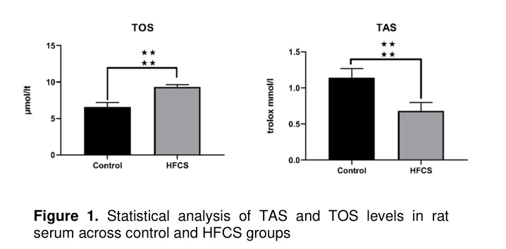
TAS and TOS Levels: In comparison to the Control group (6.605±0.5522 µmol/lt), rats in the serum HFCS group exhibited higher serum TOS levels (9.286±0.3143 µmol/lt) (p<0.0001) (95% CI: 2.157 to 3.204). Furthermore, when evaluating serum TAS levels in rats against the Control group (1.143±0.1125 trolox mmol/l), serum levels in the HFCS (0.7044±0.1172 trolox mmol/l) group were significantly reduced (p<0.0001) (95% CI: -0.5729 to -0.3053) (Figure 1).
Immunohistochemical Findings
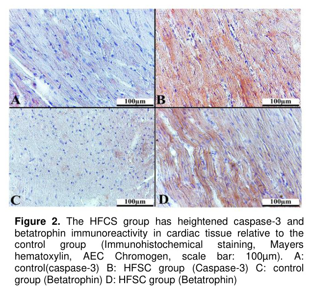
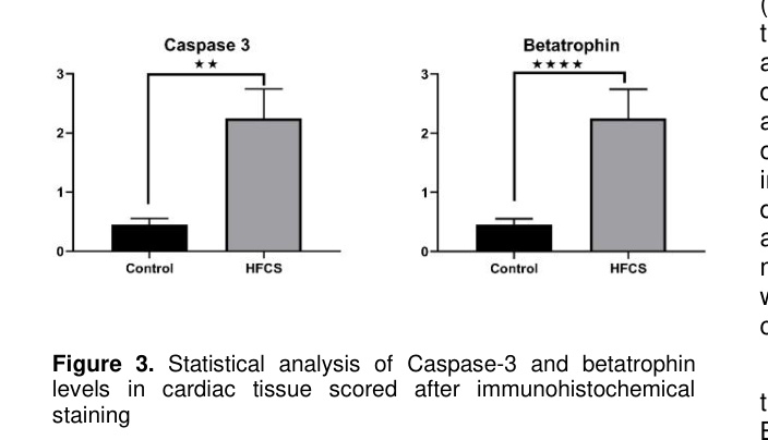
Tissue caspase-3 and betatrophin levels: Immunohistochemical staining for Caspase-3 and Betatrophin immunoreactivity was evaluated and quantified under a light microscope and subsequently analyzed statistically (Figure 2). Betatrophin levels in the HFCS group(2.029±0.7387) were considerably elevated compared to the control group (0.4429±0.9775) (p<0.0001) (95% CI: 0.9721 to 2.199). Likewise, levels of caspase-3 were substantially higher in the HFCS group (median:1.8) when contrasted with the Control group (median:0.4) (Hodges-Lehmann: 1.5) (p=0.0022) (Figure 3).
Tartışma
The rise in the use of high-fructose foods, particularly high-fructose corn syrup (HFCS), has resulted in a notable increase in obesity, insulin resistance, metabolic syndrome, and cardiovascular illnesses. Research on the metabolic implications of fructose indicates that this monosaccharide influences energy metabolism, oxidative stress, inflammation, lipogenesis, and cellular apoptotic pathways [12,13]. This study's findings indicate that HFCS consumption elevates oxidative stress, diminishes antioxidant capability, and initiates apoptotic processes in heart tissue. Moreover, the elevation of betatrophin levels is believed to correlate with the metabolic and cardiac alterations prompted by fructose intake.Our investigation revealed a significant increase in serum TOS levels in the HFCS group, accompanied by a drop in TAS levels. These findings align with existing data suggesting that elevated fructose intake enhances oxidative stress production. The swift exhaustion of ATP and the heightened synthesis of uric acid during fructose metabolism initiate the generation of reactive oxygen species (ROS) [14]. Fructose, when processed in the liver via fructokinase, results in a fast reduction of ATP, mitochondrial impairment, and subsequently an elevation in oxidative stress [15].
A strong correlation exists between oxidative stress and cardiovascular disorders. The elevation of reactive oxygen species (ROS) results in structural and functional impairments in cardiomyocytes due to lipid peroxidation, protein oxidation, and DNA damage [16]. Prior research has demonstrated that a high-fructose diet elevates oxidative stress indicators and diminishes antioxidant enzyme activity in experimental animals [17]. In our investigation, TAS levels were shown to decline, which was interpreted as HFCS consumption inhibiting endogenous antioxidant defense mechanisms. Consequently, it substantiates the notion that fructose might induce not only metabolic abnormalities but also oxidative damage in heart tissue.
The results of our investigation indicated a considerable increase in Caspase-3 immunoreactivity in the HFCS group. Caspase-3 is typically regarded as a significant indicator of apoptotic cell death mechanisms [18]. The elevated expression of Caspase-3 in cardiac tissue indicates that HFCS induces cardiomyocyte apoptosis. The rise in oxidative stress, along with the disruption of mitochondrial membrane permeability, the augmented release of cytochrome c, and the activation of intrinsic apoptotic pathways, culminates in the induction of Caspase-3 [19]. Prior research has demonstrated that elevated fructose intake induces apoptotic alterations in heart tissue and exacerbates myocardial injury [20]. The elevation of Caspase-3 in our work constitutes a significant discovery corroborating the cardiotoxic effects of HFCS.
The substantial rise in betatrophin levels is one of the most remarkable discoveries of our investigation. Betatrophin is a hormone linked to lipid metabolism and insulin resistance, produced in the liver and adipose tissue [21]. Prior research indicated that betatrophin might enhance glucose homeostasis through the promotion of pancreatic β-cell proliferation; however, newer investigations have revealed that elevated betatrophin levels correlate with metabolic syndrome, obesity, and insulin resistance [22]. Our data indicated that the intake of HFCS elevates betatrophin levels, potentially as a component of the metabolic stress response.
Dihydroxyacetone phosphate and glyceraldehyde, products of fructose metabolism, serve as possible substrates for triglyceride synthesis. Elevated fructose intake enhances lipogenesis and diminishes fatty acid oxidation [23]. Enhanced lipogenesis facilitates the onset of hepatic steatosis, hypertriglyceridemia, and insulin resistance. The observed increase in betatrophin in our study may be linked to lipid metabolism disorders generated by HFCS, aligning with existing literature on the association between betatrophin and triglyceride metabolism [24,25].
The link between oxidative stress and betatrophin remains incompletely understood; nonetheless, existing evidence indicate that oxidative stress may elevate betatrophin expression. In the context of insulin resistance and inflammation, elevated oxidative stress can stimulate betatrophin production via hepatic metabolic pathways [26]. Our data indicates that the elevation of TOS levels and the increase in betatrophin expression suggest a potential correlation between these two mechanisms. The elevation of betatrophin levels, along with increased Caspase-3 in cardiac tissue, indicates that betatrophin may serve as both a metabolic regulator and a contributor to the heart stress response.
The detrimental impacts of HFCS intake on the cardiovascular system extend beyond oxidative stress and apoptosis. High fructose also contributes to endothelial dysfunction, inflammation, hypertension, and cardiac fibrosis [27,28]. Long-term fructose consumption has been associated with heart hypertrophy and diastolic dysfunction [29]. Consequently, the prolonged intake of HFCS may be regarded as a significant risk factor for the onset of metabolic syndrome as well as direct alterations in heart structure.
This research possesses certain limitations. The primary limitation of the study is the omission of supplementary oxidative stress and inflammation markers, which would enhance the understanding of oxidative stress and cardiac damage, along with the absence of a comparison between the acute and chronic effects of HFCS consumption. Future investigations, bolstered by molecular, biochemical, and functional analyses, will elucidate the consequences of fructose ingestion on heart tissue in greater detail.
Sonuç
In conclusion, our research indicates that the use of HFCS elevates blood oxidant levels and diminishes antioxidant capacity, hence activating apoptotic mechanisms in cardiac tissue and potentially resulting in cardiometabolic disorders. It indicates that betatrophin may serve as a potential biomarker or therapeutic target in this context. Nonetheless, sophisticated experimental and clinical investigations are required to elucidate the effects of betatrophin on cardiac tissue and its molecular mechanisms.References
- White JS. Straight talk about high-fructose corn syrup: what it is and what it ain't. The American Journal of Clinical Nutrition 2008;88(6):1716S-1721S.
- Ma J, McKeown NM, Hwang S-J, et al. Sugar-sweetened beverage consumption is associated with change of visceral adipose tissue over 6 years of follow-up. Circulation 2016;133(4):370-377.
- Hernández-Díazcouder A, Romero-Nava R, Carbó R, Sánchez-Lozada LG, Sánchez-Muñoz F. High fructose intake and adipogenesis. International journal of molecular sciences 2019;20(11):2787.
- DeBosch BJ, Chen Z, Saben JL, Finck BN, Moley KH. Glucose transporter 8 (GLUT8) mediates fructose-induced de novo lipogenesis and macrosteatosis. Journal of Biological Chemistry 2014;289(16):10989-10998.
- Jung S, Bae H, Song W-S, Jang C. Dietary fructose and fructose-induced pathologies. Annual Review of Nutrition 2022;42:45-66.
- Guo Q, Cao S, Wang X. Betatrophin and insulin resistance. Metabolites 2022;12(10):925.
- Guo XR, Wang XL, Chen Y, et al. ANGPTL8/betatrophin alleviates insulin resistance via the Akt-GSK3β or Akt-FoxO1 pathway in HepG2 cells. Experimental Cell Research 2016;345(2):158-167.
- Wu S, Gao H, Ma Y, et al. Characterisation of betatrophin concentrations in childhood and adolescent obesity and insulin resistance. Pediatric Diabetes 2016;17(1):53-60.
- Hao Q, Zheng A, Zhang H, Cao H. Down-regulation of betatrophin enhances insulin sensitivity in type 2 diabetes mellitus through activation of the GSK-3β/PGC-1α signaling pathway. Journal of Endocrinological Investigation 2021;44(9):1857-1868.
- Karaman ME, Arslan C, Gürsu MF. Effects of different exercise loads on serum betatrophin (ANGPTL-8/lipasin) and cartonectin (CTRP-3) levels in metabolic syndrome. Turkish Journal of Biochemistry 2022;47(1):71-78.
- Türk A, Karadağ A, Zencirci B, Özay Y, Güler O. Investigation of the effects of ghrelin and kisspeptin levels in liver tissue of rats fed with high fructose diet-A histological study. Ahi Evran Medical Journal 2023;7(3):346-352.
- Johnson RJ, Nakagawa T, Sanchez-Lozada LG, et al. Sugar, uric acid, and the etiology of diabetes and obesity. Diabetes 2013;62(10):3307-3315.
- Lanaspa MA, Sanchez-Lozada LG, Choi Y-J, et al. Uric acid induces hepatic steatosis by generation of mitochondrial oxidative stress: potential role in fructose-dependent and-independent fatty liver. Journal of Biological Chemistry 2012;287(48):40732-40744.
- Sautin YY, Johnson RJ. Uric acid: The oxidant-antioxidant paradox. Nucleosides, Nucleotides, and Nucleic Acids 2008;27(6-7):608-619.
- Nakagawa T, Hu H, Zharikov S, et al. A causal role for uric acid in fructose-induced metabolic syndrome. American Journal of Physiology-Renal Physiology 2006;290(3):F625-F31.
- Ogrunc M, Di Micco R, Liontos M, et al. Oncogene-induced reactive oxygen species fuel hyperproliferation and DNA damage response activation. Cell Death & Differentiation 2014;21(6):998-1012.
- Madlala HP, Maarman GJ, Ojuka E. Uric acid and transforming growth factor in fructose-induced production of reactive oxygen species in skeletal muscle. Nutrition Reviews 2016;74(4):259-266.
- Elmore S. Apoptosis: A review of programmed cell death. Toxicologic Pathology 2007;35(4):495-516.
- Kroemer G, Galluzzi L, Brenner C. Mitochondrial membrane permeabilization in cell death. Physiological Reviews 2007;87(1):99-163.
- Mellor K, Ritchie RH, Meredith G, et al. High-fructose diet elevates myocardial superoxide generation in mice in the absence of cardiac hypertrophy. Nutrition 2010;26(7-8):842-848.
- Abu-Farha M, Al Madhoun A, Abubaker J. The Rise and the Fall of Betatrophin/ANGPTL8 as an Inducer of β‐Cell Proliferation. Journal of diabetes research. 2016;2016(1):4860595.
- Fu Z, Berhane F, Fite A, et al. Elevated circulating lipasin/betatrophin in human type 2 diabetes and obesity. Scientific Reports 2014;4(1):5013.
- Jensen T, Abdelmalek MF, Sullivan S, et al. Fructose and sugar: A major mediator of non-alcoholic fatty liver disease. Journal of Hepatology 2018;68(5):1063-1075.
- Iskender H, Yenice G, Terim Kapakin KA, et al. Effects of high fructose diet on lipid metabolism and the hepatic NF-κB/SIRT-1 pathway. Biotechnic & Histochemistry 2022;97(1):30-38.
- de Farias Lelis D, Andrade JMO, Almenara CCP, et al. High fructose intake and the route towards cardiometabolic diseases. Life Sciences 2020;259:118235.
- Ebert T, Kralisch S, Hoffmann A, et al. Circulating angiopoietin-like protein 8 is independently associated with fasting plasma glucose and type 2 diabetes mellitus. The Journal of Clinical Endocrinology & Metabolism 2014;99(12):E2510-E7.
- Zhang DM, Jiao RQ, Kong LD. High Dietary Fructose: Direct or Indirect Dangerous Factors Disturbing Tissue and Organ Functions. Nutrients 2017;9(4).
- Xu C, Yu J. Pathophysiological mechanisms of hypertension development induced by fructose consumption. Food & Function 2022;13(4):1702-1717.
- Daniels LJ, Annandale M, Koutsifeli P, et al. Elevated myocardial fructose and sorbitol levels are associated with diastolic dysfunction in diabetic patients, and cardiomyocyte lipid inclusions in vitro. Nutrition & Diabetes 2021;11(1):8.