
Setilpiridinyum Klorürün Yumurtalık ve Prostat Kanseri Hücre Hatlarında Sitotoksik ve Genotoksik Potansiyelinin Değerlendirilmesi
Cilt 40, Sayı 3
Kasım 2026 · Sayfalar 223-231
Setilpiridinyum Klorürün Yumurtalık ve Prostat Kanseri Hücre Hatlarında Sitotoksik ve Genotoksik Potansiyelinin Değerlendirilmesi
Evaluation of the Cytotoxic and Genotoxic Potential of Cetylpyridinium Chloride in Ovarian and Prostate Cancer Cell Lines
Esmanur BOZOĞLU1, Samet ÖZ2, Suat TEKİN3
2Osmaniye Korkut Ata University, Vocational School of Health Services, Department of Veterinary Medicine, Osmaniye, TÜRKİYE
3İnönü University, Faculty of Medicine, Department of Physiology, Malatya, TÜRKİYE
Amaç: Setilpiridinyum klorür (CPC), kuaterner amonyum yapısına sahip katyonik bir yüzey aktif madde olup ağız hijyeni ürünlerinde yaygın olarak kullanılmaktadır. Son yıllarda yapılan çalışmalar, CPC’nin çeşitli kanser hücre tipleri üzerinde sitotoksik etkiler gösterebileceğini ortaya koymuştur. Bu çalışmanın amacı, CPC’nin insan over kanseri (A2780) ve prostat kanseri (LNCaP) hücre hatları üzerindeki sitotoksik ve genotoksik etkilerini in vitro koşullarda değerlendirmektir.
Gereç ve Yöntem: Hücre canlılığı, hücrelerin 1, 10, 100 ve 1000 µM konsantrasyonlarında CPC’ye 24 saat süreyle maruz bırakılmasının ardından MTT yöntemi kullanılarak belirlenmiştir. Genotoksik etkiler ise tek hücre jel elektroforezi (Comet) testi ile değerlendirilmiştir.
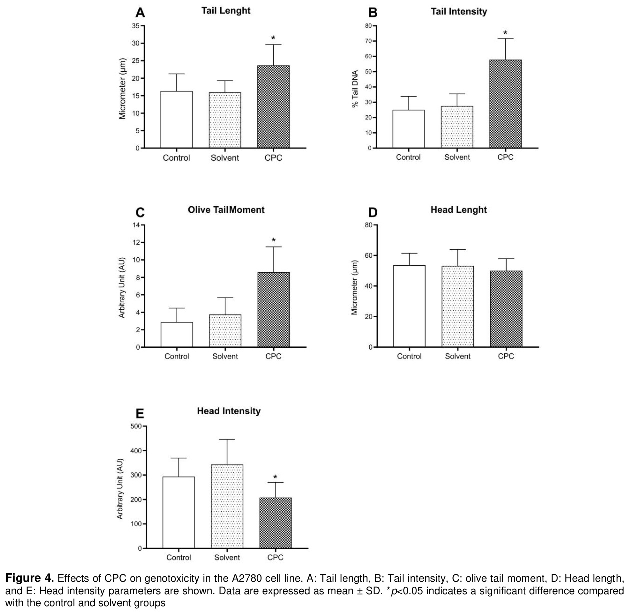
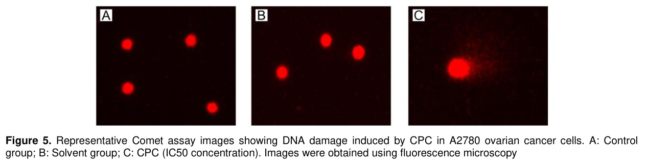
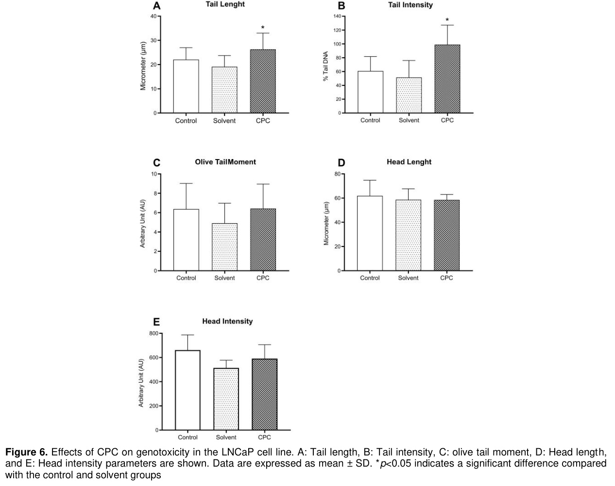
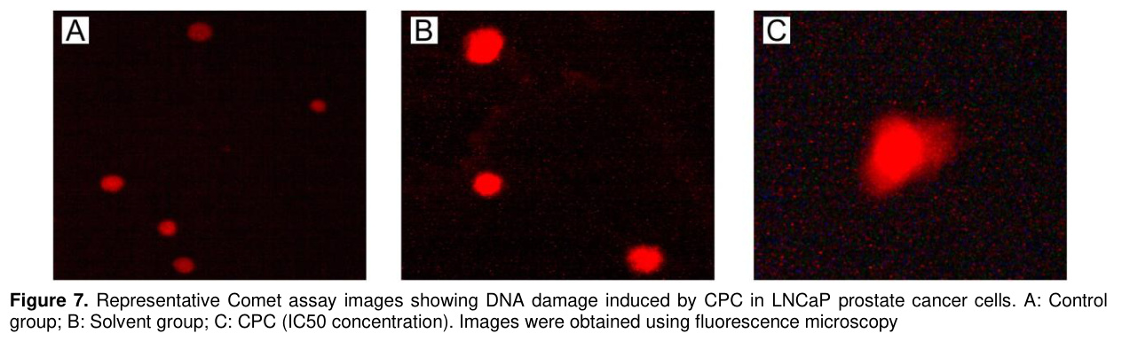
Bulgular: Elde edilen sonuçlar, CPC’nin her iki hücre hattında da konsantrasyona bağlı olarak hücre canlılığını anlamlı derecede azalttığını göstermiştir (p<0.05). Özellikle 10 µM ve üzerindeki konsantrasyonlarda belirgin sitotoksik etki gözlenmiş ve daha yüksek dozlarda hücre canlılığında dramatik bir azalma meydana gelmiştir. Comet testi sonuçları, A2780 hücre hattında kuyruk uzunluğu, kuyruk yoğunluğu ve olive tail moment parametrelerinde anlamlı artışlar olduğunu göstermiştir. LNCaP hücre hattında ise kuyruk uzunluğu ve kuyruk yoğunluğunda artışlar gözlenmiş olup bu durum DNA hasarının varlığına işaret etmektedir. Ayrıca floresan mikroskopi görüntülerinde, CPC ile muamele edilen hücrelerde belirgin Comet kuyruğu oluşumu ve artmış DNA migrasyonu saptanmıştır.
Sonuç: Sonuç olarak elde edilen bulgular, CPC’nin A2780 ve LNCaP kanser hücre hatlarında konsantrasyona bağlı sitotoksik ve genotoksik etkiler oluşturduğunu göstermektedir. Bu sonuçlar, CPC’nin kanser hücreleri üzerindeki biyolojik etkilerinin daha ayrıntılı moleküler çalışmalar ve in vivo araştırmalarla ileri düzeyde incelenmesi gerektiğini düşündürmektedir.
Objective: Cetylpyridinium chloride (CPC) is a cationic surfactant with a quaternary ammonium structure that is widely used in oral hygiene products. Recent studies have suggested that CPC may exert cytotoxic effects on various cancer cell types. The aim of the present study was to evaluate the cytotoxic and genotoxic effects of CPC on human ovarian cancer (A2780) and prostate cancer (LNCaP) cell lines under in vitro conditions.
Materials and Methods: Cell viability was determined using the MTT assay following 24-hour exposure to CPC at concentrations of 1, 10, 100, and 1000 µM. Genotoxic effects were assessed using the single-cell gel electrophoresis (Comet) assay.
Results: The results demonstrated that CPC significantly decreased cell viability in both cell lines in a concentration-dependent manner (p<0.05). A marked cytotoxic effect was observed particularly at concentrations of 10 µM and above, with a dramatic reduction in cell viability at higher doses. Comet assay results revealed significant increases in tail length, tail intensity, and olive tail moment parameters in the A2780 cell line. In the LNCaP cell line, increases in tail length and tail intensity were observed, indicating the presence of DNA damage. In addition, fluorescence microscopy images showed prominent Comet tail formation and increased DNA migration in CPC-treated cells.
Conclusion: In conclusion, findings indicate that CPC induces concentration-dependent cytotoxic and genotoxic effects in A2780 and LNCaP cancer cell lines. These results suggest that the biological effects of CPC on cancer cells should be further investigated through more detailed molecular and in vivo studies.
Giriş
Cancer is a heterogeneous group of diseases characterized by uncontrolled cell proliferation, insensitivity to growth-suppressive signals, and evasion of programmed cell death, arising from the disruption of mechanisms that regulate cellular growth and proliferation [1,2]. The process of carcinogenesis is defined as a multistep biological process that develops as a result of the accumulation of genetic and epigenetic alterations in cells over time and progresses through sequential stages [3,4]. During this process, the loss of cell cycle checkpoints, impairment of DNA damage response mechanisms, and dysregulation of apoptotic signaling pathways contribute to tumor development [2,5].Cancer remains one of the most significant public health challenges worldwide. According to data based on 2022 estimates and updated in early 2024, approximately 20 million new cancer cases were diagnosed globally, and nearly 9.7 million people died from cancer-related causes [6]. Due to population aging and changes in lifestyle-related factors, the number of new cancer cases is projected to increase by 77%, reaching approximately 35 million annually by 2050 [6]. Prostate cancer, one of the gender-specific malignancies, is among the most frequently diagnosed cancers in men and represents a significant global health burden [7]. According to GLOBOCAN 2022 data, prostate cancer ranks among the top five cancers in terms of incidence across both sexes, with approximately 1.47 million new cases reported in 2022 [7]. Ovarian cancer, an important gynecological malignancy in women, is characterized by relatively lower incidence rates but notably high mortality [7] According to global data, approximately 325,000 new cases of ovarian cancer were diagnosed worldwide in 2022, and more than 206,000 deaths were attributed to ovarian cancer [7]. The systemic toxicity associated with current chemotherapeutic protocols, the emergence of drug resistance mechanisms, and the high cost of treatment have directed oncology research toward safer and more cost-effective alternatives. In this context, investigating the anticancer effects of compounds that are already in clinical use for different medical purposes and whose safety profiles are well established (drug repurposing) has emerged as a strategic approach that offers significant advantages in terms of time and cost in the drug development process [8,9].
Cetylpyridinium chloride (CPC), a cationic quaternary ammonium compound, is widely used in products such as mouthwashes and lozenges due to its broad-spectrum antimicrobial properties [10,11]. The biological activity of CPC is primarily attributed to mechanisms such as its ability to bind to negatively charged cell membranes due to its positively charged structure, thereby disrupting membrane integrity and inducing apoptosis [11,12]. Recent studies have reported that CPC can exert cytotoxic effects in various cancer cell models [13,14]; however, studies evaluating these effects across different cancer cell types remain limited in the literature. In light of this information, this study aimed to evaluate the potential cytotoxic and genotoxic effects of CPC, a compound known for its broad-spectrum antimicrobial activity, on human ovarian (A2780) and prostate (LNCaP) cancer cell lines representing gynecological and urological malignancy models under in vitro conditions.
Gereç ve Yöntem
Research and Publication Ethics: This study was conducted using established human cancer cell lines and did not involve human participants or animals; therefore, ethical committee approval was not required.Chemicals: Cetylpyridinium chloride (CPC; Sigma-Aldrich, CAS No: 6004-24-6), fetal bovine serum (FBS; Serox, Germany), penicillin–streptomycin (Gibco, USA), dimethyl sulfoxide (DMSO; Merck, Germany), Minimum Essential Medium (MEM) non-essential amino acids (Biological Industries, Israel), insulin (Novorapid, Denmark), 3-(4,5-dimethyl-2-thiazolyl)-2,5-diphenyl-2H-tetrazolium bromide (MTT; Serva, Germany), RPMI-1640 medium, and other chemicals (Biowest, USA) were used in the study. CPC was dissolved in distilled water to prepare working concentrations of 1, 10, 100, and 1000 µM. The CPC concentrations used in this study were selected based on concentration ranges previously employed in in vitro toxicity and genotoxicity studies reported in the literature [13].
Cell Culture: This study was conducted in the laboratories of the Department of Physiology, Faculty of Medicine, İnönü University. A2780 and LNCaP cancer cell lines were used in the study. The A2780 and LNCaP cells were cultured in RPMI-1640 medium supplemented with 10% fetal bovine serum (FBS), 100 U/mL penicillin, 0.1 mg/mL streptomycin, and 5 mL MEM non-essential amino acid solution. All cells were incubated in 75 cm2 culture flasks in an incubator (ESCO, Egaa, Denmark), and the culture medium was replaced twice a week. To assess cell viability, the cells were stained with 0.4% trypan blue and counted under a microscope. Cells with a viability greater than 90% were used for the MTT assay analyses [15,16]. For MTT assay analyses, once the cells reached confluence, they were detached from the culture flasks using a trypsin-EDTA solution, seeded into 96-well plates at a density of 15x103 cells per well, and incubated for 24 hours [17]. After the initial incubation period, CPC was added to the 96-well plates at concentrations of 1, 10, 100, and 1000 µM, and the cells were incubated for an additional 24 hours. All incubation steps were carried out in an incubator maintained at 37 °C with 5% CO2.
MTT Assay: Cytotoxicity was evaluated using the MTT assay, a widely used enzymatic test method. This method is based on the reduction of the tetrazolium ring of the MTT dye. MTT is actively taken up by viable cells and is reduced by mitochondrial succinate dehydrogenase to form formazan, a blue-purple, water-insoluble product. Formazan formation occurs only in cells containing active mitochondria and is therefore considered an indicator of cell viability, correlating with the number of living cells [18]. First, a working MTT solution (0.5 mg/mL) was prepared from the stock MTT solution in sterile phosphate-buffered saline. After 24 hours of incubation with CPC, 50 µL of the MTT solution was added to each well containing A2780 and LNCaP cells. The plates were then incubated in an incubator for
3 hours. Following incubation, the MTT solution wasremoved from the plates, and 100 µL of DMSO was added to each well to dissolve the formazan crystals formed in the cells. The optical density of the cells was measured at 550 nm using an ELISA microplate reader (Thermo MultiskanGo, USA) [19,20]. The mean absorbance values obtained from the control wells were considered to represent 100% cell viability. The values obtained from solvent- and CPC-treated wells were compared with those of the control wells to calculate the percentage of cell viability [21,22]. These experiments were repeated at least ten times on different days. Each experiment represented an independent biological replicate performed on different days.
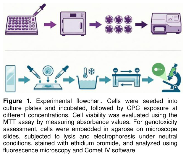
Comet Assay: The Comet assay, also known as single-cell gel electrophoresis, is a sensitive and reliable method widely used for the evaluation of DNA damage and genotoxic effects [23,24]. The Comet assay was performed under neutral electrophoresis conditions to evaluate DNA strand damage. First, a 0.65% (w/v) high melting point agarose (HMA) solution was prepared in phosphate-buffered saline (PBS) and spread onto pre-chilled microscope slides, which were then allowed to dry in the dark for 24 hours. After determining the IC₅₀ value of CPC for each cell line, the cells were exposed to this concentration for 1 hour. Following incubation, CPC was removed, and the cells were suspended in low melting point agarose (LMA) and transferred onto HMA-precoated slides. The preparations were covered with coverslips and incubated in the dark at +4 °C for 15-20 minutes to allow agarose polymerization. After solidification of the agarose, the coverslips were removed and the slides were immersed in cold lysis solution and incubated at 4 °C in the dark for 1 hour. The lysis buffer was freshly prepared by adding 1% Triton X-100 and 1% DMSO to the stock solution. [25]. Following the lysis step, the slides were placed in a horizontal electrophoresis tank (Bio-Rad, USA) filled with pre-chilled neutral electrophoresis buffer. Electrophoresis was performed for 20 minutes under constant conditions of 25 V and 300 mA, allowing negatively charged DNA fragments to migrate toward the positive electrode. After electrophoresis, the slides were neutralized by immersing them in 0.4 M Tris buffer (pH 7.5) at 4 °C for three consecutive washes of 5 minutes each to prevent further DNA migration. Subsequently, the preparations were stained with 50 µL of ethidium bromide and incubated in the dark at +4 °C for 20-30 minutes. Fluorescence images were obtained using a Leica fluorescence microscope, and quantitative analysis of DNA damage was performed using Comet IV software. For each slide, at least 100 randomly selected cells were analyzed to evaluate tail length, tail intensity, olive tail moment, head length, and head intensity parameters [16]. Changes observed in these parameters were used to evaluate both the extent of DNA damage formation and the magnitude of the damage.
The experimental flowchart is illustrated in Figure 1.
Statistical Analysis: Statistical analysis was performed using the IBM SPSS software program for Windows (SPSS Inc., Chicago, IL, USA). Data are presented as mean ± standard deviation (SD). Comparisons of the experimental results among groups were conducted using the Kruskal-Wallis H test. When statistically significant differences were detected among groups, multiple comparisons were performed using the Mann-Whitney U test with Bonferroni correction (p<0.05 was considered statistically significant). The half-maximal inhibitory concentration (IC50) and logarithmic half-maximal inhibitory concentration (logIC50) values were calculated using GraphPad Prism 8 software.
Bulgular
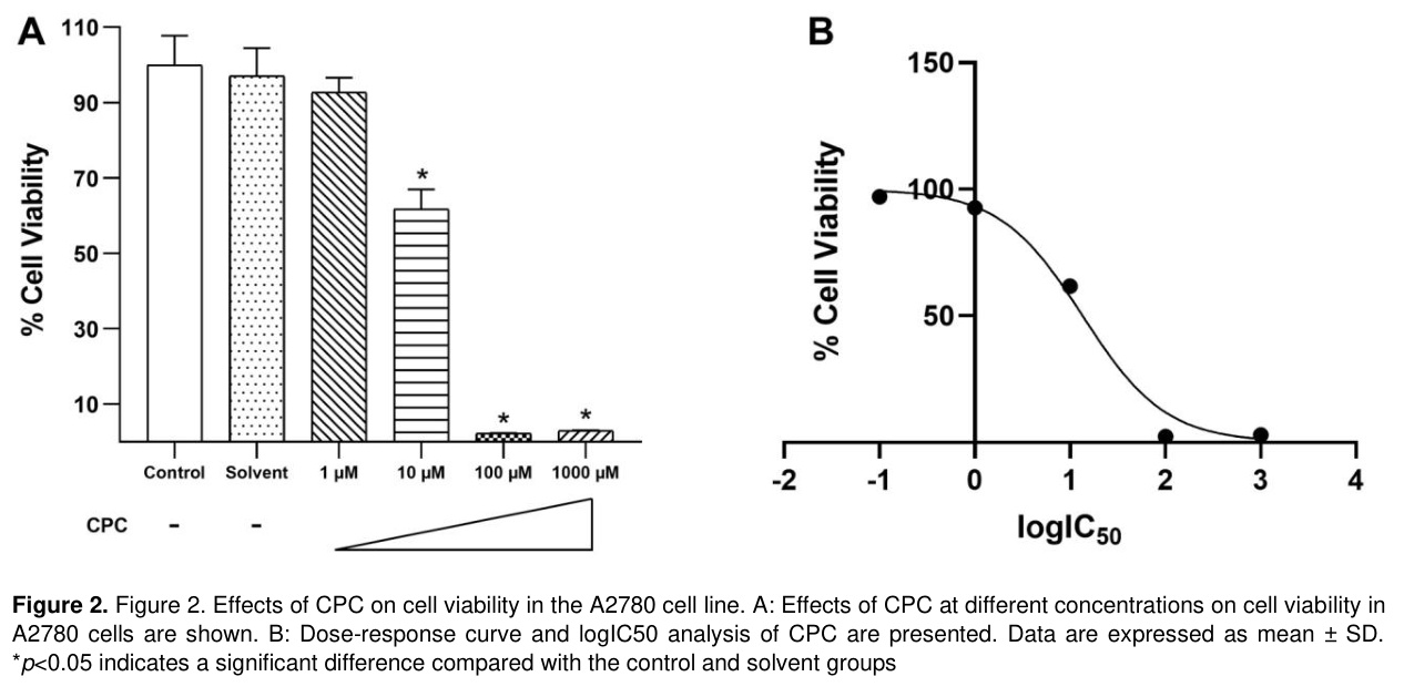
Effects of Cetylpyridinium Chloride on Cell Viability in the A2780 Cell Line. Figure 2 shows cell viability (Figure 2A) and the dose–response curve (Figure 2B) in the A2780 ovarian cancer cell line. The cell viability analysis revealed that CPC induced a pronounced and concentration-dependent cytotoxic effect. No significant difference in cell viability was observed between the control and solvent groups, whereas treatment with 10 µM CPC significantly reduced cell viability compared with both the control and solvent groups (p<0.05). At higher concentrations (100 and 1000 µM), CPC caused a marked decrease in cell viability, and the majority of cells lost viability (p<0.05).
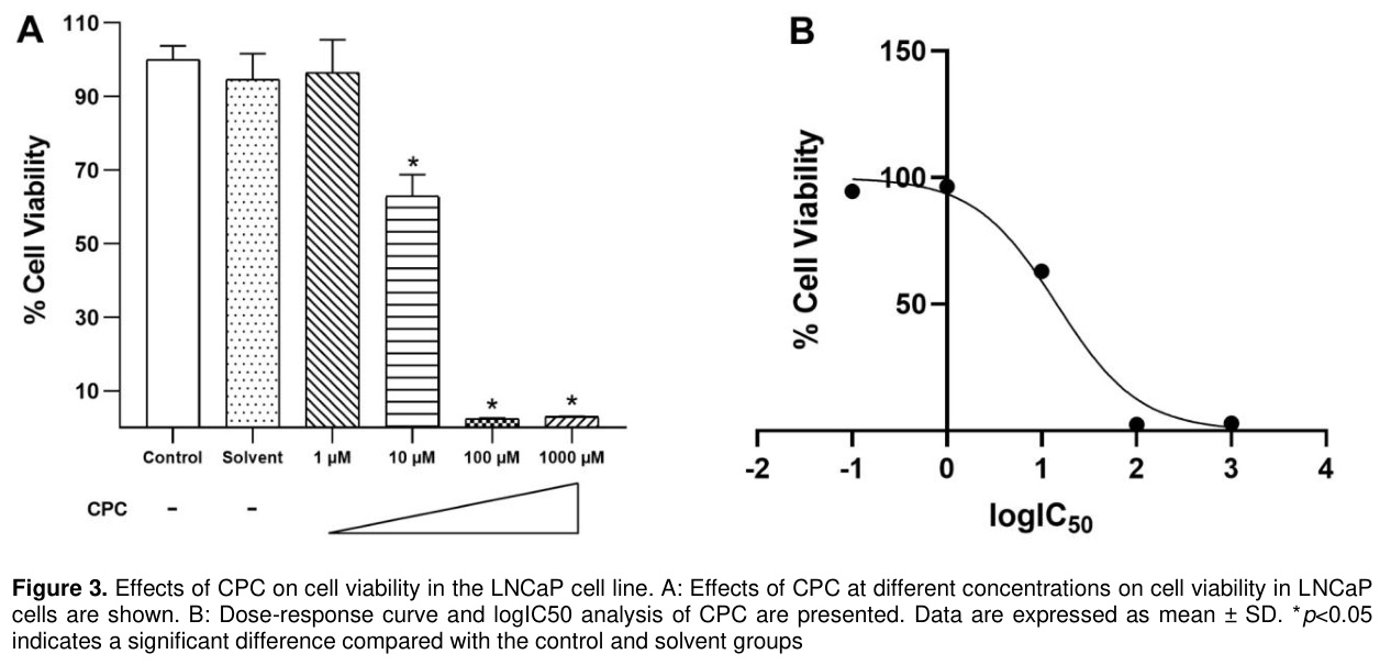
Effects of Cetylpyridinium Chloride on Cell Viability in the LNCaP Cell Line: Figure 3 shows cell viability (Figure 3A) and the dose-response curve (Figure 3B) in the LNCaP prostate cancer cell line. The cell viability analysis revealed that CPC induced a pronounced and concentration-dependent cytotoxic effect. No significant difference in cell viability was observed between the control and solvent groups, whereas treatment with 10 µM CPC significantly reduced cell viability compared with both the control and solvent groups (p<0.05). At higher concentrations (100 and 1000 µM), CPC caused a marked decrease in cell viability, and the majority of cells lost viability (p<0.05).
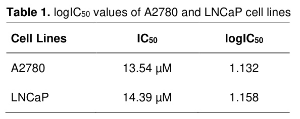
In addition, the logIC50 and corresponding IC50 values (13.54 µM for A2780 and 14.39 µM for LNCaP) obtained from the dose-response curves of CPC are presented in Table 1 to enable the quantitative evaluation of its cytotoxic effect.
Effects of Cetylpyridinium Chloride on Genotoxicity in the A2780 Cell Line: Figure 4 presents the results of the Comet assay performed in the A2780 ovarian cancer cell line. CPC exposure significantly increased tail length (Figure 4A), tail intensity (Figure 4B), and olive tail moment (Figure 4C) compared with both the control and solvent groups (p<0.05). In contrast, no significant difference was observed among the groups in terms of head length (Figure 4D) (p>0.05). However, head intensity was significantly decreased following CPC exposure (Figure 4E) (p<0.05). No significant differences were detected between the control and solvent groups for the evaluated parameters (p>0.05). In control and solvent groups, minimal DNA migration was observed and nuclear integrity was largely preserved (Figure 5A–B). In contrast, CPC-treated cells showed pronounced Comet tail formation and increased DNA migration (Figure 5C). These morphological changes indicate that CPC exposure induces DNA strand breaks and genotoxic damage in the cells.
Effects of Cetylpyridinium Chloride on Genotoxicity in the LNCaP Cell Line: Figure 6 presents the results of the Comet assay performed in the LNCaP prostate cancer cell line. CPC exposure significantly increased tail length (Figure 6A) and tail intensity (Figure 6B) compared with both the control and solvent groups (p<0.05). In contrast, no significant differences were observed among the groups in terms of olive tail moment (Figure 6C), head length (Figure 6D), and head intensity (Figure 6E) (p>0.05). No significant differences were detected between the control and solvent groups for any of the evaluated parameters (p>0.05). In the control and solvent groups, DNA migration was minimal and nuclear integrity was largely preserved (Figure 7A–B). In contrast, CPC-treated cells showed pronounced Comet tail formation and increased DNA migration (Figure 7C). These morphological changes indicate that CPC exposure induces DNA strand breaks and genotoxic damage in the cells.
Tartışma
CPC is a cationic surfactant with a quaternary ammonium structure that is widely used in personal oral hygiene products such as toothpaste and mouthwashes [26]. There is substantial scientific evidence supporting the beneficial effects of CPC in reducing gingival inflammation and controlling dental plaque formation [27-29]. In addition, this compound has been reported to exhibit antiviral properties. It has been shown to inhibit the replication of herpes simplex virus in gingival fibroblasts [30] and to interfere with the assembly of capsid proteins in hepatitis B virus [31]. However, it is well known that the biological effects of bioactive agents may vary depending on the cell type and cellular characteristics. Considering its widespread exposure potential, the possible cytotoxic and genotoxic effects of CPC on cancer cells have not yet been sufficiently elucidated. Therefore, in the present study, we aimed to evaluate the potential cytotoxic and genotoxic effects of CPC in ovarian and prostate cancer cell lines.In this study, CPC was shown to induce concentration-dependent cytotoxic effects in both A2780 and LNCaP cell lines. In both cell lines, a significant reduction in cell viability was observed particularly at concentrations of 10 µM and above, indicating that CPC exhibits notable biological activity against proliferative cancer cells. The dramatic loss of viability observed at higher concentrations suggests that the compound has the potential to induce strong cellular stress in cancer cells. Genotoxicity analyses further indicate that the cytotoxic effect may be associated with DNA damage. In the A2780 cell line, the increases observed in tail length, tail intensity, and olive tail moment parameters, together with the decrease in head intensity, clearly demonstrate that CPC disrupts DNA integrity. In the LNCaP cell line, although increases in tail length and tail intensity support the presence of DNA damage, the absence of significant changes in olive tail moment and other head parameters suggests a cell line-specific sensitivity profile. These findings indicate that the genotoxic effects of CPC may vary depending on the cell type.
In a study conducted by García-Cuellar et al., CPC was reported to significantly inhibit cell proliferation in human lung cancer A549 cells and to strongly suppress tumor cell growth, particularly when combined with nanoparticles [13]. In this study, CPC treatment was shown to reduce tumor cell viability starting from low micromolar concentrations, indicating a high sensitivity of cancer cells to this compound. In addition, a recent study reported that CPC suppresses tumor growth in pancreatic ductal adenocarcinoma cells and induces paraptotic cell death associated with endoplasmic reticulum stress rather than classical apoptosis. In this process, activation of the ERN1–MAP3K5–p38 MAPK signaling pathway was observed, and CPC treatment was reported to significantly reduce tumor progression in both in vitro and in vivo models [32]. These findings, demonstrating that CPC can activate cell death mechanisms in cancer cells by triggering cellular stress responses, are consistent with the decrease in cell viability and the DNA damage observed in our study.
Cationic surfactants with a quaternary ammonium structure are known to affect cell membrane integrity, alter membrane permeability, and disrupt mitochondrial functions [33]. Such effects may lead to an increase in reactive oxygen species (ROS), resulting in oxidative stress and ultimately DNA damage. The Comet assay findings observed in our study suggest that the cytotoxic effect of CPC may be associated with DNA damage. Although CPC is commonly used at safe concentrations in oral hygiene products, it should be considered that biologically active agents may elicit different responses in different cell types. In particular, cancer cells may exhibit varying sensitivities to genotoxic agents due to their increased proliferation rate and altered redox balance. In this context, our findings reveal the potential of CPC to induce DNA damage in cancer cells and suggest that its cellular effects should be further investigated through more detailed mechanistic studies.
Limitations
Although cell culture models provide important advantages for investigating cellular mechanisms, they cannot fully reflect in vivo conditions such as immune responses and systemic metabolic effects. In addition, the possible molecular mechanisms underlying the genotoxic effects of CPC were not investigated in detail in this study. Further molecular analyses are required to determine potential mechanisms such as oxidative stress, mitochondrial dysfunction, or the activation of cell death pathways. Moreover, only cancer cell lines were evaluated in this study, and the lack of assessment of CPC effects on normal cells represents a limitation in terms of selectivity. Therefore, future studies conducted using more comprehensive in vitro models involving different cell types as well as in vivo experimental systems would contribute to a better understanding of the biological effects of CPC.
References
- Vogelstein B, Kinzler KW. Cancer genes and the pathways they control. Nature Medicine 2004;10(8):789-799.
- Evan GI, Vousden KH. Proliferation, cell cycle and apoptosis in cancer. Nature 2001;411(6835):342-348.
- Hanahan D, Weinberg RA. The hallmarks of cancer. Cell 2000;100(1):57-70.
- Hanahan D, Weinberg RA. Hallmarks of cancer: The next generation. Cell 2011;144(5):646-674.
- Lowe SW, Cepero E, Evan G. Intrinsic tumour suppression. Nature 2004;432(7015):307-315.
- Bray F, Laversanne M, Sung H, et al. Global cancer statistics 2022: GLOBOCAN estimates of incidence and mortality worldwide for 36 cancers in 185 countries. CA: A Cancer Journal for Clinicians 2024;74(3):229-263.
- Siegel RL, Miller KD, Fuchs HE, Jemal A. Cancer statistics, 2022. CA: A Cancer Journal for Clinicians 2022;72(1):7-33.
- Vasan N, Baselga J, Hyman DM. A view on drug resistance in cancer. Nature 2019;575(7782):299-309.
- Holohan C, Van Schaeybroeck S, Longley DB, Johnston PG. Cancer drug resistance: an evolving paradigm. Nature Reviews Cancer 2013;13(10):714-726.
- Gilbert P, Moore LE. Cationic antiseptics: Diversity of action under a common epithet. Journal of Applied Microbiology 2005;99(4):703-715.
- Mao X, Auer DL, Buchalla W, et al. Cetylpyridinium chloride: mechanism of action, antimicrobial efficacy in biofilms, and potential risks of resistance. Antimicrobial Agents and Chemotherapy 2020;64(8):10-1128.
- Narwal E, Choudhary J, Kumar M, et al. Botanicals as promising antimicrobial agents for enhancing oral health: A comprehensive review. Critical Reviews in Microbiology 2025;51(1):84-107.
- García-Cuellar CM, Hernández-Delgadillo R, Solis-Soto JM, et al. Cetylpyridinium chloride inhibits human breast tumor cells growth in a no-selective way. Journal of Applied Biomaterials & Functional Materials 2022;20:22808000221092157.
- Cai K, Fang Y, Zhang Y, et al. Cetylpyridinium chloride inhibits hepatocellular carcinoma growth and metastasis through regulating epithelial-mesenchymal transition and apoptosis. Plos One 2024;19(9):e0310391.
- Küçükbay H, Mumcu A, Tekin S, Sandal S. Synthesis and evaluation of novel $ N, N'$-disubstituted benzimidazolium bromides salts as antitumor agents. Turkish Journal of Chemistry 2016;40(3):393-401.
- Oz S, Sekerci G, Yuksel F, Tekin S. Cytotoxic and genotoxic effects of nateglinide on human ovarian, prostate, and colon cancer cell lines. 2023.
- Şekerci G, Keskin T, Tekin S. In vitro evaluation of the cytotoxic effects of rupatadine on various cancer cell lines. Anatolian Journal of Pharmaceutical Sciences 2026;5(1):1-7.
- Denizot F, Lang R. Rapid colorimetric assay for cell growth and survival: Modifications to the tetrazolium dye procedure giving improved sensitivity and reliability. Journal of Immunological Methods 1986;89(2):271-277.
- Mosmann T. Rapid colorimetric assay for cellular growth and survival: Application to proliferation and cytotoxicity assays. Journal of Immunological Methods 1983;65(1-2):55-63.
- Şahal H, Öz S, Keskin T, et al. New sulfa drug derivatives and their zinc (II) complexes: Synthesis, spectroscopic properties and in vitro cytotoxic activities. Journal of Biomolecular Structure and Dynamics 2024;42(1):134-147.
- Koran K, Tekin Ç, Çalışkan E, et al. Synthesis, structural and thermal characterizations and in vitro cytotoxic activities of new cyclotriphosphazene derivatives. Phosphorus, Sulfur, and Silicon and the Related Elements 2017;192(9):1002-1011.
- Doğan H, Bahar MR, Çalışkan E, et al. Synthesis and spectroscopic characterizations of hexakis [(1-(4′-oxyphenyl)-3-(substituted-phenyl) prop-2-en-1-one)] cyclotriphosphazenes: Their in vitro cytotoxic activity, theoretical analysis and molecular docking studies. Journal of Biomolecular Structure and Dynamics 2022;40(7):3258-3272.
- Klaude M, Eriksson S, Nygren J, Ahnström G. The comet assay: Mechanisms and technical considerations. Mutation Research/DNA Repair 1996;363(2):89-96.
- Devlin H-L, Mack PC, Burich RA, et al. Impairment of the DNA repair and growth arrest pathways by p53R2 silencing enhances DNA damage–induced apoptosis in a p53-dependent manner in prostate cancer cells. Molecular Cancer Research 2008;6(5):808-818.
- Öz S, Bahar MR, Özkaya SN, Tekin S. Investigation of the cytotoxic and genotoxic effects of paroxetine on human ovarian, prostate and colon cancer cell lines. İnönü Üniversitesi Sağlık Hizmetleri Meslek Yüksek Okulu Dergisi 2026;14(1):138-48.
- Kim H, Yoo J, Lim YM, et al. Comprehensive pulmonary toxicity assessment of cetylpyridinium chloride using A549 cells and Sprague–Dawley rats. Journal of Applied Toxicology 2021;41(3):470-482.
- Haps S, Slot DE, Berchier CE, Van der Weijden GA. The effect of cetylpyridinium chloride‐containing mouth rinses as adjuncts to toothbrushing on plaque and parameters of gingival inflammation: A systematic review. International Journal of Dental Hygiene 2008;6(4):290-303.
- Herrera D, Escudero N, Pérez L, et al. Clinical and microbiological effects of the use of a cetylpyridinium chloride dentifrice and mouth rinse in orthodontic patients: A 3-month randomized clinical trial. European Journal of Orthodontics 2018;40(5):465-474.
- Teng F, He T, Huang S, et al. Cetylpyridinium chloride mouth rinses alleviate experimental gingivitis by inhibiting dental plaque maturation. International Journal of Oral Science 2016;8(3):182-190.
- Alvarez DM, Duarte LF, Corrales N, Smith PC, González PA. Cetylpyridinium chloride blocks herpes simplex virus replication in gingival fibroblasts. Antiviral Research 2020;179:104818.
- Seo HW, Seo JP, Cho Y, et al. Cetylpyridinium chloride interaction with the hepatitis B virus core protein inhibits capsid assembly. Virus Research 2019;263:102-111.
- Tang H, Chen F, Gao W, et al. Cetylpyridinium chloride triggers paraptosis to suppress pancreatic tumor growth via the ERN1-MAP3K5-p38 pathway. Iscience 2024;27(8).
- Inácio ÂS, Costa GN, Domingues NS, et al. Mitochondrial dysfunction is the focus of quaternary ammonium surfactant toxicity to mammalian epithelial cells. Antimicrob Agents Chemother 2013;57(6):2631-9.