 |
 |
| [ Ana Sayfa | Editörler | Danışma Kurulu | Dergi Hakkında | İçindekiler | Arşiv | Yayın Arama | Yazarlara Bilgi | E-Posta ] | |
| Fırat Üniversitesi Sağlık Bilimleri Tıp Dergisi | |||||
| 2021, Cilt 35, Sayı 1, Sayfa(lar) 077-079 | |||||
| [ Özet ] [ PDF ] [ Benzer Makaleler ] [ Yazara E-Posta ] [ Editöre E-Posta ] | |||||
| Kafa İçi Kitle Lezyonları ile İlişkili Beyin Ödemine Bağlı Kuadripareziye İkincil Rabdomyoliz | |||||
| Ali GÜREL1, Fatih GENÇ1, Ferhat BALGETİR2 | |||||
| 1Fırat University, Faculty of Medicine, Nephrology Department, Elazig, TURKIYE 2Fırat University, Faculty of Medicine, Neurology Department, Elazig, TURKIYE |
|||||
| Anahtar Kelimeler: Rabdomiyoliz, kuadriparezi, beyin ödemi, kafa içi kitle | |||||
| Özet | |||||
Giriş: Rabdomiyoliz, hasarlanmış iskelet kası hücre içeriğinin dolaşıma salınması sonucu gelişen ve bazen hayatı tehdit eden sonuçları olan karmaşık bir tıbbi durumdur. Travma, ilaçlar, toksinler, enfeksiyonlar, metabolik anormallikler, ısı ilişkili durumlar, uzun süreli hareketsizlik ve aşırı egzersiz nedensel faktörler arasında olabilir. Olgu Sunumu: Nadir bir nedenden dolayı rabdomiyoliz gelişen ileri yaşta bir kadın hasta sunuyoruz: intrakraniyal kitle lezyonlarıyla ilişkili beyin ödemine bağlı akut kuadripareze sekonder rabdomiyoliz. Sonuç: Rabdomiyoliz nörolojik patolojiler sonucu gelişen akut kuadripareziye bağlı nadir görülen bir komplikasyon olabilir. Rabdomiyoliz hayatı tehdit edebilecek sonuçlar doğurabileceği için klinisyenlerce erken tanı ve tedavisi önemlidir. |
|||||
| Giriş | |||||
Rhabdomyolysis is a complex medical condition that involves rapid dissolution of damaged or injured skeletal muscle. Disruption of skeletal muscle integrity causes direct release of intracellular muscle components, including myoglobin, creatine kinase (CK), aldolase and lactate dehydrogenase (LDH), as well as electrolytes, into the blood stream and extracellular space 1. Rhabdomyolysis can range from an asymptomatic Picture with an elevated CK level to a life-threatening condition associated with excessive increases in CK, electrolyte imbalances, acute renal failure and diffuse intravascular coagulation 2. Although rhabdomyolysis is most commonly caused by direct traumatic injury; drugs, toxins, infections, muscle ischemia, electrolyte and metabolic abnormalities, genetic disorders, heavy exercises, prolonged bed dependence, heat-related conditions such as neuroleptic malignant syndrome and malignant hyperthermia may also be causative factor. Decreased limb muscle strength, myalgia, swelling, massive necrosis that manifests as gross pigmenturia without hematuria are common features of both traumatic and non-traumatic rhabdomyolysis 3. In this article, we present a case in which rhabdomyolysis occurs due to an uncommon cause: Rhabdomyolysis secondary to acute quadriparesis due to brain edema associated with intracranial mass lesions. |
|||||
| Olgu Sunusu | |||||
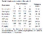
A 66-year-old female patient was admitted to the emergency department because of weakness in four extremity proximal muscles, more prominent in the legs, after fever that started 3 days ago. On physical examination; confusion, disorientation, discooperation and decreased muscle strength in all extremities were found. Vital signs except 37.4 ⁰C body temperature and other system examinations were normal. Labaratory results were as follows; CK: 71560 U/L, LDH: 1095U/L, aspartate aminotransferase (AST): 1198 U/L, Urea: 47 mg/dL, creatinine: 1.04 mg/dL and other parameters were generally in normal range (Table 1). In urinalysis; dark urine with positive blood rection but normal range erythrocyte count was observed.
The patient was hospitalized in the Nephrology service with a pre-diagnosis of rhabdomyolysis and hydration treatment was started. The patients CK enzyme level started to decrease. However because of decreased muscle strength, impaired consciousness, not being able to recognize his relatives and having meaningless speech, she was consulted with the neurology department. Brain computed tomography and diffusion MR images revealed multiple intracranial mass lesions and brain edema due to these lesions (Figure 1). During 3 days, 1000 mg/day intravenous methylprednisolone treatment was administered. Partial improvement was observed in the confusion and, in the light of clinical and radiological diagnosis, biopsy was planned for pathologic diagnosis. In this case; there was no traumatic, pharmacologic, toxic, infectious factor and electrolyte disorder that may cause rhabdomyolysis other than quadriparesis due to brain edema associated with intracranial mass lesions. This is an exceptional state that causes rhabdomyolysis in the light of the literature.
|
|||||
| Tartışma | |||||
There was no history of any chronic medical problem in our patient. She did not have traumatic, pharmacologic, toxic, infectious factor and electrolyte disorder that may cause rhabdomyolysis. In our patient, quadriparesis and rhabdomyolysis were attributed to brain edema associated with intracranial mass lesions. MRI and CT images supported this. Quadriparesis improved after regressed cerebral edema with corticosteroid treatment, and additionally, with appropriate hydration, a significant improvement was observed in rhabdomyolysis. The most common causes of rhabdomyolysis are drug or alcohol abuse, drug use, trauma, excessive exercise, infections, electrolyte imbalances, neuromuscular causes, and inactivity. Rhabdomyolysis may also be caused by tightening of the muscles and being in the same position for a long time. The pathogenesis of rhabdomyolysis is related to direct sarcolemic injury or depletion of ATP in myocytes and leads to irregular leakage of calcium ions into cells 4. Sarcoplasmic calcium is strictly regulated by energy-dependent ion pumps such as Na+/ K+ATPase and Ca+2 ATPase in sarcolemma. These pumps keep calcium levels in resting muscle low, but allow actin-myosin binding and increase in calcium levels when muscle contraction is required. Regardless of the underlying mechanism, muscle injury increases sarcoplasmic calcium and causes persistent contraction. Finally, there is muscle fiber necrosis after activation of the cell protease. Then, potassium, phosphate, myoglobin, CK and uric acid are released out of the cell into the systemic circulation 5. Rhabdomyolysis is clinically characterized by myalgia, red-brown urine due to myoglobinuria, and increased serum muscle enzymes 6. Taştekin et al. 7, presented two rhabdomyolysis cases with rare etiologic factors. One developed acute renal failure associated with rhabdomyolysis after epileptic seizure and other developed acute renal failure associated with rhabdomyolysis after swimming. With these rare cases, they emphasized that epileptic seizures and excessive exercise such as swimming can cause rhabdomyolysis. Statins are well decsribed and frequent cause of drug- induced rhabdomyolysis 8. On the other hand systemic diseases such as Henoch- Schonlein purpura may be a rare and surprising cause of rhabdomyolysis. In their report, Turan MI et al 9, treated HSP caused rhabdomyolysis case successfully with appropriate fluid and steroid treatment. In the present rhabdomyolysis case we represent here, all other causes of etiologic factors excluded, and it was caused by a very exceptional etiology due to quadriparesis caused by brain edema associated with intracranial mass lesions. |
|||||
| Kaynaklar | |||||
1) Sauret JM, Marinides G, Wang GK. Rhabdomyolysis. Am Fam Physician 2002; 65: 907-912.
2) Huerta-Alard ́ın AL, Varon J, Marik PE. Bench-to-bedside review: Rhabdomyolysis - an overview for clinicians. Crit Care 2005; 9: 158-169.
3) Lima RS, da Silva Junior GB, Liborio AB. Acute kidney injury due to rhabdomyolysis. Saudi J Kidney Dis Transpl 2008; 19: 721-729.
4) Giannoglou GD, Chatzizisis YS, Misirli G. The syndrome of rhabdomyolysis: Pathophysiology and diagnosis. European Journal of Internal Medicine 2007;18: 90-100.
5) Khan. FY. Rhabdomyolysis: A review of the literature. Neth J Med 2009; 67: 272-283.
6) Akbaş EM, Özlü C, Ünver E (Editörler). Gürel A. Rabdomiyoliz. Dahili Aciller. 1. Baskı, İstanbul: Akademisyen Kitabevi, 2020.
7) Taştekin F, Sırmatel P, Şahin G. Rabdomiyolizin nadir sebepleri; Epileptik nöbet ve yüzmeden sonra rabdomiyoliz. Turk Neph Dial Transpl 2018; 27: 93-95.
|
|||||
| [ Başa Dön ] [ Özet ] [ PDF ] [ Benzer Makaleler ] [ Yazara E-Posta ] [ Editöre E-Posta ] | |||||
 |
| [ Ana Sayfa | Editörler | Danışma Kurulu | Dergi Hakkında | İçindekiler | Arşiv | Yayın Arama | Yazarlara Bilgi | E-Posta ] |

