
Canlı Karaciğer Donörlerinde Portal Ven Varyasyonlarının BT Anjiyografi ile Değerlendirilmesi
Cilt 40, Sayı 3
Kasım 2026 · Sayfalar 261-266
Canlı Karaciğer Donörlerinde Portal Ven Varyasyonlarının BT Anjiyografi ile Değerlendirilmesi
Evaluation of Portal Vein Variations in Living Hepatic Donors Using CT Angiography
Murat OYNAK1, Kağan ÇEKEN2, Adnan KABAALİOĞLU3
2AkdenizUniversity, Faculty of Medicine Department of Radiology, Antalya, TÜRKİYE
3Varisson Radiology Center Merdivenköy, İstanbul, TÜRKİYE
Amaç: Karaciğer nakli, son dönem karaciğer yetmezliği olan bireyler için hayati bir tedavi yöntemi olarak kullanılmaktadır. Bu araştırma, bilgisayarlı tomografi anjiyografi (BT anjiyografi) görüntüleme yöntemi kullanılarak canlı karaciğer nakli donörlerinde portal ven varyasyonlarını incelemiştir.
Gereç ve Yöntem: Temmuz 2014 ile Aralık 2015 tarihleri arasında Akdeniz Üniversitesi Hastanesinde, canlı karaciğer donörü olan 100 hastanın ameliyat öncesi BTA görüntüleri retrospektif olarak incelendi. Portal ven varyasyonları Atasoy kriterlerine göre sınıflandırıldı (Tip I: normal bifurkasyon; Tip II: trifurkasyon; Tip III: sağ posterior dalın doğrudan ana PV gövdesinden ayrılması). Sağ PV segmental varyasyonları da detaylı olarak değerlendirildi.
Bulgular: Olguların %80.0’inde Tip I anatomi saptanırken, Tip II %8.0 ve Tip III varyasyon %12.0 oranında izlendi. Tip I anatomiye sahip donörlerin %11.25’inde ikincil sağ portal ven segmental varyasyonları tespit edildi; bunlar arasında segment 7’nin bağımsız çıkışı (%3.75) ve sağ portal ven trifurkasyonu (%3.75) en sık görülen varyasyonlardı. Varyasyon tipleri ile cinsiyet (p=0.923) veya yaş (p=0.755) arasında istatistiksel olarak anlamlı bir ilişki saptanmadı. Çok düzlemli rekonstrüksiyon ve maksimum yoğunluk projeksiyonu (MIP) teknikleriyle desteklenmiş BTA, vasküler haritalamayı yüksek doğrulukla sağladı.
Sonuç: Türk donör adaylarında saptanan %20’lik PV varyasyon oranı küresel literatürle uyumlu olup, bu anatomik paternlerin bölgesel etnisite, yaş ve cinsiyetten bağımsız olduğunu göstermektedir. BTA ile yapılan preoperatif haritalama; cerrahi planlamayı kolaylaştıran, postoperatif vasküler komplikasyonları azaltabilen ve transplantasyon sürecinde donör ile alıcı güvenliğini en üst düzeye çıkaran non-invaziv faydalı bir yaklaşımdır.
Objective: Liver transplantation is a vital treatment method for individuals with end-stage hepatic failure. This study investigated portal vein variations in living liver transplant donors using computed tomography angiography (CTA) imaging.
Materials and Methods: Preoperative CTA images of 100 patients who were living hepatic donors at Akdeniz University Hospital between July 2014 and December 2015 were retrospectively reviewed. Portal vein variations were classified according to Atasoy criteria (Type I: normal bifurcation; Type II: trifurcation; Type III: direct separation of the right posterior branch from the main portal vein trunk). Right portal vein segmental variations were also evaluated in detail.
Results: Type I anatomy was detected in 80.0% of cases, while Type II was observed in 8.0% and Type III variation in 12.0%. Secondary right portal vein segmental variations were detected in 11.25% of donors with Type I anatomy; among these, separate origin of the segment 7 branch (3.75%) and right portal vein trifurcation (3.75%) were the most frequently observed variations. No statistically significant relationship was found between variation types and gender (p=0.923) or age (p=0.755). CTA supported by multiplanar reconstruction and maximum intensity projection (MIP) techniques provided vascular mapping with high accuracy.
Conclusion: The 20% variation rate detected in Turkish donor candidates is consistent with the global literature and demonstrates that these anatomical patterns are independent of regional ethnicity, age, and gender. Preoperative CTA mapping is a beneficial non-invasive approach that facilitates surgical planning, reduces postoperative vascular complications, and maximizes donor and recipient safety during the transplantation process.
Giriş
The surgical procedure of liver transplantation provides life-saving benefits to patients suffering from end-stage liver failure. The worldwide adoption of living donor liver transplantation (LDLT) has grown because the supply of cadaveric donors remains insufficient [1]. Consequently, a detailed evaluation of hepatic vascular anatomy stands as an essential requirement to achieve safe and successful outcomes for both the donor and the recipient. The portal vein plays a vital role in hepatobiliary surgery, as it delivers approximately 75% of the total hepatic blood circulation [2]. Prior research indicates that portal vein variations affect roughly 20% of the general population, primarily encompassing portal vein trifurcation and anomalies of the right posterior portal vein branch arising directly from the main portal vein trunk [3]. Therefore, a thorough preoperative assessment of these variations remains vital to minimize surgical risks and optimize procedural planning.While the current literature contains extensive data on the frequencies and types of portal vein variations, few studies have specifically investigated these anatomical patterns within the Turkish living liver donor population, as most existing cohorts focus on broader general populations or other distinct ethnic groups [4]. Furthermore, there is a lack of standardized data regarding the gender distribution of these variations and their potential correlation with patient age [5]. Although the efficacy of non-invasive imaging modalities like computed tomography angiography (CTA) for evaluating portal venous anatomy is well-established, further data are required to clarify its specific diagnostic accuracy in screening living liver donor candidates [1,2,6]. Hence, this study aims to investigate the incidence, specific types, and demographic associations of portal vein variations in living liver donors utilizing high-resolution CTA imaging.
Gereç ve Yöntem
Research and Publication Ethics: The study protocol was approved by the Institutional Ethics Committee of Akdeniz University Hospital University Faculty of Medicine (Date: February 25, 2015; Decision Number: 85). Due to the retrospective nature of the data review, the requirement for informed consent was waived. Patient data were fully anonymized to maintain strict confidentiality. The study was conducted in accordance with the principles of the Helsinki Declaration.Study Population and Sample: One hundred living organ donor candidates who underwent routine preoperative liver CTA at our center between July 2014 and December 2015 were retrospectively reviewed from the transplant donor pool of Akdeniz University Hospital University Faculty of Medicine. A post-hoc power analysis performed using G*Power 3.1 software indicated that with an estimated portal vein variation prevalence of 20-25% in the general population, our sample size of 100 cases was sufficient to detect an effect size of 8.5% with a 5% alpha error and 80% power. Given that this study represents an initial regional baseline for the Mediterranean region, these prevalence data will serve as a foundation for future large-scale multi-center investigations.
Inclusion criteria were defined as follows: age between 19 and 70 years, completion of a full living liver donor evaluation, preoperative diagnostic CTA performed at our center, and availability of complete postoperative follow-up records. Candidates were excluded if they did not proceed to surgery due to insufficient left lobe volume or if their CTA scans were of sub-diagnostic quality for accurate portal vein assessment.
Study Procedures: Preoperative hepatic CTA is routinely performed for all donor candidates at our institution. Scans were acquired using either a 64-slice (Toshiba Aquilion 64, Tokyo, Japan) or a 128-slice Siemens SOMATOM Definition Edge (Siemens, Erlangen, Germany) device, based on equipment availability and clinical urgency. In all protocols, unenhanced baseline images were obtained first, followed by the intravenous administration of 120 mL of non-ionic contrast medium at an injection rate of 5 mL/s. Bolus tracking was utilized with the region of interest (ROI) placed on the abdominal aorta, employing an automated triggering threshold of 120 HU. Contrast-enhanced phases were captured during the arterial, portal, and late venous phases at 15 seconds, 45 seconds, and 160 seconds, respectively (Table 1).
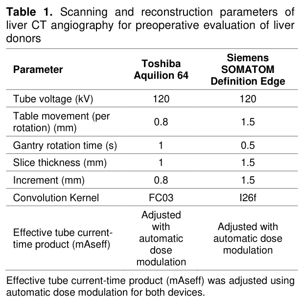
Portal vein morphology was evaluated exclusively during the portal venous phase. All datasets were transferred to the hospital picture archiving and communication system (PACS) and independently reviewed on a Sectra Workstation IDS7 (v17.3; Sectra AB, Linköping, Sweden) by a single radiologist with three years of specialized experience in abdominal imaging.
Evaluation of Portal Vein Variations: Anatomical variations of the portal venous system were classified according to the criteria defined by Atasoy et al. Under this system, normal anatomy (Type I) is defined as the main portal vein bifurcating into the left and right portal veins, followed by the conventional division of the right portal vein into right anterior and right posterior branches. Type II (trifurcation) is defined as the simultaneous trifurcation of the main portal vein into the left, right anterior, and right posterior portal veins at the same level. Type III consists of an anomalous branching pattern where the right posterior portal vein arises as the first branch directly from the main portal vein trunk, before the origination of the common trunk for the left and right anterior portal veins [7]. Additionally, right portal vein segmental variations—including the separate origin of segment 6, separate origin of segment 7, concurrent separate origins of segments 6 and 7, and right portal vein trifurcation/quadrifurcation—were meticulously documented within each primary portal vein type [8].
Statistical Analysis. Statistical analyses were performed using SPSS software (v15.0.0; SPSS Inc., Chicago, IL, USA). Continuous variables were expressed as mean±standard deviation (SD) and categorical variables as frequencies and percentages. Normality of data distribution was verified using the Shapiro-Wilk test. The independent samples t-test was employed to compare continuous variables across groups, while the chi-square test was used for categorical data comparison. Statistical significance was established at p<0.05.
Bulgular
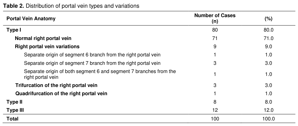
A total of 100 patient datasets were evaluated. The mean age of the cohort was 35.7±9.0 years (range: 20–69 years), comprising 54 males (54.0%) and 46 females (46.0%). The mean age was 35.5±8.9 years (range: 20–69 years) for male patients and 36.0±9.2 years (range: 20–54 years) for female patients, demonstrating no statistically significant age difference between genders (p=0.817).Conventional Type I portal vein anatomy was observed in 80 patients (80.0%), while Type II and Type III variations were detected in 8 (8.0%) and 12 (12.0%) patients, respectively. Among the individuals with Type I anatomy (n=80), secondary variations within the right portal vein branches were identified in 9 cases (11.25%). The breakdown of these right segmental variations was as follows: right portal vein quadrifurcation in 1 patient (1.25%), right portal vein trifurcation in 3 patients (3.75%), an isolated separate origin of the segment 6 branch in 1 patient (1.25%), an isolated separate origin of the segment 7 branch in 3 patients (3.75%), and concurrent separate origins of both segment 6 and 7 branches in 1 patient (1.25%) (Table 2).
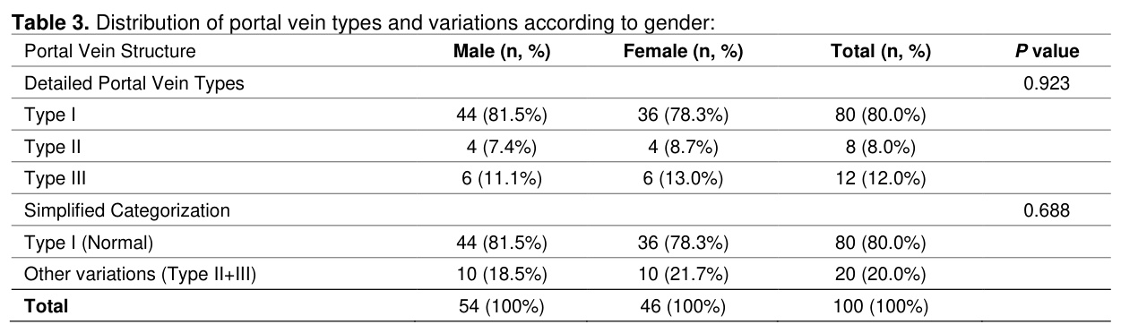
When analyzing portal vein classifications relative to gender, no statistically significant difference was observed (p=0.923). Type I anatomy was present in 81.5% of males and 78.3% of females; Type II was found in 7.4% of males and 8.7% of females; and Type III was observed in 11.1% of males and 13.0% of females. Furthermore, when grouping patients dichotomously into normal (Type I) versus variant anatomy (Types II and III), no gender-specific correlation was established (p=0.688) (Table 3). Similarly, no significant difference in mean age was noted between the variant and normal anatomy cohorts (p=0.755).
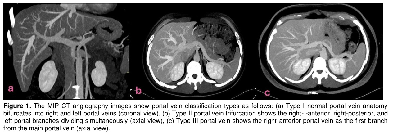
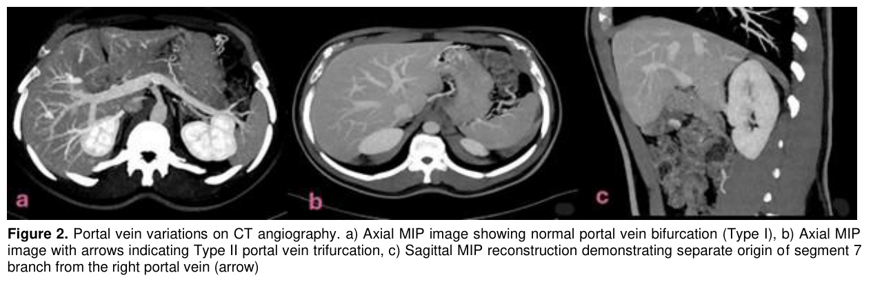
High-resolution CTA allowed clear multiplanar visualization of these distinct patterns. Coronal maximum intensity projection (MIP) reconstructions effectively demonstrated the bifurcation of the main portal vein into the left and right branches in Type I cases. In contrast, axial imaging clearly delineated Type II trifurcation (simultaneous branching of the right anterior, right posterior, and left branches) and Type III morphology, where the right posterior portal vein uniquely presented as the first branch arising from the main portal trunk (Figure 1). For sub-segmental right portal vein variants, a combination of axial MIP and sagittal MIP reconstructions successfully demonstrated configurations such as the isolated separate origin of the segment 7 branch (Figure 2).
Tartışma
This study aimed to delineate the incidence, specific morphological types, and demographic distributions of portal vein variations among candidates presenting for LDLT at a high-volume transplant center.The precise evaluation of portal vein anatomy heavily relies on advanced post-processing imaging techniques. As reported by Marciuc et al., post-processing algorithms greatly enhance the visualization of complex vascular beds [9]. In our cohort, the implementation of multiplanar reconstruction (MPR) and MIP protocols enabled a comprehensive, multi-angle assessment of portal venous architecture. This detailed analysis was facilitated by high-resolution datasets acquired from 64- and 128-slice CT scanners. Technical parameters, specifically thin-slice collimation (1–1.5 mm), precise contrast bolus timing, and 3D reconstructions, proved critical for resolving fine right-sided segmental branches. Crucially, automated dose modulation technology minimized patient radiation exposure without sacrificing diagnostic image quality. Our results are in precise agreement with the findings of Sotomayor et al., who reported a matching 20% portal vein variation rate utilizing contrast-enhanced, thin-slice triple-phase abdominal CT imaging [10].
Furthermore, recent literature underscores the diagnostic value of high-resolution vascular mapping. Ali et al. demonstrated that high spatial resolution cone-beam CT angiography yields exceptional visualization of sub-segmental portal branches, reinforcing our observation that multiplanar 3D datasets are vital for understanding complex anatomy [11]. Similarly, Machry et al. utilized automated segmentation algorithms to accurately define portal boundaries, even on thicker (3 mm) multi-phase CT scans [12]. Preoperative detection remains paramount; Goldaracena et al. observed that vascular variations occur in more than half of living liver donors and emphasized the strict necessity of comprehensive profiling prior to final surgical planning [13]. Yagi et al. also noted that portal vein anomalies directly alter surgical dissection lines and subsequent vascular reconstruction approaches, corroborating the global consensus that detailed preoperative knowledge of these variations is fundamental to transplant success [14].
The 20% overall variation rate and 80% Type I prevalence noted in our study sit comfortably within the 10% to 35% range reported in the comprehensive review by Borhani et al. [15]. Our Type I prevalence is slightly lower than the 88.5% incidence of Type A (equivalent to Type I) anatomy reported by Khalid et al., a discrepancy likely attributable to differences in sample sizes, geographic cohorts, or classification nuances [5]. Regarding specific variants, we observed a Type II (trifurcation) rate of 8%, whereas Shehta et al. reported a slightly higher rate of 13.5% [16].
Notably, our study provides valuable data regarding right-sided segmental variations in candidates who otherwise possess a standard main trunk (Type I) configuration (11.25%). Among these, the separate origin of the segment 7 branch (3.75%) and right portal vein trifurcation (3.75%) were the most common, followed by less frequent configurations like isolated segment 6 anomalies (1.25%) or right portal trunk quadrifurcation (1.25%). These sub-types represent high-stakes anatomical landmarks that must be carefully managed during right lobe donor hepatectomies to avoid accidental transection or compromise of the remaining sectorial flow [16].
Our findings also revealed that Type III variations (12%) occurred with a slightly higher frequency than Type II variations (8%). This distribution is consistent with several regional studies; for instance, Kirimker et al. evaluated 203 Turkish living liver donors and found a comparable predominance of Type III variations (11.3%) over Type II (3.4%) [17]. Similarly, Masood et al. documented a Type III rate of 13.9% versus a Type II rate of 7.9% in a Pakistani population [18]. Identifying a Type III variation is surgically critical because the anomalous early takeoff of the right posterior portal vein from the main trunk necessitates meticulous dissection and often mandates complex, multi-orifice vascular reconstructions or separate graft venoplasties.
Finally, our analysis confirmed that portal vein variations occur independently of gender (p=0.923), with variations found in 21.7% of female and 18.5% of male candidates.
The primary limitation of this study is its relatively modest sample size from a single center. While larger epidemiological studies exist in the literature, they typically include heterogeneous patient populations undergoing abdominal CT for any clinical indication. In contrast, our cohort was restricted strictly to highly screened liver donor candidates, providing a highly homogeneous study group at the expense of a larger sample volume. Additionally, due to the retrospective imaging-focused design, our dataset lacks correlation with postoperative donor outcomes, vascular complications, or long-term graft survival. Future prospective, larger-scale studies combining preoperative anatomical mapping with clinical follow-up are warranted to definitively establish the prognostic impact of these specific variations.
In our study, the occurrence and distribution of portal vein variations among Turkish living liver donor candidates (20%) appear consistent with global literature findings, suggesting that regional or ethnic differences may not significantly alter these anatomical patterns.
Although portal vein variations are less frequent than hepatic artery or biliary tract anomalies, their meticulous preoperative identification is considered a beneficial approach.
In this context, CTA enhanced with multiplanar reconstruction and MIP techniques stands out as a reliable, non-invasive imaging method. Mapping of portal vein variations can also play a significant role in preventing postoperative vascular complications, facilitating surgical success, and maximizing safety throughout the transplantation process.
References
- Cai L, Yeh BM, Westphalen AC, Roberts JP, Wang ZJ. Adult living donor liver imaging. Diagn Interv Radiol 2016;22(3):207-214.
- Sureka B, Patidar Y, Bansal K, et al. Portal vein variations in 1000 patients: surgical and radiological importance. Br J Radiol 2015;88(1054):20150326.
- Jhaveri KS, Guo L, Guimarães L. Current state-of-the-art MRI for comprehensive evaluation of potential living liver donors. AJR Am J Roentgenol 2017;209(1):55-65.
- Ulusoy M, Bolatlı G, Koplay M, Acar M, Zararsız İ. Evaluation of portal vein variations in multidetector CT. J Surg Med 2020;4(11):978-981.
- Khalid A, Saleem MA, Haq I, et al. Anatomical variations in living donors for liver transplantation - prevalence and correlation. Langenbecks Arch Surg 2023;408:387.
- Hecht EM, Kambadakone A, Griesemer AD, et al. Living donor liver transplantation: Overview, imaging technique, and diagnostic considerations. AJR Am J Roentgenol 2019;213(1):54-64.
- Atasoy C, Ozyürek E. Prevalence and types of main and right portal vein branching variations on MDCT. AJR Am J Roentgenol 2006;187(3):676-681.
- Carneiro C, Brito J, Bilreiro C, et al. All about portal vein: A pictorial display to anatomy, variants and physiopathology. Insights Imaging 2019;10(1):38.
- Marciuc EA, Popescu RM, et al. Postprocessed computed tomography angiography images - an underused modality of better understanding aneurysms. Med Surg J - Rev Med Chir Soc Med Nat Iasi 2020;124(3):412-418.
- Sotomayor C, Garcia D, Rebolledo P, et al. Staged preoperative evaluation of potential living donors for liver transplantation. Transl Gastroenterol Hepatol 2025;10:2.
- Ali H, Weinstein J, Sarwar A, et al. Angiography with cone-beam CT versus contrast-enhanced MRI for living donor transplant imaging: Is MRI enough? Clin Anat 2024;37(2):185-192.
- Machry M, Ferreira LF, Lucchese AM, Kalil AN, Feier FH. Liver volumetric and anatomic assessment in living donor liver transplantation: The role of modern imaging and artificial intelligence. World J Transplant 2023;13(6):290-298.
- Goldaracena N, Vargas PA, McCormack L. Pre-operative assessment of living liver donors' liver anatomy and volumes. Updates Surg. 2024.
- Yagi S, Singhal A, Jung DH, Hashimoto K. Living-donor liver transplantation: Right versus left. Int J Surg 2020;82:128-33.
- Borhani AA, Elsayes KM, Catania R, et al. Imaging evaluation of living liver donor candidates: Techniques, protocols, and anatomy. Radiographics 2021;41(6):1572-1591.
- Shehta A, Elshobari M, Salah T, et al. Feasibility and outcomes of living-donor liver transplantation utilizing the right hemi-liver graft with portal vein anatomical variations. Langenbecks Arch Surg 2023;408:387.
- Kirimker EO, Kologlu M, Celik SU, et al. Living liver donor hilar anatomical variations and impact of variant anatomy on transplant outcomes. Medicine (Baltimore) 2022;101(37).
- Masood L, Sayeed S, Aslam S, Faiz BY. Relationship between variant biliary and vascular anatomy in living liver donors in Pakistani population. J Dow Univ Health Sci 2022;16(1):34-40.