
Pediatrik Uykuyla İlişkili Hipermotor Epilepside Klinik Özellikler ve Karbamazepine Erken Yanıt
Volume 40, Issue 3
November 2026 · Pages 277-281
Pediatrik Uykuyla İlişkili Hipermotor Epilepside Klinik Özellikler ve Karbamazepine Erken Yanıt
Clinical Characteristics and Early Carbamazepine Responsiveness in Pediatric Sleep-Related Hypermotor Epilepsy
Yavuz ATAŞ1, Nihal SOYLU1
Amaç: Uyku ile ilişkili hipermotor epilepsi (sleep-related hypermotor (hyperkinetic) epilepsy-SHE), ağırlıklı olarak uykuda ortaya çıkan kısa süreli, stereotipik hipermotor nöbetlerle karakterize nadir bir fokal epilepsi sendromudur. Pediatrik hastalarda interiktal elektroensefalografi (EEG), nörogörüntüleme ve genetik incelemelerin sıklıkla tanısal açıdan yetersiz kalması nedeniyle tanı güçleşebilmektedir. Bu çalışmada SHE tanılı pediatrik hastaların klinik, EEG, nörogörüntüleme, genetik ve tedavi özelliklerinin değerlendirilmesi amaçlandı.
Gereç ve Yöntem: Uluslararası Epilepsi ile Savaş Birliği tanı kriterlerine göre SHE tanısı konulan pediatrik hastalar retrospektif olarak analiz edildi. Demografik özellikler, nöbet semiyolojisi, EEG bulguları, nörogörüntüleme sonuçları, genetik analizler, tedavi yanıtı ve izlem sonuçları değerlendirildi.
Bulgular: Çalışma kohortu 7 kız ve 4 erkek hastadan oluşmaktaydı. Nöbet başlangıç yaşı ortancası 9 yıl (dağılım: 7–13 yıl), ortanca izlem süresi ise 21 ay (dağılım: 18–26 ay) idi. En belirgin nöbet semiyolojileri hipermotor davranışlar, epileptik dolaşma, iktal korku/anksiyete ve asimetrik tonik/distonik postürlerdi. İnteriktal skalp EEG'si 9 hastada (%81.8), beyin manyetik rezonans görüntülemesi ise 10 hastada (%90.9) normal bulundu. Bir hastada kortikal displazi saptandı. Genetik analizlerde patojenik veya olası patojenik bir varyant tespit edilmedi. İdiyopatik SHE tanılı tüm hastalarda, çalışma izlem süresi boyunca 10–15 mg/kg/gün karbamazepin monoterapisi ile tam nöbet kontrolü sağlandı. Tam nöbet kontrolü sağlanamayan tek hastada yapısal bir beyin lezyonu bulunmakta olup, bu hasta çoklu antinöbet ilacı tedavisi gerektirdi.
Sonuç: Karbamazepin tedavisi, idiyopatik pediatrik SHE olgularında erken nöbet kontrolünün sağlanmasında oldukça etkili görünmektedir. Bulgularımız, SHE'nin tanı ve klinik yönetiminde ayrıntılı nöbet semiyolojisinin değerlendirilmesinin kritik önemini ortaya koymaktadır.
Objective: Sleep-related hypermotor epilepsy (SHE) is a rare focal epilepsy syndrome characterized by brief, stereotyped hypermotor seizures predominantly occurring during sleep. Diagnosis in pediatric patients may be challenging because interictal electroencephalography (EEG), neuroimaging, and genetic investigations are often diagnostically uninformative. This study aimed to evaluate the clinical, electroencephalographic, neuroimaging, genetic, and treatment characteristics of pediatric patients with SHE.
Materials and Methods: Pediatric patients diagnosed with SHE according to the diagnostic criteria of the International League Against Epilepsy were retrospectively analyzed. Demographic characteristics, seizure semiology, EEG findings, neuroimaging results, genetic analyses, treatment response, and follow-up outcomes were reviewed.
Results: The cohort included 7 girls and 4 boys, with a median seizure onset age of 9 years (range: 7–13 years). Median follow-up duration was 21 months (range: 18–26 months). Prominent seizure semiologies included hypermotor behaviors, wandering behavior, ictal fear/anxiety, and asymmetric tonic/dystonic posturing. Interictal scalp EEG was normal in 9 patients (81.8%), while brain magnetic resonance imaging was normal in 10 patients (90.9%). Cortical dysplasia was identified in one patient. Genetic analyses did not reveal pathogenic or likely pathogenic variants. Complete seizure freedom was observed in all idiopathic SHE cases during the study follow-up period while receiving carbamazepine monotherapy (10–15 mg/kg/day). The only patient without complete seizure control had a structural lesion and required polytherapy.
Conclusion: Carbamazepine treatment appears highly effective for early seizure control in idiopathic pediatric SHE cases. Our findings support the importance of detailed semiological assessment in the diagnosis and management of SHE.
Introduction
Sleep-related hypermotor epilepsy (SHE) is a focal epilepsy syndrome characterized by stereotyped hypermotor seizures that predominantly occur during sleep, usually last less than 2 minutes, and have an abrupt onset and termination [1].Previously referred to as “nocturnal frontal lobe epilepsy,” this condition is currently defined as SHE because it is not exclusively confined to the frontal lobe [2]. Seizures typically arise in clusters during non-rapid eye movement (NREM) sleep and are characterized by sudden arousal, dystonic-postural movements, vocalization, and complex hyperkinetic behaviors [3]. SHE is a rare epilepsy syndrome with an estimated prevalence of approximately 1.8 per 100,000 individuals; however, its true prevalence may be higher because of its clinical resemblance to parasomnias [4]. Structural, genetic, and genetic-structural factors may play a role in the etiology. However, the underlying etiology cannot be determined in a significant proportion of patients, and drug resistance has been reported in approximately 30% of cases [4]. The current literature has largely focused on drug-resistant and surgically treated patient groups, whereas studies comprehensively evaluating the clinical characteristics and treatment responses of idiopathic SHE cases in the pediatric age group remain limited [2,4-6]. SHE presents significant diagnostic challenges because of its marked clinical overlap with parasomnias and the frequently normal or nonspecific findings of interictal and ictal electroencephalography (EEG) and magnetic resonance imaging (MRI) [4,7-9]. This situation may lead to delayed diagnosis and postponement of appropriate antiseizure treatment, particularly in the pediatric age group [4]. Because seizures predominantly occur during sleep and are often not directly witnessed, the diagnostic process relies largely on detailed history-taking and seizure semiology [2,4]. This study aimed to evaluate the clinical characteristics, electrophysiological and neuroimaging findings, genetic investigation results, and response to antiseizure treatment in pediatric patients diagnosed with SHE, a rare epileptic syndrome, at a tertiary care center.
Material and Method
Research and Publication Ethics: This study was approved by the Clinical Research Ethics Committee of the University of Health Sciences Van Training and Research Hospital (Decision No: 2026/03/29; Date: March 6, 2026).This single-center retrospective case series included patients diagnosed with SHE who were followed up at the pediatric neurology clinic of a tertiary care center. The diagnosis of SHE was established in accordance with the recommendations of the International League Against Epilepsy (ILAE) based on clinical history, seizure semiology, the temporal relationship of seizures with sleep, home video recordings, and EEG findings[1]. Home video recordings were available for all patients and were independently reviewed by two experienced pediatric neurologists as part of the diagnostic evaluation. Patients who did not fulfill the ILAE diagnostic criteria for SHE, including those with seizures occurring exclusively during wakefulness or generalized-onset seizures, as well as those with insufficient clinical records or lacking regular follow-up data, were excluded from the study.
Demographic and clinical data were obtained from patient medical records. Age, sex, age at seizure onset, age at diagnosis, disease duration, clinical course, sleep-related seizure characteristics, presence of daytime seizures, family history of epilepsy, neurological examination findings, and diagnostic evaluations performed before admission to our clinic were recorded. Seizure semiology was assessed according to the ILAE classification, with particular attention to sudden arousal, dystonic/tonic posturing, complex hypermotor behaviors, vocalization, and stereotyped nocturnal motor manifestations. Routine interictal EEG was performed in all patients. EEG recordings were evaluated for the presence of epileptiform discharges, focal slowing, nonspecific abnormalities, and the localization/lateralization characteristics of epileptiform activity.
All patients underwent brain MRI using an epilepsy-specific imaging protocol. MRI findings were evaluated for structural abnormalities, including cortical developmental malformations, focal lesions, and other structural pathologies. Genetic testing was available in six patients and was performed using clinical exome sequencing (CES). Detected variants were classified as pathogenic, likely pathogenic, variant of uncertain significance (VUS), likely benign, or benign according to the American College of Medical Genetics and Genomics (ACMG) criteria. Antiseizure treatments administered during the follow-up period were recorded. Treatment response was classified as complete seizure freedom, ≥50% reduction in seizure frequency, <50% reduction, or no treatment response after at least 6 months of follow-up.
Statistical analyses were performed using IBM SPSS Statistics for Windows, Version 29.0 (IBM Corp., Armonk, NY, USA). The distribution of continuous variables was assessed using the Shapiro–Wilk test. Normally distributed variables were presented as mean ± standard deviation, whereas non-normally distributed variables were expressed as median (minimum–maximum). Categorical variables were presented as numbers and percentages (%). Owing to the limited sample size, analyses were primarily descriptive in nature.
Results
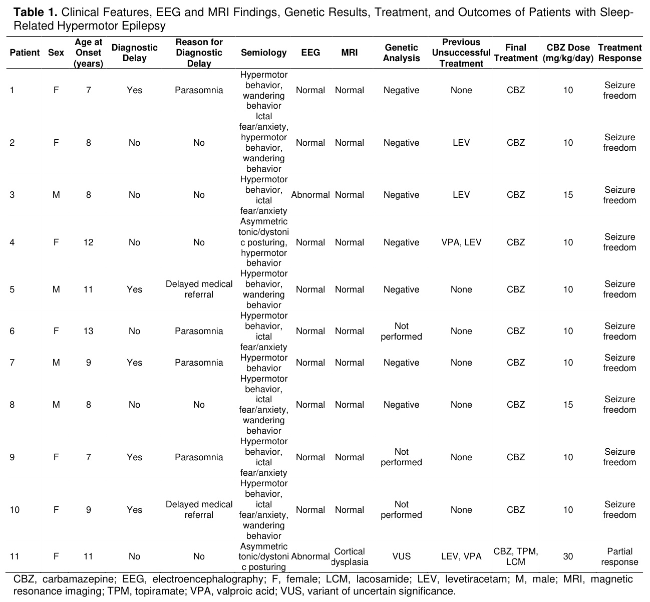
A total of 11 patients diagnosed with SHE (7 females and 4 males) were included in the study. The median follow-up duration was 21 months (minimum–maximum: 18–26 months). The median age at seizure onset was 9 years (minimum–maximum: 7–13 years). Diagnostic delay was observed in six patients, primarily due to delayed medical referral (n=2) and previous follow-up with a diagnosis of sleep-related behavioral disorder/parasomnia (n=4). The median duration of diagnostic delay was 4 months (minimum–maximum: 2–8 months). In the majority of patients, seizures were exclusively sleep-related (81.8%), whereas two patients (18.2%) had a history of at least one seizure episode during wakefulness. Only one patient (9.1%) had a second-degree relative with a history of epilepsy, which was not associated with SHE. Idiopathic SHE accounted for 90.9% of the cases (n=10). Brain MRI findings were normal in 10 patients (90.9%), while one patient (9.1%) had a lesion consistent with focal cortical dysplasia. Genetic analysis was performed in six patients; clinical exome sequencing (CES) was performed in four patients, and an epilepsy gene panel was applied in two patients. No pathogenic or likely pathogenic variants were identified in any of the investigated cases. The most common semiological feature was hypermotor behavior, observed in 10 patients (90.9%). Wandering behavior and ictal fear/anxiety were observed in five patients each (45.5%), whereas asymmetric tonic/dystonic posturing was identified in two patients (18.2%). At the final clinical evaluation, complete seizure freedom was achieved in 90.9% of patients with carbamazepine monotherapy at doses ranging from 10–15 mg/kg/day during a follow-up period of at least 6 months. All of these patients belonged to the idiopathic group. In contrast, complete seizure control could not be achieved despite multiple antiseizure medications in the patient with focal cortical dysplasia. Among treatment responders, the median time to seizure freedom was 10 days (minimum–maximum: 2–21 days). Clinical features, EEG and MRI findings, genetic results, treatment characteristics, and clinical outcomes of the patients are presented in Table 1. During follow-up, seizure recurrence was observed in three patients within the first week after carbamazepine treatment had been discontinued by their families without medical recommendation. After treatment was restarted, complete seizure control was regained within one week in all three patients. All patients had normal neurological examination findings and age-appropriate neurodevelopmental milestones. One patient had a diagnosis of attention-deficit/hyperactivity disorder.
Discussion
The principal finding of our study was that carbamazepine monotherapy, a sodium channel blocker, at doses ranging from 10–15 mg/kg/day, achieved complete seizure freedom throughout a follow-up period of at least 6 months, particularly in patients with idiopathic SHE. The current literature on SHE has largely focused on drug-resistant cases, and patients considered candidates for epilepsy surgery, whereas data regarding pharmacologically responsive non-lesional pediatric SHE cases remain limited [10,11]. Prognostic studies have reported that seizure freedom can be achieved in more than 70% of patients during follow-up periods of at least 24 months [4,10,11]. In contrast, a study including a broader clinical spectrum demonstrated that long-term spontaneous remission rates were limited in both lesional and non-lesional SHE cases, with the cumulative probability of remission ranging from 20.4% at 10 years to 28.4% at 40 years after disease onset [5]. In the same study, the absence of structural brain pathology and seizures occurring exclusively during sleep were identified among the strongest favorable prognostic factors [5]. Nevertheless, treatment-resistant seizures have been reported to persist in approximately one-third of patients [12,13]. In our study, complete seizure control was achieved following carbamazepine treatment in all patients with idiopathic SHE. In contrast, only a partial response was obtained despite multiple antiseizure medications in the single patient with a structural etiology associated with cortical dysplasia. Taken together, the current literature and our findings suggest that advanced neuroimaging investigations may be warranted to exclude underlying structural lesions in SHE cases showing an inadequate response to sodium channel blocker therapy [13]. Furthermore, the findings of our study were largely consistent with the clinical profile associated with favorable prognostic features reported in the literature. The absence of structural lesions or evident secondary etiologies in most of our cases, together with the predominantly sleep-related nature of seizures and the relatively short follow-up period, may have contributed to the favorable treatment outcomes observed in our cohort. The limited lateralizing and localizing value of interictal and ictal EEG findings, together with the frequently normal MRI findings, further increases the diagnostic importance of seizure semiology [7,14]. In our series, MRI findings were normal in 90.9% of patients, with focal cortical dysplasia identified in only one patient. Normal interictal scalp EEG findings were observed in 81.8% of our cases, which was consistent with the literature. In our study, seizure semiology mainly consisted of wandering behaviour, hypermotor behaviours, ictal fear/anxiety, and tonic/dystonic posturing. The occurrence of seizures during sleep and the repetitive nature of similar motor patterns within the same patient were consistent with the typical semiological features of SHE [15].Marked emotional behaviors, complex aggressive behavioral patterns, or prolonged postictal confusion were not observed in our cases. Therefore, careful evaluation of clinical history and seizure semiology is critical to the diagnostic process. In recent years, SHE is not exclusively a frontal lobe epilepsy, but rather a heterogeneous network disorder associated with frontal, temporal, insular, and more widespread epileptic networks [16]. The semiological diversity observed in our study may also be related to this heterogeneous organization of the epileptic network. Because SHE seizures may present as brief, abrupt-onset hypermotor attacks with a tendency to occur in clusters, they are known to be confused with parasomnias, psychogenic nonepileptic seizures, and other sleep-related movement disorders [3,17]. Previous studies have reported that diagnostic delay is common in patients with SHE, and that one of the major causes is misdiagnosis as parasomnia [17]. Indeed, a large cohort study demonstrated that 53.7% of cases had a diagnostic delay of more than 12 years on average, with parasomnia being the most frequent misdiagnosis (55.5%) [5]. Similarly, diagnostic delay was identified in six patients in our series, four of whom had initially received a preliminary diagnosis of parasomnia, consistent with previous literature findings [5].
In addition, families may interpret paroxysmal events occurring during sleep as normal sleep behaviors, which appears to be another factor contributing to diagnostic delay. In our study, the median duration of diagnostic delay was 4 months, which was remarkably shorter than that reported in previous series [5]. This finding may reflect increased clinical awareness of sleep-related paroxysmal events in childhood, earlier referral to pediatric neurology clinics, and the contribution of caregiver-recorded home videos to the assessment of seizure semiology.
In a study by Laura Licchetta et al. [18], the overall frequency of pathogenic variants in SHE cohorts was reported to be approximately 8.7%, increasing to 19% in familial cases and remaining around 7% in sporadic cases. The absence of pathogenic or likely pathogenic variants in our study suggests that genetic investigations may have limited diagnostic yield, particularly in sporadic/non-lesional pediatric SHE cases.
Our findings suggest that EEG, neuroimaging, and genetic investigations may have limited diagnostic yield in pediatric SHE. Similarly, Bisulli et al. [2] emphasized that ancillary investigations, including EEG, neuroimaging, and genetic testing, primarily serve to support the diagnosis and exclude alternative etiologies, whereas careful clinical history and detailed seizure semiology remain the cornerstone of SHE diagnosis. In addition, the rapid and marked clinical response to low-dose carbamazepine treatment in patients with an appropriate clinical phenotype may be considered a supportive clinical feature favoring the diagnosis of SHE.
Our study has several limitations, including the limited sample size, single-center design, relatively short follow-up period, and the lack of genetic testing in all patients.
Another limitation of the present study is the absence of video-EEG monitoring. However, all patients had caregiver-recorded home videos demonstrating characteristic sleep-related hypermotor seizures, and all fulfilled the ILAE diagnostic criteria for SHE. The strengths of our study include the predominance of idiopathic cases within the cohort, allowing treatment outcomes to be evaluated in a relatively homogeneous clinical phenotype. In addition, the favorable response to carbamazepine and the recurrence of seizures following treatment discontinuation in some patients are consistent with the characteristic treatment responsiveness of SHE and support the clinical relevance of our findings.
Conclusion
In conclusion, our findings highlight the central role of careful seizure semiology in the diagnosis of pediatric SHE, particularly when EEG, neuroimaging, and genetic investigations are non-contributory. Increased clinical awareness of SHE and early recognition of its characteristic semiological features may facilitate timely diagnosis and appropriate treatment. Carbamazepine appears to be highly effective for early seizure control, particularly in idiopathic pediatric SHE cases.References
- Riney K, Bogacz A, Somerville E, et al. International League Against Epilepsy classification and definition of epilepsy syndromes with onset at a variable age: position statement by the ILAE Task Force on Nosology and Definitions. Epilepsia. 2022;63(6):1443–74.
- Bisulli F, Berkovic SF, Scheffer IE, et al. Sleep-related hypermotor epilepsy-No longer controversial. Epilepsia. 2025;66(7):2182–9.
- Tinuper P, Bisulli F, Cross JH, et al. Definition and diagnostic criteria of sleep-related hypermotor epilepsy. Neurology. 2016;86(19):1834–42.
- Menghi V, Bisulli F, Tinuper P, Nobili L. Sleep-related hypermotor epilepsy: prevalence, impact and management strategies. Nat Sci Sleep. 2018;10:317.
- Licchetta L, Bisulli F, Vignatelli L, et al. Sleep-related hypermotor epilepsy: Long-term outcome in a large cohort. Neurology. 2017;88(1):70–7.
- Xu J, Jin B, Zhang L, Wang S. Advances in sleep-related hypermotor epilepsy. Zhejiang Da Xue Xue Bao Yi Xue Ban. 2020;49(4):425–30.
- Tinuper P, Bisulli F. From nocturnal frontal lobe epilepsy to Sleep-Related Hypermotor Epilepsy: A 35-year diagnostic challenge. Seizure. 2017;44:87–92.
- Scheffer IE, Berkovic SF, Bhatia KP, et al. Autosomal dominant frontal epilepsy misdiagnosed as sleep disorder. The Lancet. 1994;343(8896):515–7.
- Weinstock A, Giglio P, Kerr SL, Duffner PK, Cohen ME. Hyperkinetic seizures in children. J Child Neurol. 2003;18(8):517–24.
- Losurdo A, Proserpio P, Cardinale F, et al. Drug-resistant focal sleep-related epilepsy: Results and predictors of surgical outcome. Epilepsy Res. 2014;108(5):953–62.
- Nobili L, Francione S, Mai R, et al. Surgical treatment of drug-resistant nocturnal frontal lobe epilepsy. Brain. 2007;130(Pt 2):561–73.
- Gibbs SA, Proserpio P, Francione S, et al. Clinical features of sleep-related hypermotor epilepsy in relation to the seizure-onset zone: A review of 135 surgically treated cases. Epilepsia. 2019;60(4):707–17.
- Asioli GM, Rossi S, Bisulli F, et al Therapy in Sleep-Related Hypermotor Epilepsy (SHE). Curr Treat Options Neurol. 2020;22(1).
- Tan X, Liu H, Li Y, et al. Clinical and EEG characteristics of sleep-related hypermotor epilepsy in children. Epilepsy Res. 2025;218.
- McGonigal A. Frontal lobe seizures: overview and update. J Neurol. 2022;269(6):3363–71.
- Bonini F, McGonigal A, Trébuchon A, et al. Frontal lobe seizures: from clinical semiology to localization. Epilepsia. 2014;55(2):264–77.
- Loddo G, Baldassarri L, Zenesini C, et al. Seizures with paroxysmal arousals in sleep-related hypermotor epilepsy (SHE): Dissecting epilepsy from NREM parasomnias. Epilepsia. 2020;61(10):2194–202.
- Licchetta L, Pippucci T, Baldassari S, et al. Sleep-related hypermotor epilepsy (SHE): Contribution of known genes in 103 patients. Seizure. 2020;74:60–4.