
Benign Trakeal Stenozda Trakeal Rezeksiyon ve Uç Uca Anastomoz: Cerrahi Sonuçlar
Volume 40, Issue 3
November 2026 · Pages 247-252
Benign Trakeal Stenozda Trakeal Rezeksiyon ve Uç Uca Anastomoz: Cerrahi Sonuçlar
Tracheal Resection and End-to-End Anastomosis in Benign Tracheal Stenosis: Surgical Outcomes
Mehmet AĞAR1, İlham GÜLÇEK2, Muhammed KALKAN3, Hakkı ULUTAŞ4, Muhammet Reha ÇELİK5
2Sanko University, Faculty of Medicine, Department of Thoracic Surgery, Gaziantep, TÜRKİYE
3Malatya Malatya Training and Research Hospital, Department of Thoracic Surgery, Malatya, TÜRKİYE
4İzmir Ekonomi University, Faculty of Medicine, Department of Thoracic Surgery, İzmir, TÜRKİYE
5Atılım University, Faculty of Medicine, Department of Thoracic Surgery, Ankara, TÜRKİYE
Amaç: Benign trakeal stenoz, genellikle uzun süreli endotrakeal entübasyon veya trakeostomi sonrası gelişen ve ciddi hava yolu obstrüksiyonuna yol açabilen önemli bir klinik durumdur. Bu çalışmada, benign trakeal stenozlu hastalarda trakeal rezeksiyon ve uç uca anastomozun etkinliği, güvenliği ve cerrahi sonuçlarının değerlendirilmesi amaçlanmıştır.
Gereç ve Yöntem: 2010–2023 yılları arasında tek merkezde post-entübasyon benign trakeal stenoz nedeniyle trakeal rezeksiyon uygulanan 26 hasta retrospektif olarak analiz edildi. Hastaların demografik özellikleri, entübasyon süresi, stenoz uzunluğu, cerrahi teknik, perioperatif ve postoperatif sonuçlar, hastanede yatış süresi ve postoperatif komplikasyonlar değerlendirildi.
Bulgular: Hastaların %65.4’ü erkek, %34.6’sı kadındı. Ortalama yaş 37.6±17.6 yıl olarak bulundu. Ortalama stenoz uzunluğu 2.37±0.5 cm, ortalama hastanede yatış süresi 10.68±3.45 gün ve cerrahi öncesi ortalama entübasyon süresi 23.5±8.5 gün idi. Hastaların %38.5’ine (26 hastanın 10’u) cerrahi öncesi endoskopik dilatasyon uygulandı. Bu hastaların %40’ında (10 hastanın 4’ü) ilk işlem sonrasında ek girişim gereksinimi oldu. Buna karşılık, preoperatif dilatasyon uygulanmayan hastalarda postoperatif dilatasyon ihtiyacı görülmedi (p=0.014). Cerrahi sonrası majör komplikasyon veya mortalite izlenmezken, hastaların %7.7’sinde minör yara enfeksiyonu gelişti.
Sonuç: Elde edilen bulgular, trakeal rezeksiyonun benign trakeal stenoz tedavisinde yüksek başarı oranı, düşük komplikasyon riski ve kalıcı kür sağlaması nedeniyle en etkili tedavi yöntemi olduğunu güçlü şekilde desteklemektedir.
Objective: Benign tracheal stenosis is a significant clinical condition that typically arises from prolonged endotracheal intubation or tracheostomy, potentially resulting in considerable airway obstruction. The current study aimed to assess the efficacy, safety, and surgical outcomes of tracheal resection with end-to-end anastomosis in patients with benign tracheal stenosis.
Materials and Methods: A retrospective analysis was conducted on 26 patients who underwent tracheal resection for post-intubation benign tracheal stenosis at a single center between 2010 and 2023. We examined the following: demographic information, duration of intubation, duration of stenosis, surgical procedure, events before and after surgery, duration of hospitalization, and complications after surgery.
Results: Of the patients, 65.4% were men and 34.6% were women. The average age was 37.6±17.6 years old. The average length of stenosis was 2.37±0.5 cm, the average length of time spent in the hospital was 10.68±3.45 days, and the average length of time spent intubated before surgery was 23.5±8.5 days. Endoscopic dilation was performed before surgery in 38.5% of patients (10 out of 26). Of these patients, 40% (4 out of 10) required additional surgery after the first surgery. Conversely, patients who did not receive preoperative dilation did not require postoperative dilation (p=0.014). There were no major complications or deaths after surgery; however, 7.7% of patients had minor wound infections.
Conclusion: Our results strongly support tracheal resection as the best treatment for benign tracheal stenosis because it has a high success rate, few complications, and can provide a long-lasting permanent cure.
Introduction
Benign tracheal stenosis (BTS) is a pathological condition characterized by the anatomical narrowing of the tracheal lumen due to non-malignant causes, significantly endangering respiratory function and quality of life. When tracheal obstruction reaches 50% or more of the luminal diameter, it usually causes clinically significant disease. This can lead to severe hypoxemia [1].The most common reason for benign tracheal stenosis is narrowing of the trachea after surgery to insert a tube or tracheostomy. Patients requiring extended mechanical ventilation exhibit a significant increase in the incidence of post-intubation and post-tracheostomy tracheal stenosis [2]. The annual incidence of iatrogenic tracheal stenosis in adults is approximately 1 per 200,000 individuals. Idiopathic subglottic stenosis, primarily affecting middle-aged women with no prior trauma history, along with autoimmune diseases and infections, represents significant etiological factors [2].
Benign tracheal stenosis begins with a wound-healing response that occurs when the tracheal mucosa and the underlying cartilage are injured. The capillary perfusion pressure in the tracheal lumen is approximately 20–30 mmHg. When the cuff pressure of an endotracheal tube surpasses this limit, mucosal microcirculation is impaired, resulting in ischemic necrosis and subsequent scar tissue development [3].
People often mistake benign tracheal stenosis for asthma or chronic obstructive pulmonary disease (COPD) because the symptoms develop slowly and are not very specific. This can make it take a long time to get the right diagnosis. Patients typically exhibit exertional dyspnea, wheezing, and coughing. Inspiratory stridor, the most common clinical sign, occurs when the airway lumen diameter drops below 5-6 mm [4]. Multidetector computed tomography is the main imaging method used in diagnostic assessment, whereas bronchoscopy is the best method to examine the shape and stage of a lesion [4].
A group of specialists must work together to treat patients with benign tracheal stenosis. The location, length, and clinical severity of the stenotic segment determine how they work together. Interventional bronchoscopic techniques, such as mechanical dilation, laser resection, and electrocautery, are commonly employed for uncomplicated, web-like, and short stenoses (<1 cm). For complex lesions, extensive segmental stenoses, or significant cartilaginous injury, surgical resection with end-to-end anastomosis is considered the most effective treatment [5].
This study emphasizes that tracheal resection is a definitive and curative procedure for benign tracheal stenosis in patients unresponsive to bronchoscopic dilation or for whom dilation is considered inappropriate.
Material and Method
Research and Publication Ethics: This research was conducted in accordance with the Ethics Committee of the Firat University Scientific Research and Publication Ethics Board (2026/02-28) (approval date: 05 February 2026).Study Design and Patient Selection: This retrospective study included 26 patients who underwent tracheal resection for benign post-intubation tracheal stenosis between 2010 and 2023. Patients with malignant tracheal stenosis and those without complete clinical data were excluded.
Demographic characteristics, indications for intubation, duration of preoperative intubation, length of the stenotic segment, surgical approach, length of hospital stay, preoperative and postoperative endoscopic interventions, and postoperative complications were systematically recorded.
Statistical Analysis: All statistical analyses were performed using IBM SPSS Statistics for Windows, version 25. The distribution of continuous variables was assessed using the Shapiro-Wilk test. Continuous variables were expressed as mean±standard deviation or median (minimum-maximum), according to data distribution, whereas categorical variables were presented as numbers and percentages. Comparisons between two independent groups were performed using the Mann-Whitney U test. Associations between categorical variables were evaluated using Fisher’s exact test because of the small sample size. The correlations between continuous variables were analyzed using Spearman’s rank correlation coefficient. Statistical significance was set at p<0.05.
Results
There were 17 men (65.4%) and 9 women (34.6%) in the study. The average age was 37.6±17.6 years, with males averaging 35.6±16.4 years and females averaging 41.3±20.0 years. The most common reasons for intubation were car accidents (11/26, 42.3%) and cardiac arrest (6/26, 23.1%). Other reasons included chronic obstructive pulmonary disease (COPD) (2/26, 7.7%), traffic accidents outside the vehicle (2/26, 7.7%), intoxication (1/26, 3.8%), aortic dissection (1/26, 3.8%), cerebrovascular events (1/26, 3.8%), pneumonia (1/26, 3.8%), and gunshot injury (1/26, 3.8%) (Table 1). In terms of the surgical approach, 25 patients (96.2%) underwent a cervical approach (collar incision), and one patient (3.8%) required a thoracotomy.The Shapiro-Wilk test showed that the distributions of age (W=0.896; p=0.013), length of hospital stay (W=0.820; p<0.001), and stenosis length (W=0.744; p<0.001) were not normally distributed. However, the preoperative duration of intubation followed a normal distribution (W=0.970; p=0.620) (Table 2).
The Cotton-Myer grading system (Grade I: <50%, Grade II: 51–70%, Grade III: 71–99%, Grade IV: complete obstruction) was used to figure out how bad the tracheal stenosis was and how bad it was. Three patients were classified as Grade I, 13 as Grade II, and ten as Grade III.
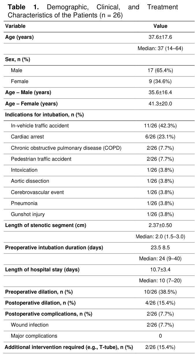
The mean stenosis length was 2.37 ± 0.50 cm, the mean length of hospital stay was 10.68 ± 3.45 days, and the mean preoperative intubation duration was 23.5 ± 8.5 days (Table 1). Ten patients underwent tracheal dilation before surgery (once for one patient, twice for six patients, three times for one patient, four times for one patient, and eight times for one patient). After surgery, four patients required tracheal dilation (once for two patients, twice for one patient, and four times for one patient). Furthermore, T-tube placement was conducted in two patients, with one patient maintaining the T-tube for two years and the other for three years, after which it was successfully removed.
There was a strong link between the need for dilation after surgery and the need for dilation before surgery. None of the patients who did not receive preoperative dilation required postoperative dilation (0/16); however, 4 four of ten patients (40%) who underwent preoperative dilation required postoperative dilation (Fisher’s exact test, p=0.014). After surgery, only two patients (7.7%) developed infections at the wound site.
Spearman's correlation analysis revealed no significant correlations between age and length of hospital stay (ρ=0.146, p=0.487), preoperative intubation duration and length of hospital stay (ρ=−0.128, p= 0.541), or age and preoperative intubation duration (ρ= 0.155, p=0.448) (Table 3). A positive correlation was noted between stenosis length and duration of hospitalization; however, this association did not reach statistical significance (ρ=0.277; p=0.180).
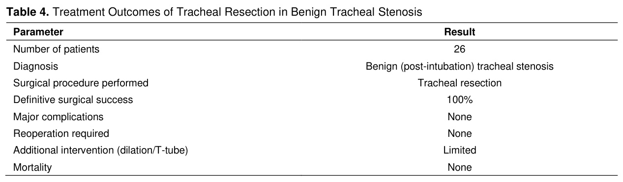
In nonparametric comparisons, there was a borderline trend indicating a difference in the length of hospital stay between female and male patients (p=0.052), with females having a median stay of 8 days and males having a median stay of 10 days. Patients who underwent preoperative dilation exhibited a reduced preoperative intubation duration (p=0.064, 18.5-26.5 days). Nonetheless, no substantial difference in hospital stay duration was noted concerning the presence of preoperative dilation (p=0.116, 8-10 days). Table 4 shows the surgical results for patients who underwent tracheal resection.


Discussion
Benign tracheal stenosis (BTS) is a pathological condition that necessitates a multidisciplinary management approach owing to its extensive etiological range and intricate morphological features. The first step in making a treatment plan is to determine what is wrong and how to classify the lesion so that surgery or an endoscopic procedure can be performed. Approximately 90% of BTS cases are caused by iatrogenic injury after endotracheal intubation or tracheostomy [1].Our study results support tracheal resection as the best treatment for BTS because it has a high success rate, few complications, and can provide a long-lasting and permanent solution. The fact that all patients in our series were treated with tracheal resection and did not have any major complications or deaths shows how well this method works. In addition, the fact that patients who had preoperative dilation required more interventions after surgery suggests that endoscopic procedures are often only temporary solutions that depend on how severe and complicated the disease is, rather than permanent solutions.
The implementation of low-pressure, high-volume cuffs has diminished the occurrence of post-intubation tracheal stenosis to approximately 1%. However, stenosis associated with extended tracheostomy cannula utilization persists as relatively prevalent, with reported rates reaching up to 30% [2]. The most important pathogenic factor is raising the pressure in the endotracheal tube cuff above the tracheal mucosal perfusion threshold of 30 cmH₂O (or 20-30 mmHg). This causes regional ischemia, leading to cartilage necrosis and the formation of fibrotic scars [3]. In our study, consistent with the existing literature, all 26 patients demonstrated tracheal stenosis after prolonged intubation. Although endotracheal tube cuff pressures could not be measured directly or verified systematically because of the retrospective nature of the study, a review of clinical records, intensive care follow-up notes, and discharge summaries suggested that cuff-related mucosal injury was the most likely underlying mechanism in the majority of patients.
Morphological and anatomical classification of BTS is crucial for predicting the natural progression of the disease and identifying the most effective treatment strategy. The most common method for grading stenosis in the literature is the Cotton-Myer system. It splits stenosis into four grades based on how much the lumen narrows: Grade I: <50%, Grade II: 51-70%, Grade III: 71-99%, and Grade IV: complete obstruction [6]. Surgical decision-making must consider not only the degree of luminal narrowing, but also the vertical length of the stenosis and the integrity of the cartilaginous support. In our study, we used the Cotton–Myer grading system. Three patients were graded as I, 13 as II, and 10 as III. There were only two cases of postoperative complications among all patients who underwent resection. Both were classified as Grade III stenoses.
In clinical settings, the most efficient way to distinguish stenosis is by categorizing it as either "simple" or "complex." Simple stenoses are characterized by lesions that are <1 cm in length, have a web-like appearance, do not encompass the entire circumference, and preserve the cartilage [2]. Conversely, complex stenoses are identified by lesions exceeding 1 cm in length, often resulting from tracheomalacia or compromised cartilage integrity [7]. The McCaffrey and Lano classification systems are also beneficial for assessing the likelihood of successful decannulation [8].
These comprehensive classification methods are essential for identifying patients who may benefit from interventional bronchoscopic techniques and those who should be referred for surgery. We assessed the length of the stenotic segment in all patients prior to bronchoscopic dilation or tracheal resection. All patients were categorized as having complex stenosis, with an average stenotic segment length of 2.37 ± 0.5 cm, and underwent surgery.
To effectively manage benign tracheal stenosis, a comprehensive multidisciplinary assessment is required to meticulously outline the etiology, anatomical characteristics, and functional implications of stenosis [1,2].
The initial step in the diagnostic process involves the use of noninvasive imaging techniques. Multidetector computed tomography (MDCT) and three-dimensional (3D) reconstruction methods are essential for precisely assessing the craniocaudal extent of stenosis, its luminal size, and its spatial relationship with nearby structures [2,3,9]. Virtual bronchoscopy provides a non-invasive approach to examine the distal airway beyond the narrowed segment, especially when the lumen is too narrow for rigid instruments, and serves as an important surgical "road map" for planning operations [3,10].
In pulmonary function tests (PFTs), the examination of flow-volume loops reveals distinct patterns of inspiratory and expiratory plateaus that indicate central airway obstruction, suggesting functional limitations. However, these patterns may only become apparent when the airway lumen diameter is reduced to approximately 8 mm [10-12].
Bronchoscopy is the best method to determine whether a patient has BTS [13-14]. During bronchoscopy, the vocal cord function, stenosis morphology (circumferential, eccentric, or web-like), and degree of stenosis according to the Cotton-Myer grading system were evaluated [15,16]. Doctors can use radial endobronchial ultrasound (r-EBUS) to examine the histological layers of the tracheal wall and the strength of the cartilage framework. This helps them determine whether the stenosis is simple or not [14]. All patients in our study underwent bronchoscopy before surgery. During this procedure, the length of the stenosis was measured, the lesions were classified as complex and staged according to the Cotton-Myer scale, and the patients were sent for surgery.
Surgical reconstruction is one of the most effective treatment approaches for benign tracheal stenosis. In stenoses that do not respond to simple dilation procedures or have complex morphology (such as damage to the cartilage or a length greater than 1 cm), surgical resection with end-to-end anastomosis is the best treatment. The optimal length of resection is typically restricted to 1-4 cm (approximately 50% of the adult tracheal length); for longer segments, supplementary procedures such as laryngeal release or mediastinal mobilization are necessary to alleviate tension at the anastomotic site [3]. When the stenotic segment is longer than 4-6 cm or more than 50% of the total tracheal length, primary reconstructive techniques become difficult because of excessive anastomotic tension, which leads to a significant increase in complication rates [5,17].
To obtain good surgical results, it is important to ensure that the anastomosis is free of tension [8]. Excessive tension at the anastomotic line is the primary factor contributing to compromised vascular perfusion, granulation tissue formation, and anastomotic dehiscence [4,18].
Interventional bronchoscopy is the first-line treatment for patients who cannot undergo surgery or who have simple stenoses (web-like lesions that are less than 1 cm long and maintain their cartilage structure). Other treatment options include laser-assisted mechanical dilation, balloon dilation, mitomycin C application, intralesional steroid injection, silicone stents (Dumon), and Montgomery T-tube placement [9-15].
In proficient institutions, mortality rates subsequent to tracheal surgery are less than 2% [6,20]. However, complications may arise, including anastomotic dehiscence and leakage due to excessive tension or devascularization [20,21], restenosis resulting from granulation tissue or fibrosis at the anastomotic site (3-10%) [16,22], recurrent laryngeal nerve injury [6,23], and dysphagia and aspiration [3,6]. In our study, only two patients (7.7%) developed wound site infections after tracheal resection, and no significant complications were observed.
The principal strength of this study lies in the assessment of a homogeneous patient cohort comprising only benign (post-intubation) tracheal stenosis cases. The results were more consistent and trustworthy because all patients were treated at the same center with the same surgical techniques and standardized perioperative care protocols. The low complication rate, absence of significant complications and mortality, and minimal necessity for additional surgical interventions are pivotal findings that validate the efficacy and safety of tracheal resection. A thorough evaluation of preoperative and postoperative interventions enhanced the clinical significance of this study.
Despite these strengths, some weaknesses need to be recognized. The primary limitations of this study include its single-center, retrospective design, relatively small sample size, and the absence of assessment of long-term functional outcomes and quality-of-life metrics.
Tracheal resection appears to be a highly effective treatment option for appropriately selected patients with benign tracheal stenosis owing to its high success rate, low complication risk, and ability to provide durable long-term outcomes. In our study, all patients underwent definitive treatment through tracheal resection, resulting in no significant complications or mortality and eliminating the need for alternative definitive surgical interventions, thereby demonstrating the clinical efficacy of this method. Tracheal resection is a safe and effective treatment for benign tracheal stenosis when performed in the right patients and in experienced centers. This can lead to long-term improvements in these patients.
References
- Cusumano G, Paternò DS, Terminella A, Sorbello M, La Via L. "We were already there where the narrow path …" Benign tracheal stenosis treatment: narrative review on the state of the art and future direction. Updates Surg 2026; 78: 429-441.
- Galluccio G, Lucantoni G, Battistoni P, et al. Interventional endoscopy in the management of benign tracheal stenoses: Definitive treatment at long-term follow-up. Eur J Cardiothorac Surg 2009; 35: 429-433.
- Ramesh C, Dehadaray A, Kaushik M, Mishra P, Sidhu S. Use of silicon T-tube for subglottic stenosis and tracheal stenosis. Iran J Otorhinolaryngol 2023; 35: 125-131.
- Hu HH, Wu FJ, Zhang JS, Chen E. Treatment of secondary benign airway stenosis after tracheotomy with Montgomery T-tube. Math Biosci Eng 2019; 16: 7839-7849.
- Mark Z, Bajzik G, Nagy A, et al. Comparison of virtual and fiberoptic bronchoscopy in the management of airway stenosis. Pathol Oncol Res 2008; 14: 313-319.
- Wierzbicka M, Tokarski M, Puszczewicz M, Szyfter W. The efficacy of submucosal corticosteroid injection and dilatation in subglottic stenosis of different aetiology. J Laryngol Otol 2016; 130: 674-679.
- Messina G, Di Filippo V, Capasso F, et al. Modified blade: an interventional option in rigid bronchoscopy for non-resectable benign tracheal stenosis. J Cardiothorac Surg 2024; 19: 73.
- Oskam LC, Honings J, van Ipenburg JA, et al. Benign subglottic stenosis from a rheumatologist' perspective: A narrative review. Clin Rheumatol 2025; 44: 4785-4796.
- Anagnostopoulos N, Dawood H, Koukaki E, et al. Choosing the right stent for each type of benign tracheal stenosis may be associated with reduced stent-related complications. Respiration 2025; 1-9.
- Özdemir C, Kocatürk CI, Sökücü SN, et al. Endoscopic and surgical treatment of benign tracheal stenosis: A multidisciplinary team approach. Ann Thorac Cardiovasc Surg 2018; 24: 288-295.
- Rahman NA, Fruchter O, Shitrit D, Fox BD, Kramer MR. Flexible bronchoscopic management of benign tracheal stenosis: long term follow-up of 115 patients. J Cardiothorac Surg 2010; 5: 2.
- Kapadia D, Simoff MJ, Diaz-Mendoza J. Benign tracheal stenosis and subglottic stenosis. Clin Chest Med 2025; 46: 349-357.
- Agrawal A, Baird BJ, Madariaga MLL, Blair EA, Murgu S. Multi-disciplinary management of patients with benign airway strictures: A review. Respir Med 2021; 187: 106582.
- Axtell AL, Mathisen DJ. Idiopathic subglottic stenosis: Techniques and results. Ann Cardiothorac Surg 2018; 7: 299-305.
- Khan A, Nath A, Mangla L, Paul M, Neyaz Z. Use of dedicated optical tracheal dilator for postintubation tracheal stenosis: First report from India. Lung India 2018; 35: 417-420.
- Uğur Chousein EG, Turan D, Tanrıverdi E, et al. The efficacy of topical mitomycin-C in the treatment of benign tracheal stenosis: A comparative study. Turk Gogus Kalp Damar Cerrahisi Derg 2025; 33: 217-225.
- Gulilat D, Genetu A, Kejela S, et al. Nonmalignant tracheal stenosis: Presentation, management and outcome in limited resources setting. J Cardiothorac Surg 2024; 19: 21.
- Chen DF, Chen Y, Zhong CH, Chen XB, Li SY. Long-term efficacy and safety of the Dumon stent for benign tracheal stenosis: A meta-analysis. J Thorac Dis 2021; 13: 82-91.
- Aydogmus U, Kis A, Ugurlu E, Ozturk G. Superior strategy in benign tracheal stenosis treatment: Surgery or endoscopy? Thorac Cardiovasc Surg 2021; 69: 756-763.
- Ming W, Zuo J, Han J, Chen J. The efficiency of endoscopic versus open surgical interventions in adult benign laryngotracheal stenosis: A meta-analysis. Eur Arch Otorhinolaryngol 2023; 280: 2421-2433.
- Viveiros F, Gomes J, Oliveira A, et al. Topical application of mitomycin-C as an adjuvant treatment to bronchoscopic procedures in post-intubation tracheal stenosis. Rev Port Pneumol 2013; 19: 276-280.
- Soumagne T, Guibert N, Atallah I, et al. Dilation versus laser resection in subglottic stenosis: Protocol for a prospective international multicentre randomised controlled trial (AERATE trial). BMJ Open 2022; 12: e053730.
- Fortin M, MacEachern P, Hergott CA, et al. Self-expandable metallic stents in nonmalignant large airway disease. Can Respir J 2015; 22: 235-236.