
MRI'de Vertebral Endplate Yüzey Morfolojisi ile Lomber Disk Dejenerasyonu Arasındaki İlişki
Cilt 40, Sayı 3
Kasım 2026 · Sayfalar 267-276
MRI'de Vertebral Endplate Yüzey Morfolojisi ile Lomber Disk Dejenerasyonu Arasındaki İlişki
Association Between Vertebral Endplate Surface Morphology and Lumbar Disc Degeneration on MRI
Arzu DİNÇ YAVAŞ1, Ayşe Serap AKGÜN2, Sümeyye AKÇAY3
2İstanbul Medipol University, Faculty of Medicine Department of Radiology, İstanbul, TÜRKİYE
3Fenerbahçe University, Faculty of Health Sciences Department of Ergotherapy, İstanbul, TÜRKİYE
Amaç: Lomber disk dejenerasyonu, bel ağrısının başlıca nedenlerinden biridir ve yaygın olarak Pfirrmann sınıflamasıyla değerlendirilmektedir. Disk dejenerasyonu ile ilişkili daha objektif ve klinik uygulamada kullanılabilir görüntüleme özelliklerine ihtiyaç vardır. Vertebral endplate morfolojisi, özellikle üst ve alt endplateler arasındaki simetri, disk dejenerasyonunun patogenezi hakkında değerli bilgiler sağlayabilir. Bu çalışmanın amacı, lomber disk dejenerasyonu ile vertebral endplate yüzey morfolojisi arasındaki ilişkiyi araştırmak ve vertebral endplate asimetrisi ile lomber disk dejenerasyonu arasındaki ilişkiyi değerlendirmektir.
Gereç ve Yöntem: Bu retrospektif çalışmada, bel ağrısı bulunan 157 hasta ile dejenerasyon saptanmayan 100 kişiden oluşan kontrol grubu lomber MRG ile değerlendirildi. Disk dejenerasyonu Pfirrmann sınıflamasına göre derecelendirildi. Endplate morfolojisi konkav, düz veya düzensiz olarak sınıflandırıldı; segmentler ayrıca simetrik veya asimetrik olarak değerlendirildi. Gruplar arasında karşılaştırmalı analizler yapıldı.
Bulgular: Endplate morfolojik özellikleri, tüm lomber segmentlerde hasta grubu ile dejenerasyon bulunmayan kontrol grubu arasında anlamlı farklılık göstermiştir (p<0.001). Asimetrik endplateler, disk dejenerasyonu bulunan hastalarda anlamlı olarak daha sık görülmüştür. Konkavite kaybı ve düzensiz endplate morfolojisi, daha yüksek derecede disk dejenerasyonu ile ilişkili bulunmuştur. Bu bulgular, endplate asimetrisi ile lomber disk dejenerasyonu arasında anlamlı bir ilişki olduğunu göstermektedir.
Sonuç: Vertebral endplate morfolojisi, MRG'de lomber disk dejenerasyonu ile anlamlı olarak ilişkilidir. Endplate morfolojisinin değerlendirilmesi, disk dejenerasyonu ile ilişkili basit ve klinik uygulamada kullanılabilir bir morfolojik özellik sağlayabilir ve mevcut MRG tabanlı derecelendirme sistemlerini tamamlayabilir. Endplate morfolojik değişiklikleri, ileri Pfirrmann dejenerasyonu bulunmayan semptomatik bireylerde de saptanabilir; bu durum, lomber disk dejenerasyonu ile ilişkili erken yapısal değişikliklerin belirlenmesinde potansiyel bir rolü olabileceğini düşündürmektedir.
Objective: Lumbar disc degeneration is a major contributor to low back pain and is commonly assessed using the Pfirrmann Classification. There is a need for more objective, clinically applicable imaging features associated with disc degeneration. Vertebral endplate morphology, particularly the symmetry between superior and inferior endplates, may provide valuable insights into the pathogenesis of disc degeneration. This study aimed to investigate the relationship between lumbar disc degeneration and vertebral endplate surface morphology and to evaluate the association between vertebral endplate asymmetry and lumbar disc degeneration.
Materials and Methods: In this retrospective study, 157 patients with low back pain and 100 individuals in a non-degenerated control group underwent lumbar MRI evaluation. Disc degeneration was graded using the Pfirrmann Classification. Endplate morphology was categorized as concave, flat, or irregular, and segments were further classified as symmetric or asymmetric. Comparative analyses were performed between groups.
Results: Endplate morphological characteristics differed significantly between the patient and non-degenerated control groups across all lumbar segments (p<0.001). Asymmetric endplates were significantly more frequent in patients with disc degeneration. Loss of concavity and irregular endplate morphology were associated with higher grades of disc degeneration. These findings demonstrate a significant association between endplate asymmetry and lumbar disc degeneration.
Conclusion: Vertebral endplate morphology is significantly associated with lumbar disc degeneration on MRI. Assessment of endplate morphology may provide a simple, clinically applicable morphologic feature associated with disc degeneration and complement existing MRI-based grading systems. Endplate morphological alterations may also be detectable in symptomatic individuals without advanced Pfirrmann degeneration.
Giriş
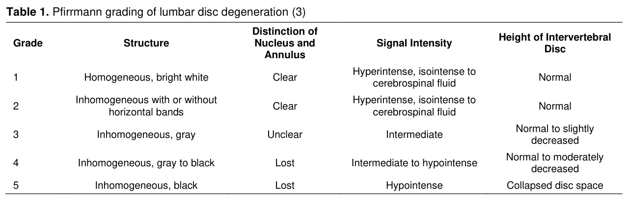
Low back pain is a common symptom encountered across all age groups, with a higher incidence among the elderly. With rising life expectancy, the incidence of low back pain also increases each year. As a result of this situation, disability, economic burden, and labor loss are experienced [1]. The most important cause of low back pain is degeneration of the lumbar discs, and the severity of these degenerative changes also determines the severity of symptoms and the treatment approach. The Pfirrmann Classification is a commonly used method of grading the severity of intervertebral disc degeneration [2]. In this method, the severity of degeneration is determined based on various magnetic resonance imaging (MRI) characteristics on a 5-grade scale [3]. This classification takes into account disc height and decreased signal intensity on MRI [3]. Several well-defined risk factors have been identified for the development of disc degeneration. Among these, vertebral endplate pathologies are thought to facilitate lumbar disc degeneration [4].Many studies have examined the relationship between disc degeneration and Modic changes, defects, and shape differences in the vertebral endplate [5,6]; however, most of these perform evaluations based on a single endplate [7-9]. In general, concavity has been evaluated in various ways, and decreased concavity has been reported to be associated with disc degeneration. There are also cadaver or animal studies on this topic that report similar results, but they are often affected by the same limitations [7-10]. Studies evaluating the superior and inferior vertebral endplates together have been reported previously [11,12]. One previous MRI-based study specifically investigated endplate asymmetry as a potential risk factor for lumbar disc degeneration [13]. However, that study primarily focused on lumbar disc herniation and asymmetry at selected lumbar levels, whereas the present study evaluates endplate morphology and symmetry patterns comprehensively across all lumbar segments in relation to the Pfirrmann Classification. Therefore, rather than introducing endplate asymmetry as a completely new concept, the present study aims to extend existing evidence by providing a comprehensive segment-level evaluation of vertebral endplate morphology and symmetry patterns across the entire lumbar spine and across different stages of disc degeneration.
The Pfirrmann Classification is an important method to determine the level of disc degeneration. However, this classification may be influenced by subjectivity in interpretation; therefore, it is important to seek a more consistent, inexpensive, repeatable, and simple method that provides relatively objective results. For this purpose, the shape characteristics of the endplates may be associated with the pathogenesis and severity of disc degeneration and may contribute to the development of more reproducible imaging assessments. The aim of this study was to evaluate the relationships between disc degeneration and superior/inferior endplate shape differences in lumbar segments.
Gereç ve Yöntem
Research and Publication Ethics: The study protocol was approved by the Istanbul Medipol University Ethics Committee (date: 30/09/2019, no: 10840098-604.01.01-E.53525). The study was conducted in accordance with the principles of the Declaration of Helsinki. Patient data were retrospectively reviewed and analyzed anonymously.In this study, 157 patients aged 18 to 60 years who underwent lumbar vertebra MRI with a 1.5 T (Tesla) Phillips Interra MRI system for low back pain between January 2016 and August 2019 were evaluated retrospectively. The exclusion criteria included a history of spinal surgery, scoliosis, lumbar fractures, tuberculosis, tumors, and infections. The MRI images were evaluated by a radiologist experienced in spine MRI. In all cases, T1W, T2W sagittal, T2W axial sequences with a slice thickness of 4 mm and interslice distance 0.8 mm were obtained. Images were reviewed using the digital measurement tools available in the PACS system. One hundred individuals without radiological evidence of lumbar disc degeneration in the same age group who underwent lumbar MRI were included in the study as a control group (Pfirrmann grades <3).
Disc degeneration was classified using the Pfirrmann Classification (Table 1).
Participants were categorized into three groups according to clinical status and Pfirrmann Classification:
Group AI (Symptomatic non-degenerated group): patients with low back pain and Pfirrmann grades 1–2 discs
Group AII (Degenerated disc group): patients with Pfirrmann grades 3–5 discs
Group B (Non-degenerated control group): individuals with Pfirrmann grades <3 used as the comparison group
This grouping strategy was designed to distinguish symptomatic patients without advanced radiological degeneration from patients with established degeneration and from the comparison group.
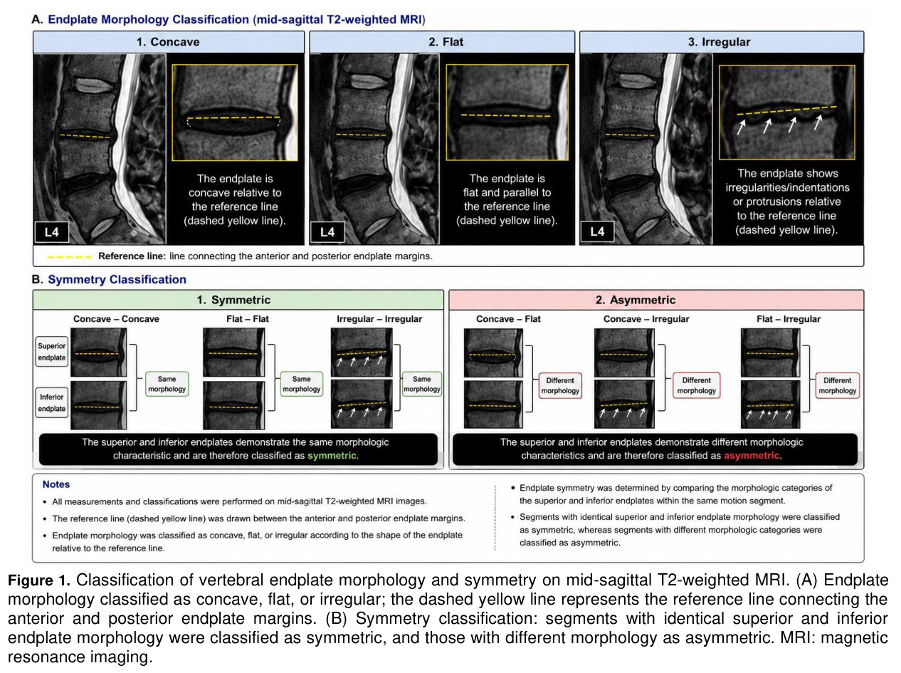
Vertebral endplate surfaces were classified into three categories: concave, flat, and irregular [13-15]. In the mid-sagittal T2WI view, a line was drawn connecting the ends of the endplates. Endplates were grouped as “flat” if the edge of the endplate could be overlapped with this line or if there was a slight depression (maximum 1 mm). Cases in which the maximum depth exceeded 1 mm were grouped as “concave”. The term “irregular” was used if the edge of the endplate exhibited significant defects or if a protrusion that crossed the connecting line was present. Six groups were formed according to the superior and inferior endplate surface shape in each segment: “concave–concave,” “concave–flat,” “concave–irregular,” “flat–flat,” “flat–irregular,” and “irregular–irregular”. Finally, two groups were identified based on these evaluations: “symmetric group” if the shapes of the superior and inferior endplates in the same segment showed the same characteristic, and “asymmetric group” if they differed.
Representative examples of concave, flat, and irregular endplates, as well as symmetric and asymmetric endplate configurations, are provided in Figure 1. The figure also illustrates the reference line drawn between the anterior and posterior endplate margins that was used for morphological classification on mid-sagittal T2-weighted MRI images.
Although both the symptomatic non-degenerated group and the control group consisted of discs with Pfirrmann grades 1–2, the groups differed in clinical status. The symptomatic non-degenerated group included symptomatic individuals presenting with low back pain, whereas the comparison group consisted of individuals without radiological evidence of lumbar disc degeneration who were used as the reference population. This distinction was introduced to investigate whether endplate morphology and symmetry patterns may differ in symptomatic patients even when advanced radiological degeneration is not yet present.
Statistical Analysis: The SPSS 22.0 software for Windows was used for statistical analysis. Categorical variables were presented as numbers and percentages, and numerical variables were presented as mean and standard deviation. Comparisons of numerical variables between two independent groups were performed using the Mann–Whitney U test because the numerical variables did not meet parametric assumptions.
For categorical variables, Pearson’s chi-square test was initially used, and expected cell counts were examined for each comparison. When the assumptions of the chi-square test were not met due to low expected cell counts, exact methods were used. Monte Carlo exact tests were used for global comparisons in contingency tables larger than 2×2, whereas Fisher’s exact test was used for 2×2 pairwise comparisons.
Subgroup analyses were performed using pairwise comparisons, and a Bonferroni correction was applied based on the number of pairwise comparisons. For comparisons among the three study groups, raw p-values were multiplied by three, and values exceeding 1.000 were reported as 1.000. The p-values presented for subgroup comparisons in the tables are Bonferroni-adjusted p-values.
Because multiple lumbar disc levels were evaluated in each participant, segment-level observations may not be fully independent. Therefore, segment-based analyses were interpreted with caution, considering the potential effect of within-patient correlation. Statistical approaches that account for clustered observations, such as mixed-effects models or generalized estimating equations, were considered as alternatives; however, the present analyses were conducted as level-specific categorical comparisons.
Effect sizes were also reported in addition to p-values. Cramer’s V was used as an effect-size measure for categorical comparisons, and odds ratios with 95% confidence intervals were reported for 2×2 comparisons when applicable. Statistical significance was accepted as p<0.05.
Bulgular
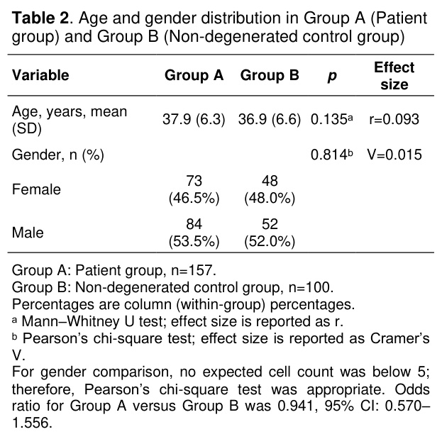
There was no statistically significant difference between Group A (symptomatic non-degenerated group and degenerated disc group) and Group B (non-degenerated control group) in terms of mean age or sex distribution. The mean age was 37.9±6.3 years in Group A and 36.9±6.6 years in Group B (p=0.135, effect size r=0.093). The female-to-male distribution was also comparable between the two groups (p=0.814, Cramer’s V=0.015) (Table 2).
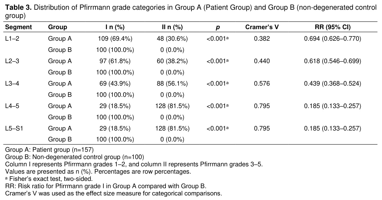
The distribution of Pfirrmann Classification grades differed significantly between Group A and Group B across all lumbar segments. In Group B, all segments were classified as Pfirrmann grade I, whereas the proportion of Pfirrmann grade II segments increased progressively in Group A from the upper to lower lumbar levels. The difference was statistically significant for all segments (p<0.001). The magnitude of association increased from L1–2 to L4–5/L5–S1, with Cramer’s V values ranging from 0.382 to 0.795 (Table 3).
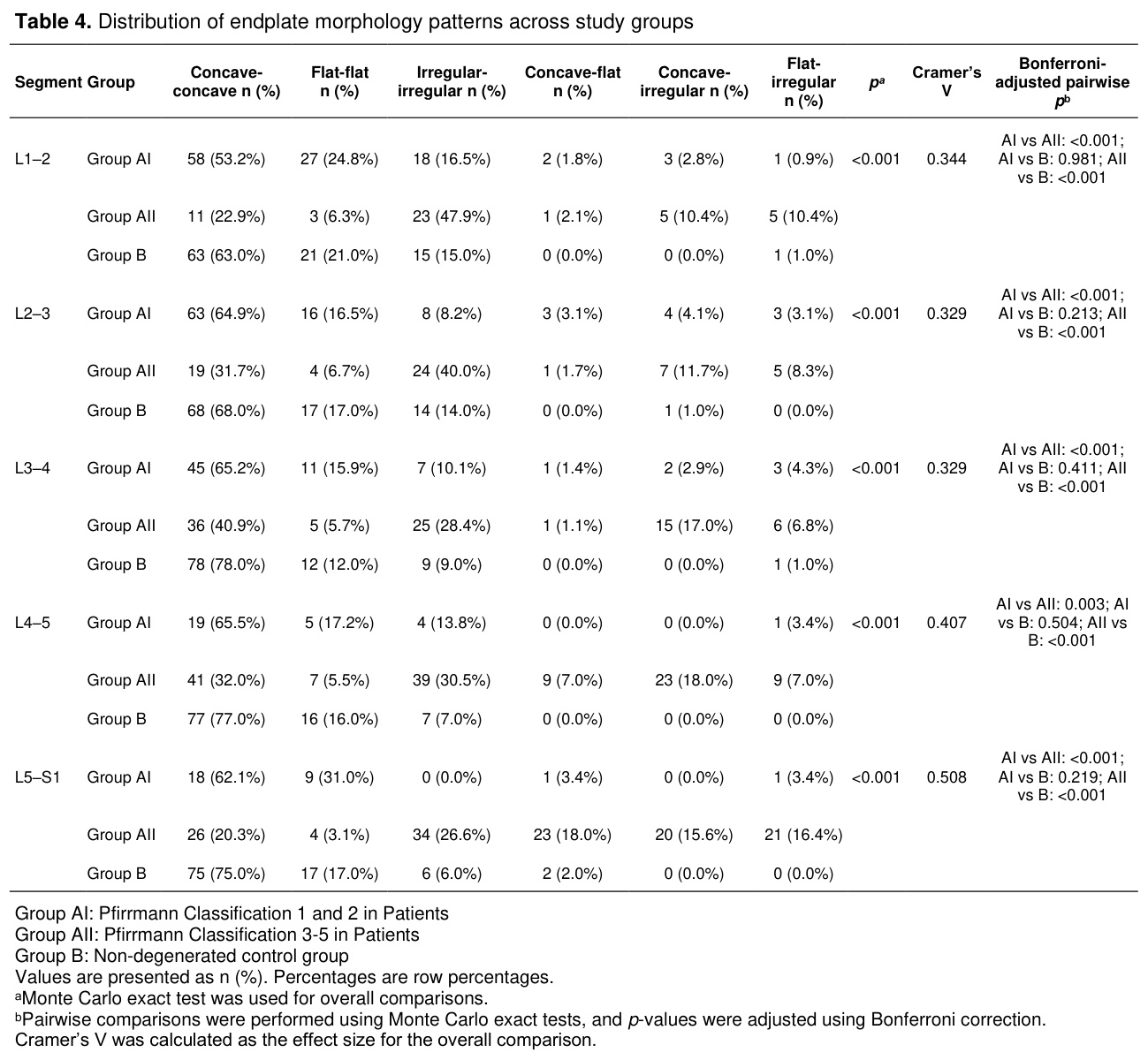
Endplate morphology patterns differed significantly among Group AI, Group AII, and Group B across all lumbar segments (p<0.001 for all). Cramer’s V values indicated a moderate association between study group and endplate morphology pattern, ranging from 0.329 to 0.508. In general, concave–concave morphology was more frequently observed in Group B, whereas irregular and asymmetric morphology combinations were more frequent in Group AII. Pairwise comparisons showed that Group AII differed significantly from Group B at all lumbar segments (p<0.001 for all). Group AI and Group B did not differ significantly in any segment, whereas Group AI and Group AII showed significant differences at all lumbar segments (Table 4).
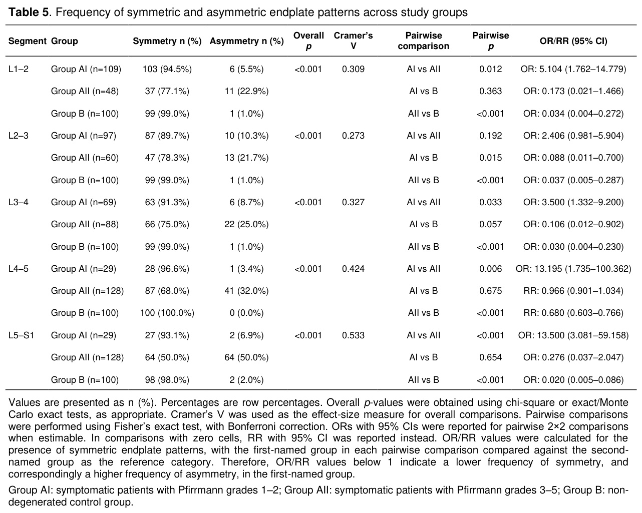
When endplate patterns were classified as symmetric or asymmetric, significant differences were observed among Group AI, Group AII, and Group B across all lumbar segments (p<0.001 for all). The overall effect size ranged from small-to-moderate to moderate, with Cramer’s V values of 0.309 at L1–2, 0.273 at L2–3, 0.327 at L3–4, 0.424 at L4–5, and 0.533 at L5–S1. Asymmetric endplate patterns were generally more frequent in Group AII than in Group AI and Group B. At L1–2, asymmetry was observed in 5.5% of Group AI, 22.9% of Group AII, and 1.0% of Group B. At L2–3, the corresponding rates were 10.3%, 21.7%, and 1.0%, respectively. At L3–4, asymmetry was present in 8.7% of Group AI, 25.0% of Group AII, and 1.0% of Group B. At L4–5, asymmetry was observed in 3.4% of Group AI, 32.0% of Group AII, and 0.0% of Group B. At L5–S1, the highest asymmetry rate was observed in Group AII, with asymmetry present in 50.0% of cases, compared with 6.9% in Group AI and 2.0% in Group B (Table 5).
Pairwise comparisons further demonstrated that Group AII had significantly higher asymmetry than Group B at all lumbar levels. The difference between Group AI and Group AII was significant at L1–2, L3–4, L4–5, and L5–S1, but not at L2–3. The difference between Group AI and Group B was significant at L2–3 but not at L1–2, L3–4, L4–5, or L5–S1. Odds ratios or risk ratios with 95% confidence intervals supported these findings and demonstrated that the magnitude of association was most prominent at L4–5 and L5–S1, particularly in comparisons involving Group AII (Table 5).
Tartışma
There are many facilitating factors for disc degeneration, but the least studied variables are structural features. In this study, the relationship between endplate morphological differences and disc degeneration was investigated. In all segments, superior/inferior endplate morphology differed significantly among Group AI, Group AII, and Group B. When these characteristics were utilized to divide patients into two groups (symmetrical/asymmetrical), significant differences were observed across all lumbar segments. In general, Group AII showed a higher frequency of asymmetric endplate patterns than Group AI and Group B, particularly in the lower lumbar segments. At L3–4, Group AII also demonstrated a higher frequency of asymmetric endplate patterns than Group B, supporting the association between advanced disc degeneration and endplate asymmetry at this level. These findings suggest that altered endplate morphology and endplate asymmetry are associated with more advanced lumbar disc degeneration.The comparison between Group AI and Group B (non-degenerated comparison group) was performed because symptom status and structural degeneration do not always coincide. Although both groups had Pfirrmann grade 1–2 discs, symptomatic individuals may harbor early morphologic or biomechanical alterations that are not fully reflected by conventional MRI degeneration grading systems. Therefore, this comparison was intended to explore whether endplate morphology could identify structural differences before advanced radiological degeneration becomes apparent.
Although the Pfirrmann Classification is important in determining the level of disc degeneration, it is a subjective tool; therefore, a more objective, consistent and standard method has been sought for classification purposes. In most studies evaluating the relationship between disc degeneration and endplate morphology, only a single endplate was considered. The results of these studies show that, similar to our findings, loss of concavity increases the incidence of disc degeneration. Endplate morphology has been evaluated by MRI in various studies; flat and irregular endplates are associated with more severe disc degeneration [15]. In a study, Wang et al. found that endplate asymmetry is more frequent in patients with degenerated lumbar disc herniation in the L4-5 segment than in patients with non-degenerated lumbar disc herniation (47% vs 21%) [13]. Although Wang et al. reported that superior–inferior endplate asymmetry may be associated with lumbar disc degeneration, the present study differs in several important aspects. First, we evaluated all lumbar motion segments systematically rather than focusing primarily on selected levels associated with disc herniation. Second, our analysis incorporated detailed morphologic categorization of endplates (concave, flat, irregular) in addition to symmetry assessment. Third, degeneration severity was evaluated using the Pfirrmann Classification across a broader patient spectrum, including symptomatic patients with both low-grade and advanced degeneration, as well as a non-degenerated comparison group. Therefore, our study expands the existing literature by providing a more comprehensive MRI-based assessment of vertebral endplate morphology in relation to lumbar disc degeneration.
Many other studies have focused on morphology rather than endplate asymmetry. In a cadaveric study examining 600 vertebral endplates, irregular endplate shape was reported to increase the risk of disc degeneration by 2.62-fold compared with concave endplates. It was emphasized that increased concavity and decreased circularity may be important in the pathogenesis of disc degeneration [16]. Barim et al. found a strong linear relationship between an increased concavity index (concavity level divided by vertebral body height) and an increased Pfirrmann Classification. It was stated that the concavity index was strongly associated with disc degeneration and was reproducible, and the consistency between the two observers, which was 66% in the Pfirrmann Classification, was 96-99% in the concavity index [17]. He et al. found that, as lumbar intervertebral disc degeneration progressed, the concave angle of the vertebral endplate decreased and the endplate tended to flatten. They reported that the degree of flattening was associated with the severity of degeneration [7]. Louie and colleagues examined the relationship between the Concavity Index and disc degeneration according to the Pfirrmann Classification. They found that the progression of intervertebral disc degeneration was proportional to the increase in endplate convexity. Accordingly, current evidence suggests that vertebral endplates are more concave in disc degenerations classified as lower grade by the Pfirrmann Classification, whereas endplates are more convex in disc degenerations classified as higher grade by the Pfirrmann Classification [8]. Xiao et al. reported that the endplate concave angle correlated positively with lumbar disc degeneration. They suggest that endplate changes indicate lumbar disc degeneration and that endplate concavity may represent imaging features associated with disc degeneration in the future [9]. Consistent with all these studies, superior and inferior endplate morphology differed significantly among Group AI, Group AII, and Group B across all lumbar segments in the present study. Similarly, when endplate patterns were classified as symmetric or asymmetric, significant differences were observed across all lumbar segments. Pairwise comparisons showed that Group AII had a significantly higher frequency of asymmetric endplate patterns than Group B at all lumbar levels, whereas the difference between Group AI and Group B was significant only at L2–3.
Dudli et al. reported that endplates with wider concavity may predispose the vertebrae to degeneration. They suggested that combining endplate shape and morphometric features increased sensitivity by affecting vertebral bodies [18]. In addition, endplate Modic changes have been reported to cause disc degeneration [19]. In a study supporting this interpretation, Chunlin and colleagues found that the frequency of Modic changes in the irregular endplate was significantly higher than in concave and flat endplates. In addition, they found that flat and irregular endplates were more common in those with herniated discs [20]. In our study, irregular endplates had the highest risk, flat endplates had a medium risk, and concave endplates had the lowest risk of degenerative disc formation.
Although the cause-effect relationship between endplate morphology and disc degeneration could not be investigated due to the design of our study, asymmetrical endplates may hypothetically alter axial load transmission by shifting stress from the nucleus pulposus to the annulus fibrosus, which could contribute to degenerative changes. This issue was examined in various studies with similar results [10,11,21]. Prospective cohort studies are needed to determine the direction of this relationship.
To improve clarity and clinical applicability, the study groups were categorized by both symptom status and degeneration severity rather than by a purely numerical grouping system. This terminology was used to avoid potential confusion between group labels and Pfirrmann grading categories. This approach was intended to provide a more intuitive and clinically interpretable framework for evaluating endplate morphology in relation to lumbar disc degeneration.
In our study, Group AII showed more pronounced endplate morphologic alterations than Group AI and Group B, particularly in the lower lumbar segments. In the L4–L5 and L5–S1 segments, Group AII was less frequently represented in the “concave–concave” morphology group and showed a higher frequency of asymmetric endplate patterns. Asymmetric endplate patterns were uncommon in Group B across all lumbar levels, including L5–S1. The highest asymmetry rate was observed in Group AII at L5–S1, where half of the segments showed asymmetric endplate morphology. This finding supports an association between advanced degeneration and endplate asymmetry, particularly in the lower lumbar spine. However, because the present study was retrospective and cross-sectional, it remains unclear whether endplate asymmetry precedes disc degeneration or develops as a secondary adaptive or degenerative change. In another study, it was reported that the sizes of the inferior and superior endplates differed across segments [11].
An important aspect of the present study was the separate evaluation of symptomatic patients with Pfirrmann grade 1–2 discs (Group AI) and the non-degenerated control group (Group B). Although Pfirrmann grades 1–2 are generally considered non-degenerative, symptomatic individuals may still exhibit early structural or biomechanical alterations not fully captured by conventional degeneration grading systems. Therefore, this subgroup analysis was performed to explore whether endplate morphology and asymmetry may differ even before advanced radiological degeneration becomes apparent. In the updated analysis, Group AI was generally more similar to the non-degenerated control group (Group B) than Group AII, although some differences were still observed at specific lumbar levels. These findings suggest that endplate morphological alterations may begin before advanced radiological degeneration becomes apparent, but this interpretation should be considered exploratory.
In this study, there were no investigations of different endplate pathologies other than morphological analyses. The resulting structural changes (circularity, thickness, Modic changes, tissue disorders, etc.) may have contributed to or been associated with endplate deformities. Furthermore, endplate concavity may deteriorate with age [22] or with increased weight burden [23]. Although age was examined in our study, other features, such as body weight, that could affect the shape of the endplate were not examined.
From a practical perspective, visual assessment of endplate morphology does not require additional software or complex quantitative measurements and may therefore represent a feasible adjunctive morphologic parameter in routine lumbar MRI interpretation.
Another potential implication of the morphologic features evaluated in this study is that assessment of endplate morphology may be suitable for future integration into AI-assisted imaging analysis systems. Although the present study did not investigate artificial intelligence-based approaches, the morphological features evaluated here may represent potential imaging features for future automated image-analysis studies.
In this study, several limitations should be acknowledged to ensure appropriate interpretation of the findings:
Due to the retrospective and cross-sectional design, causal relationships between vertebral endplate morphology and disc degeneration cannot be established. Longitudinal follow-up data were not available; therefore, it remains unclear whether endplate morphological changes precede disc degeneration or represent a secondary adaptive or degenerative phenomenon.
Interobserver and intraobserver reliability analyses were not performed. The subjective nature of the Pfirrmann Classification may lead to interobserver variability. In the present study, endplate symmetry and morphology were assessed using visually identifiable structural features that may be practical for routine MRI evaluation. However, interobserver and intraobserver reliability analyses were not performed; therefore, the reproducibility and observer dependence of this approach cannot be determined from the current data.
The 1-mm threshold used to differentiate flat and concave endplate morphology was based on prior literature but was not independently validated within the present cohort. This may affect the robustness and generalizability of morphological classification.
Although statistical adjustments, such as the Bonferroni correction, were applied to subgroup analyses, the segment-level structure of the data poses an important statistical limitation. Because multiple lumbar disc levels were assessed within the same individual, observations from different segments may not be completely independent and may be affected by within-patient correlation. This may influence the precision of statistical estimates and the risk of type I error in segment-based comparisons. Although segment-based analyses provide clinically useful level-specific information, future studies using statistical approaches that account for clustered or repeated measurements, such as mixed-effects models or generalized estimating equations, may better address the correlated structure of lumbar segment data.
Another limitation is that the independent association between vertebral endplate morphology/asymmetry and disc degeneration could not be evaluated. Although age and sex distributions were comparable between groups, several potentially important confounding variables—including body mass index, occupation, physical activity level, smoking status, mechanical loading, Modic changes, disc herniation, and other degenerative spinal findings—were not available in the retrospective dataset. Therefore, multivariable analysis adjusting for these factors could not be performed. Accordingly, the present findings should be interpreted as demonstrating an association rather than an independent predictive or causal relationship. Future prospective studies incorporating comprehensive clinical and imaging covariates are required to determine whether endplate morphology or asymmetry is independently associated with lumbar disc degeneration. Biomechanical validation was not performed; therefore, the proposed relationship between endplate asymmetry and altered mechanical loading remains theoretical and requires further experimental confirmation.
Finally, there was no longitudinal follow-up or biomechanical validation in our study.
Future prospective cohort studies incorporating standardized imaging protocols, reliability testing, comprehensive clinical covariates, and biomechanical validation are needed to clarify the causal relationship between endplate morphology and lumbar disc degeneration.
The principal contribution of the present study is not the introduction of endplate asymmetry as a novel concept, but the comprehensive evaluation of both endplate morphology and symmetry patterns across all lumbar motion segments and across different stages of Pfirrmann-defined degeneration. This broader assessment provides additional information regarding the distribution and clinical associations of endplate morphological alterations throughout the lumbar spine.
Vertebral endplate morphology, particularly loss of concavity and increased superior–inferior endplate asymmetry, was significantly associated with lumbar disc degeneration on MRI. However, because this retrospective study did not include reliability testing or longitudinal validation, endplate morphology should be interpreted as an associated morphologic feature rather than a validated predictive imaging biomarker.
References
- Hartvigsen J, Hancock MJ, Kongsted A, et al. What low back pain is and why we need to pay attention. Lancet 2018; 391(10137): 2356-2367.
- Kettler A, Wilke J. Review of existing grading systems for cervical or lumbar disc and facet joint degeneration. Eur Spine J 2006; 15(6): 705-718.
- Pfirrmann CW, Metzdorf A, Zanetti M, Hodler J, Boos N. Magnetic resonance classification of lumbar intervertebral disc degeneration. Spine (Phila Pa 1976) 2001;26(17):1873-1878.
- Rade M, Määttä JH, Freidin MB, et al. Vertebral endplate defect as initiating factor in intervertebral disc degeneration: Strong association between endplate defect and disc degeneration in the general population. Spine (Phila Pa 1976) 2018; 43(6): 412-419.
- Lv B, Yuan J, Ding H, et al. Relationship between endplate defects, Modic change, disc degeneration, and facet joint degeneration in patients with low back pain. Biomed Res Int 2019; 2019: 9369853.
- Teichtahl AJ, Urquhart DM, Wang Y, et al. Lumbar disc degeneration is associated with Modic change and high paraspinal fat content - a 3.0T magnetic resonance imaging study. BMC Musculoskelet Disord 2016; 17(1): 439.
- He X, Liang A, Gao W, et al. The relationship between concave angle of vertebral endplate and lumbar intervertebral disc degeneration. Spine (Phila Pa 1976) 2012; 37(17): E1068-1073.
- Louie PK, Orías AAE, Fogg LF, et al. Changes in lumbar endplate area and concavity associated with disc degeneration. Spine (Phila Pa 1976) 2018; 43(19): E1127-e1134.
- Xiao L, Ni C, Shi J, et al. Analysis of correlation between vertebral endplate change and lumbar disc degeneration. Med Sci Monit 2017; 23: 4932-4938.
- Duran S, Cavusoglu M, Hatipoglu HG, Sozmen Cılız D, Sakman B. Association between measures of vertebral endplate morphology and lumbar ıntervertebral disc degeneration. Can Assoc Radiol J 2017;68(2):210-216.
- Wang Y, Battie MC, Videman T. A morphological study of lumbar vertebral endplates: Radiographic, visual and digital measurements. Eur Spine J 2012; 21(11): 2316-2323.
- Poureisa M, Daghighi MH, Mesbahi S, Hagigi A, Fouladi DF. End plate disproportion and degenerative disc disease: a case-control study. Asian Spine J 2014;8(4):405-411.
- Wang Y, Wang H, Lv F, et al. Asymmetry between the superior and inferior endplates is a risk factor for lumbar disc degeneration. J Orthop Res 2018; 36(9): 2469-2475.
- Lakshmanan P, Purushothaman B, Dvorak V, et al. Sagittal endplate morphology of the lower lumbar spine. Eur Spine J 2012; 21 Suppl 2: S160-164.
- Pappou IP, Cammisa FP, Girardi FP. Correlation of end plate shape on MRI and disc degeneration in surgically treated patients with degenerative disc disease and herniated nucleus pulposus. Spine J 2007; 7(1): 32-38.
- Wang Y, Videman T, Battie MC. Morphometrics and lesions of vertebral end plates are associated with lumbar disc degeneration: Evidence from cadaveric spines. J Bone Joint Surg Am 2013; 95(5): e26.
- Barim M, Sesek R, Schall M. The concavity index: A novel approach for quantifying intervertebral disc degeneration. Vol 60 2016; 953-957.
- Dudli S, Enns-Bray W, Pauchard Y, et al. Larger vertebral endplate concavities cause higher failure load and work at failure under high-rate impact loading of rabbit spinal explants. J Mech Behav Biomed Mater 2018; 80: 104-110.
- Oyinloye OI, Bamidele JO, Popoola GO. Modic changes in adults with chronic low back pain in North Central Nigeria. Journal of the West African College of Surgeons 2017; 7(2): 77-92.
- Chunlin L, Fang L, Zhicheng Z. Morphology of endplate in lumbar degenerative disc disease, a MRI study. Chinese Journal of Spine and Spinal Cord 2012; 22(5): 393-397.
- Orias E A A., Andersson GBJ, Inoue N. Influence of disc degeneration on endplate concavity, in ORS 2012 Annual Meeting. 2012: San Francisco, California.
- Twomey LT, Taylor JR. Age changes in lumbar vertebrae and intervertebral discs. Clin Orthop Relat Res 1987; 224: 97-104.
- Moorman C, Johnson DC, Pavlov H, et al. Hyperconcavity of the lumbar vertebral endplates in the elite football lineman. Am J Sports Med 2004; 32(6): 1434-1439.
- Urakal Kist: US, BT ve MRG Bulguları (Olgu Sunumu)
- Diyabetik Nefropatide Difüzyon MR Bulguları
- Parkinson Hastalığında Substantia Nigranın Difüzyon Ağırlıklı MRG Bulguları
- Spontan Spinal Epidural Hematomlar: 12 Olgunun Değerlendirilmesi
- Prostat Kanseri ile Benign Prostat Hiperplazisinin Ayırıcı Tanısında Difüzyon Ağırlıklı Manyetik Rezonans Görüntülemenin Yeri