
Glifosat-Bazlı Herbisite Maruz Kalan Fare Korteksinde Koenzim Q10 'un Nöroproteksiyon Etkisinin Histopatolojik Değerlendirmesi
Volume 40, Issue 3
November 2026 · Pages 282-287
Glifosat-Bazlı Herbisite Maruz Kalan Fare Korteksinde Koenzim Q10 'un Nöroproteksiyon Etkisinin Histopatolojik Değerlendirmesi
Histopathological Assessment of Coenzyme Q10 Mediated Neuroprotection in the Mouse Cortex Exposed to a Glyphosate-Based Herbicide
Halil İbrahim SAYGI1, Şule AYLA1, Duygu MUTLUAY2, İlknur DURSUN3
2Mehmet Akif Ersoy Medeniyet University, Faculty of Veterinary Medicine Department of Histology and Embryology, Burdur, TÜRKİYE
3İstanbul Sağlık ve Teknoloji University, Faculty of Medicine Department of Physiology, İstanbul, TÜRKİYE
Amaç: Glifosat-bazlı herbisitler (GBH) dünya genelinde yaygın olarak kullanılmakta olup, olası nörotoksik etkileri hem araştırmacılar hem de toplum için giderek artan bir merak konusudur. Koenzim Q10 (CoQ10) gibi mitokondri hedefli antioksidanların nöroprotektif etkileri iyi bilinmekle birlikte, GBH’lere karşı koruyucu özellikleri hala tam olarak bilinmemektedir. Bu çalışma, farelerde GBH maruziyetinin kortekste oluşturduğu hasarın CoQ10 ile onarılma potansiyelini değerlendirmeyi amaçlamaktadır.
Gereç ve Yöntem: 50 adet erişkin erkek ICR fare beş gruba ayrıldı: Kontrol, Sham, CoQ10 (200 mg/kg/gün), GBH (Roundup, 500 mg/kg/gün) ve GBH+CoQ10. 49 günlük oral tedavi sonrasında hayvanlar ötenazi edildi ve beyin dokusu Hematoksilen-Eozin (H&E) ve krezil viyole (Nissl) boyaları ile histolojik inceleme için alındı. Kortikal histopatolojik değerlendirme QuPath yazılımı kullanılarak gerçekleştirildi.
Bulgular: Histolojik inceleme, Kontrol, Sham ve CoQ10 gruplarında normal kortikal yapıların korunduğunu gösterilmiştir. GBH maruziyeti; küçülmüş, hiperkromatik ve piknotik nöronlar, dejeneratif değişiklikler, granülovakuoler dejenerasyon ve nöropilde belirgin vakuolizasyon ile karakterize nöronal hasara neden olduğu gözlemlenmiştir. GBH+CoQ10 grubunda ise kortikal organizasyon ve nöronal morfoloji büyük ölçüde korundu; yalnızca hafif fokal vakuolizasyon izlenmiştir.
Krezil viyole boyamasında, GBH uygulanan farelerde belirgin nöron kaybı, Nissl cisimciklerinde azalma ve sitoplazmik renk değişikliği gözlemlenirken, GBH+CoQ10 kesitlerinde daha yüksek nöronal yoğunluk ve belirgin Nissl granülleri gözlenmiştir. Nicel analizlerde, GBH uygulaması diğer tedavi gruplarına kıyasla kortikal nöron sayısını anlamlı düzeyde azalttığı gösterilmiştir. Ancak CoQ10 ile birlikte GBH verilmesi, bu azalmayı tersine çevirdiği izlenmiştir.
Sonuç: GBH maruziyeti, fare serebral korteksinde nöronal dejenerasyona neden olurken, CoQ10 desteği kortikal hücre yapısını ve nöronal yoğunluğu koruyarak GBH’ye bağlı beyin toksisitesine karşı nöroprotektif bir yardımcı ajan olarak potansiyelini desteklemektedir.
Objective: Glyphosate-based herbicides (GBHs) are widely used globally, and their potential neurotoxicity is of increasing concern to researchers and the public. Mitochondrial-targeted antioxidants such as Coenzyme Q10 (CoQ10) have established neuroprotective effects yet approaches to neuroprotection against GBHs are still being developed. This study evaluates the potential of CoQ10 to reverse cortical damage caused by GBH exposure in mice.
Materials and Methods: Fifty adult male ICR mice were assigned to five groups: control, sham, CoQ10 (200 mg/kg/day), GBH (Roundup, 500 mg/kg/day), and GBH+CoQ10. After 49 days of oral treatment, mice were euthanized and brains collected for histological examination using Hematoxylin and Eosin (H&E) and cresyl violet (Nissl) staining. Cortical histopathology was analyzed with QuPath software.
Results: Histological examination revealed that all normal cortical structures were found in the control, sham, and CoQ10 groups. Exposure to GBH produced shrunken, hyperchromatic, and pyknotic neurons with degenerative changes, as well as granulovacuolar degeneration and neuropil vacuolation. In the GBH+CoQ10 group, the cortical organization and neuronal morphology were well-preserved, with only mild focal vacuolation present.
Cresyl violet staining revealed severe neuronal loss, Nissl substance depletion, and cytoplasmic pallor in GBH-treated mice, whereas GBH+CoQ10 sections showed higher neuronal density and clear Nissl granules. Quantitatively, GBH significantly reduced cortical neuron number compared with other treatment groups. However, GBH co-treatment with CoQ10 reversed this decline.
Conclusion: GBH exposure induces neuronal degeneration in the mouse cerebral cortex, while CoQ10 supplementation preserves cortical cytoarchitecture and neuronal density, supporting its potential as a neuroprotective adjunct against GBH-induced brain toxicity.
Introduction
Over the past two decades, glyphosate-based herbicides (GBH) have become some of the most used and most widely distributed herbicides, and agrochemicals, among the global population [1]. Glyphosate (N-phosphonomethyl-glycine; GLY) is an herbicide that is widely used around the world and is one of the most frequently applied substances used for the control of weeds. Although initial toxicity studies of glyphosate theorized that the probability of glyphosate causing harm from exposure in mammals was low, the hypothesis has changed, and research studies have shown that the central nervous system may be affected during both acute and chronically prolonged exposures [2]. Herbicides that contain glyphosate could cross the blood-brain barrier and influence brain tissue [3], wherein these substances activate neuroinflammatory signaling and increase the concentration of TNF-α and other inflammatory responses [4,5]. In addition to neuroinflammation, GBHs disrupt multiple fundamental processes for neuronal survival and function. Research has shown that glyphosate and its derivatives can cause oxidative stress, disrupt mitochondrial respiration processes, disrupt glutamatergic and cholinergic neurotransmission processes, and damage hippocampal and cortical circuits [6-8]. Behavioral experiments on rodent models demonstrate that subchronic exposure to GBH induces anxiety, learning and memory deficits, and altered social behavior, which are consistent with cellular impairment [9-12]. Collectively, such changes indicate neuronal degeneration, along with loss of cognitive functions. This finding increases concern about the chronic effects of GBH exposure on the nervous system. These alterations are associated with deteriorating behavior and a decline in cognitive abilities, indicating a serious health risk.Coenzyme Q (CoQ) is found in the membranes of all cell types and is one of the constituents of the respiratory chain along with other structures of the mitochondria, performing numerous vital functions in the metabolism of the cells. As one of the antioxidants, CoQ10 is useful in the treatment and management of diverse disorders associated with the dysfunction of mitochondria [13,14]. Research indicates that CoQ10 is neuroprotective in several types of neurodegenerations and ischemic injury. Orally administering CoQ10 increases the amount of CoQ10 present in the brain and in the mitochondria. CoQ10 limits the damage of oxidative stress, preserves the integrity of the mitochondria, and controls the apoptosis pathways [15-17]. Given that glyphosate-induced neurotoxicity is characterized by oxidative stress and mitochondrial dysfunction, CoQ10 could provide neuroprotective effects against the neurotoxicity arising from GBH exposure.
The current research was designed to assess the possible protective CoQ10's effects on GBH-induced changes in mice cerebral cortex tissue. Thus, we carried out hematoxylin & eosin and Nissl staining on cortex of mice exposed to GBH and with/without CoQ10. We hypothesized that GBH exposure would induce degeneration of the cortical neurons and abnormal arrangement of cortical cells. CoQ10, on the contrary, would remedy some of these changes through its actions on oxidative and inflammatory processes in the cortex.
Material and Method
Research and Publication Ethics: This study was approved by Experimental Animals Ethics Committee of the Mehmet Akif Ersoy University, Approval No: 1390-2024, Approval No: 1390-2024, Date: 19.12.2024Animals: All experimental procedures were carried out using adult male ICR mice, aged 10–12 weeks and weighing between 30 and 40 g. The animals were housed in a controlled environment with a room temperature of 20–24 °C and a relative humidity of 45–65%, under a 12-hour light/12-hour dark cycle. Food and water were provided ad libitum throughout the study. All experimental protocols complied with the Animal Welfare Act and the Guide for the Care and Use of Laboratory Animals, as outlined by Mehmet Akif Ersoy University.
Experimental Design: Each experimental group consisted of 10 subjects including the control group (C, n=10), Sham group (S, n=10), CoQ10 group (n=10), GBH group (n=10), and GBH+CoQ10 group (n=10). For 49 consecutive days, all treatments were given once a day via oral gavage. A commercially available GBH, Roundup (manufactured by Monsanto Company in St. Louis, Missouri), has been used in the present study. This product contains 486 g/L of glyphosate in the form of its isopropylamine salt, corresponding to 360 g/L of glyphosate acid. A dosage of 500 mg/kg body weight was selected, which is below the no-observed adverse effect level (NOAEL) for glyphosate exposure in mice as established by the U.S. Environmental Protection Agency [18]. The selected dosage is determined according to corresponding previous experimental studies in mice [19]. In accordance with the protocol provided by Nyariki et al. [20], CoQ10 (Solgar®️, 30 mg/mL in rice bran oil) was administered once daily by oral gavage at a dose of 200 mg/kg body weight. The formulation was used as supplied by the manufacturer (30 mg/mL), and the gavage volume was adjusted according to the body weight of each mouse (approximately 6.7 mL/kg). As a control for vehicle effects, the sham group received the same volume of rice bran oil orally with a gavage. The GBH+CoQ10 group received Roundup (500 mg/kg BW/day) and CoQ10 (200 mg/kg BW/day) both administered through oral gavage. CoQ10 was administered two hours before Roundup each day to reduce the risk of upper gastrointestinal irritation and potential drug interactions. On the 50th day, all mice were euthanized via intraperitoneal administration of tribromoethanol (250 mg/kg) and subsequently sacrificed by cervical dislocation to confirm euthanasia.
Histopathology: After euthanizing the experimental subjects, brains were carefully removed and placed directly into 10% neutral buffered formalin (NBF). They were fixed after 48 hours at room temperature (RT). After fixation, the tissues were rinsed under running tap water and then dehydrated in a graded ethanol series to remove water, cleared in two changes of pure xylene, and infiltrated with molten paraffin wax at 60 °C. The paraffin-embedded tissues were oriented and blocked in fresh paraffin to form uniform tissue blocks. Coronal sections were obtained at a thickness of 4 µm using a rotary microtome.
Hematoxylin and Eosin (H&E) Staining: Sections were first incubated at 60 °C oven overnight and then deparaffinized in two changes of xylene (15 minutes each) and rehydrated through a descending alcohol series followed by rinsing in distilled water. Slides were then immersed in Harris hematoxylin for 15 minutes, followed by differentiation in 1% acid alcohol for a few seconds to remove excess stain. Sections were then blued in running tap water for 10 minutes. Cytoplasmic staining was achieved by immersing the slides in 1% eosin Y solution for 1 minute. The stained slides were dehydrated through ascending grades of ethanol, cleared in xylene, and mounted with a resinous mounting medium. H&E staining provided general histological information on cortical and subcortical structures and neuropil integrity.
Cresyl Violet (Nissl) Staining: Some sections were processed for Cresyl Violet staining to evaluate neuronal morphology and Nissl substance integrity. Slides were deparaffinized and rehydrated as described above, then immersed in 0.1% Cresyl Violet acetate solution for 5 minutes at RT. After that the sections were rinsed in distilled water and differentiated in 1% glacial acid - alcohol prepared by 96% ethanol. Sections were then dehydrated with absolute ethanol, cleared in xylene, and covered with mounting medium. The presence of distinct Nissl bodies and clearly defined nuclei made neurons visible.
Microscopic Imaging and Quantitative Analysis: The examination of the stained sections were done by an Olympus BX53 light microscope under 20X magnification. The images were captured using an Olympus DP21 digital camera attached to the microscope. Imaging parameters such as illumination, magnification, and exposure were standardized for all samples to ensure consistent image quality and comparability. Histopathological assessments were performed blind to experimental group to minimise potential observer bias.
Neuronal density quantification was performed using QuPath image analysis software [21]. For Cresyl Violet–stained sections, five non-overlapping microscopic fields from the cerebral cortex area were randomly selected and photographed at 20X magnification for each specimen. Average neuron numbers from the five fields of cortical area were calculated per specimen, and group averages were statistically compared.
Statistical Analysis: Statistical analysis of neuronal density within cortex area was performed using GraphPad Prism (version 8.0; GraphPad Software, Inc., San Diego, CA, USA) and data are expressed as the mean±standard deviation (SD). One-way analysis of variance (ANOVA) was used to check differences among groups, followed by Tukey’s multiple comparison tests to compare groups with each other. A p-value less than 0.05 was considered statistically significant.
Results
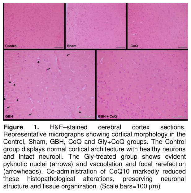
Histopathological evaluation of hematoxylin and eosin stainings revealed distinct morphological changes between the experimental groups (Figure 1). The cerebral cortex sections of the Control group showed normal histology defined by well-preserved neurons with clearly spesicific cell boundaries and centrally located nuclei. The neuropil was dense and homogeneous, with no vacuolatios, neuronal loss, or necrotic changes. Similarly Sham and CoQ groups had normal appearing neurons across the cerebral cortex. There were no prominent vacuolation and refraction in the neuropil area of these groups. In contrast, GBH treated group demonstrated significant neuropathological alterations. Neurons were seen as shrunken and hyperchromatic, displaying pyknotic nuclei (arrows), which is an indication of neuronal degeneration. The neuropil area showed prominent vacuole formations and focal rarefactions. There were many neurons displaying granulovacuolar changes (arrowheads). In the GBH + CoQ10 co-treated group, the cortical histology was relatively more preserved compared with the GBH group. Most neurons were seen morphologically normal with normal nuclei and minimal cytoplasmic condensations. The neuropil appeared largely uniform, with only occasional vacuolated regions.
Cresyl violet staining of the brain sections showed some histological differences among the experimental groups (Figure 2). In the Control, Sham and CoQ groups the cortical histology appeared normal, characterized by densely packed neurons with well-defined cell bodies, prominent Nissl substance, and centrally located nuclei. The neuropil seems intact and uniform with no evidence of neuronal loss or prominent degeneration. However the sections from the GBH group exhibited marked neurodegenerative changes. A significant reduction in neuronal density was detected and it is accompanied by extensive neuronal shrinkage and loss of Nissl substances. Also cytoplasmic paleness is noteworthy all over the cortical area. The neuropil appeared vacuolated and less compact. In GBH+CoQ group, neuronal density was higher than in the GBH group. Most of the neurons displayed preserved morphology and distinct Nissl granules. Only minimal degenerative alterations were detected.
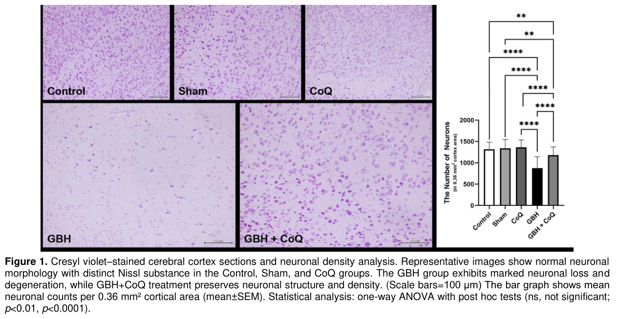
Quantitative Analysis of Neuronal Density: Cresyl Violet staining suggested a decrease in neuron numbers in the cortex region. Five photographs were taken from the five different cortex areas of the specimens and the numbers of neurons in each of them were counted. Quantitative comparasion of neuron numbers demonstrated significant differences among the groups (Figure 2, right panel). The GBH group showed a marked decrease in neuron number compared with the Control, Sham and CoQ groups (****p<0.0001). Co-treatment with CoQ10 significantly attenuated this decrease, with neuronal density in the GBH+CoQ group which is significantly much more with recpect to the GBH group (****p<0.0001). However it is not significantly different from the Control or CoQ groups (ns).
Discussion
According to this current study, subchronic oral exposure to a glyphosate-based herbicide (GBH) over a seven-week period results in structural changes to the cerebral cortex of adult male mice. The GBH group exhibited intense hyperchromatic neurons seen under the H&E and cresyl violet staining, with features such as shrunkening, pyknotic nuclei, and vacuolization of the neuropil, as well as a granulovacuolar degeneration with a notable decrease in the neuronal density. In contrast, animals that received co-treatment with coenzyme Q10 maintained a cortical architecture and cell density like the control, sham, and CoQ10 alone group with no neural loss, preserved Nissl substance, and only mild vacuolation. These findings suggest that CoQ10 provides substantial histological protection against GHB-induced cortical tissue damage.The current data support the expanding evidence on the neurotoxic effects of GBHs on the central nervous system. The effects of glyphosate on animal models has been examined in a number of studies. One of these studies reviewed scientific literature concerning glyphosate exposure effects on animals [22]. In the present review and in other related studies demonstrate a link between exposure to glyphosate and a greater degree of oxidative stress, neuroinflammation, and impaired function of mitochondria [6,8]. Along with molecular alterations, exposure to chronic or developmental GBH changes synaptic configurations and modifies the patterns of neuronal firing within the hippocampus and prefrontal cortex of rodents [23]. Moreover, other studies have demonstrated that exposure to Roundup can result in the development of neurotoxicity in the hippocampal region of immature rats, which is often accompanied by glutamatergic excitotoxicity and altered calcium regulation [24]. The long-term effects of cognitive changes, in addition to the neural alterations, indicate that the neurons located in the cortex are most likely susceptible to damage due to exposure to GBHs. In addition, exposure records have shown associations with behavioral and cognitive deficits, suggesting a potential correlation. Several studies have documented learning and memory impairments, social interaction deficits, and anxiety-like behavioral responses in rodents exposed to GBHs [9,12]. CoQ10 plays important roles both as an essential component of the mitochondrial electron transport chain and as an important lipid-soluble antioxidant of biological membranes [25]. Oral supplementation of Coenzyme Q10 has been reported to produce its higher concentration in the brain. This demonstrates the capacity of Coenzyme Q10 to cross the blood-brain barrier [26]. According to research, mitigated oxidative stress and impaired mitochondrial function in neurodegenerative disorders such as Parkinson's, Huntington's and Alzheimer's can be neuroprotectively altered by CoQ10, as evidenced by data from both preclinical and clinical studies. The neuroprotective effects of CoQ10 supplementation have been demonstrated in various models of neurodegeneration and ischemic injury, indicating the preservation of tissue architecture and a decrease in neuronal loss [27]. The results of the present study regarding neuronal morphology, Nissl substance, and the number of cortical neurons in the GBH + CoQ10 group align with previous studies. Our findings align with other studies showing CoQ10 protection of extra-neural tissues exposed to GBHs. For instance, Mutluay et al., [19] demonstrated that Glyphosate-based herbicide damaged testicular function and sperm quality by increasing oxidative stress and inflammatory signaling while suppressing antioxidant defenses. Coenzyme Q10 co-treatment largely reversed these changes, improving sperm parameters and restoring antioxidant and anti-inflammatory balance. Similarly, Nyariki et al.,[20] showed that CoQ10 is able to mitigate oxidative stress and neuroinflammation from experimental cerebral malaria. This study further adds to the evidence that CoQ10 performs a moderating ability on neuroinflammation and oxidative stress in the central nervous system. Together, these results support the antioxidant and mitochondrial regulatory properties of CoQ10, suggesting their potency despite long-term GBH exposure.
In conclusion, our work supports the view that mice exposed to GBH at a dose below the regulatory threshold for NOAEL and throughout the sub-chronic term had sustained clinically relevant histopathological changes in the cerebral cortex. It was shown that mitochondrial dysfunction and oxidative stress could be the protective pathway as the preserved neurons were also positively correlated to the CoQ10 administered that almost entirely prevented the damage. Furthermore, CoQ10 exhibits mitochondrial and antioxidant effects that make it a promising candidate for reducing these adverse effects.
Study Limitations
There are a few limitations to consider in this study. In the given experiments, only male mice were used, and therefore, were not able to assess any potential sex differences in the responses to the herbicide. Additionally, we only used one dose during a sub-chronic time period, rather than examining multiple doses and/or long-term chronic exposure.
References
- Martins-Gomes C, Silva TL, Andreani T, Silva AM. Glyphosate vs. glyphosate-based herbicides exposure: A review on their toxicity. J Xenobiot 2022;12(1):21-40.
- Costas-Ferreira C, Durán R, Faro LRF. Toxic effects of glyphosate on the nervous system: A systematic review. Int J Mol Sci 2022;23(9):4605.
- Martinez A, Al-Ahmad AJ. Effects of glyphosate and aminomethylphosphonic acid on an isogeneic model of the human blood-brain barrier. Toxicol Lett 2019;304:39-49.
- Bartholomew SK, Winslow W, Sharma R, et al. Glyphosate exposure exacerbates neuroinflammation and Alzheimer's disease-like pathology despite a 6-month recovery period in mice. J Neuroinflammation 2024;21(1):316.
- Winstone JK, Pathak KV, Winslow W, et al. Correction: Glyphosate infiltrates the brain and increases pro-inflammatory cytokine TNFα: implications for neurodegenerative disorders. J Neuroinflammation. 2024;21(1):20. https://doi.org/10.1186/s12974-023-02990-9 Erratum for: J Neuroinflammation. 2022 Jul 28;19(1):193.
- Astiz M, de Alaniz MJ, Marra CA. Effect of pesticides on cell survival in liver and brain rat tissues. Ecotoxicol Environ Saf 2009;72(7):2025-2032.
- Gallegos CE, Bartos M, Gumilar F, et al. Intranasal glyphosate-based herbicide administration alters the redox balance and the cholinergic system in the mouse brain. Neurotoxicology 2020;77:205-215.
- Strilbyska OM, Tsiumpala SA, Kozachyshyn II, et al. The effects of low-toxic herbicide Roundup and glyphosate on mitochondria. EXCLI J 2022; 21:183-196.
- Bali YA, Kaikai NE, Ba-M'hamed S, et al. Learning and memory impairments associated to acetylcholinesterase inhibition and oxidative stress following glyphosate based-herbicide exposure in mice. Toxicology 2019;415:18-25.
- Ait Bali Y, Ba-Mhamed S, Bennis M. Behavioral and Immunohistochemical Study of the Effects of Subchronic and Chronic Exposure to Glyphosate in Mice. Front Behav Neurosci 2017;8(11):146.
- Ait Bali Y, Kaikai NE, Ba-M'hamed S, et al. Anxiety and gene expression enhancement in mice exposed to glyphosate-based herbicide. Toxics 2022;10(5):226.
- Ait Bali Y, Madougou FA, Ba-M'hamed S, et al. Glyphosate-based herbicide exposure affects cognitive flexibility and social cognition in adult mice. Neurosci Lett 2024;837:137912.
- Guerra RM, Pagliarini DJ. Coenzyme Q biochemistry and biosynthesis. Trends Biochem Sci 2023;48(5):463-476.
- Wang Y, Lilienfeldt N, Hekimi S. Understanding coenzyme Q. Physiol Rev 2024;104(4): 1533-1610.
- Salama M, Yuan TF, Machado S, et al. Co-enzyme Q10 to treat neurological disorders: basic mechanisms, clinical outcomes, and future research direction. CNS Neurol Disord Drug Targets 2013;(5):641-664.
- Duberley KE, Heales SJ, Abramov AY, et al. Effect of Coenzyme Q10 supplementation on mitochondrial electron transport chain activity and mitochondrial oxidative stress in Coenzyme Q10 deficient human neuronal cells. Int J Biochem Cell Biol 2014;50:60-63.
- Bagheri S, Haddadi R, Saki S, et al. Neuroprotective effects of coenzyme Q10 on neurological diseases: A review article. Front Neurosci 2023;17:1188839.
- U.S. Environmental Protection Agency Reregistration Eligibility Decision (RED): Glyphosate, 1993; EPA-738-R-93–014.U.S. Government Printing Office, Washington, DC. (Accessed on 26 Nov 2025)
- Mutluay D, Özbek KÇ, Tenekeci GY, et al. Coenzyme Q10 protects against glyphosate-based herbicide-induced testicular and sperm toxicity by modulating Nrf2/Keap1/HO-1 and TLR4/NF-κB pathways. Naunyn Schmiedebergs Arch Pharmacol. 2025.
- Nyariki JN, Ochola LA, Jillani NE, et al. Oral administration of Coenzyme Q10 protects mice against oxidative stress and neuro-inflammation during experimental cerebral malaria. Parasitol Int 2019;71:106-120.
- Bankhead P, Loughrey MB, Fernández JA, et al. QuPath: Open source software for digital pathology image analysis. Sci Rep 2017;7(1):16878.
- Moser VC, Morris-Schaffer K, Richardson JR, et al. Glyphosate and neurological outcomes: A systematic literature review of animal studies. J Toxicol Environ Health B Crit Rev 2022;25(4):162-209.
- Solomon J, Gutierrez-Reyes CD, Chávez-Reyes J, et al. Neuroglycome alterations of hippocampus and prefrontal cortex of juvenile rats chronically exposed to glyphosate-based herbicide. Front Neurosci 2024;18:1442772.
- Cattani D, de Liz Oliveira Cavalli VL, Heinz Rieg CE, et al. Mechanisms underlying the neurotoxicity induced by glyphosate-based herbicide in immature rat hippocampus: Involvement of glutamate excitotoxicity. Toxicology 2014;320:34-45.
- Ebrahimi A, Kamyab A, Hosseini S, et al. Involvement of Coenzyme Q10 in Various Neurodegenerative and Psychiatric Diseases. Biochem Res Int 2023:5510874.
- Matthews RT, Yang L, Browne S, et al. Coenzyme Q10 administration increases brain mitochondrial concentrations and exerts neuroprotective effects. Proc Natl Acad Sci USA 1998;95(15):8892-8897.
- Fakharaldeen ZA, Al-Mudhafar A, Gany SN, et al. Neuroprotective effects of Coenzyme Q10 in ischemia-reperfusion injury via inflammation and oxidative stress reduction in adult male rats. J Med Life 2023;16(10):1534-1539.