
Erkek ve Dişi Ratlarda Kafatası Kemiklerindeki Morfometrik ve Mineral Yapı Değişikliklerinin Yaşamın Farklı Periyotlarındaki Analizi
Volume 40, Issue 3
November 2026 · Pages 253-260
Erkek ve Dişi Ratlarda Kafatası Kemiklerindeki Morfometrik ve Mineral Yapı Değişikliklerinin Yaşamın Farklı Periyotlarındaki Analizi
Analysis of Morphometric and Mineral Structure Changes in Skull Bones of Male and Female Rats at Different Periods of Life
Yücehan YILMAZ1, Yeşim ASLAN KANMAZ2, Saime Betül BAYGELDİ2, Meryem KARAN2
2Fırat University, Faculty of Veterinary Medicine, Department of Anatomy, Elazığ, TÜRKİYE
Amaç: Çalışma, dişi ve erkek ratların ergenlik öncesi, ergenlik- erişkinlik arası, genç erişkinlik dönemi ve ileri (yaşlı) erişkinlik dönemlerinde kafatası (yassı) kemiklerindeki morfometrik ve mineral değişim oranlarını ve bunlara cinsiyetin bir etkisinin olup olmadığını ortaya koymak amacıyla yapılmıştır.
Gereç ve Yöntem: Bu çalışmada 40 Wistar Albino rat (20 erkek, 20 dişi) Kullanıldı. Çalışmamızda ergenlik öncesi, ergenlik-erişkinlik arası, genç erişkin ve yaşlı erişkin olarak 4 grup oluşturuldu ve her grupta 5 dişi 5 erkek hayvan kullanıldı. Kemik mineral düzeyleri için X-ray floresan (XRF) ölçümleri yapıldı. Kafatasının metrik ölçümleri ise dijital kumpas ile; 15 adet ölçüm noktası, mandibula için ise 5 osteometrik ölçüm noktası baz alınarak yapıldı. İstatistiksel analizinde IBM SPSS Statistics Versiyon 22.0 paket programı kullanıldı.
Bulgular: Ca/P oranı yaşamın her periyodunda dişilerde erkeklerden daha yüksekti. Ca/P oranı en yüksek erkeklerde 1. Grupta, dişilerde ise 2. Grupta görüldü. Ca/Poranı gruplar ve cinsiyetler arasında istatistiksel olarak anlamlıydı (p<0.001). Znoranı her grupta da dişilerde erkeklere göre daha yüksekti, fakat 1. Gruptan 4. Grubakadar yaş ilerledikçe oranın düştüğü gözlendi. Sıçan kafatasında cinsiyetler arasındaMg, Al, Si, P, Cl, K, Ca, ve Zn (p<0.001) arasında anlamlı farklar bulundu. Kafatasıbazı ölçümlerinde (LD, BL, CBL, GBOC, BS, OZB, GBN, PB) 2. grupta (ergenlik-erişkinlik arası) dişi ratların daha yüksek değerlere sahip olduğu görüldü.
Sonuç: Sonuç olarak, rat kafatası kemiklerindeki morfometrik ölçümler ve mineral yapı değişiklikleri ergenlik ve yetişkinlik dönemleri temel alınarak her iki cinsiyette ortaya konuldu. Kafatası kemiklerindeki morfometrik ve mineral düzey farklılıklarınayaş periyotlarının ve cinsiyet faktörünün etkili olduğu belirlendi.
Objective: The study was conducted to determine the rates of morphometric and mineral changes in the skull (flat) bones of female and male rats during the pre-adolescent, adolescent-adulthood, young adult, and older adult stages, and toinvestigate whether gender has an effect on these changes.
Materials and Methods: In this study, 40 Wistar Albino rats (20 male, 20 female) wereused. Four groups were formed with 5 female 5 male animals in each group. X-ray fluorescence (XRF) measurements were performed to determine bone mineral levels. Metricmeasurements of the skull were taken using a digital caliper, based on 15 measurement points, while 5 osteometric measurement points were used for themandible. IBM SPSS Statistics Version 22.0 software was used for statisticalanalysis.
Results: The Ca/P ratio was statistically significant between groups and sexes (p<0.001). The Zn ratio was higher in females than males in all groups, but it wasobserved to decrease with age from group 1 to group 4. significant differences werefound between sexes in Mg, Al, Si, P, Cl, K, Ca, and Zn in rat skulls (p<0.001). Insome skull measurements (LD, BL, CBL, GBOC, BS, OZB, GBN, PB), female rats inGroup 2 (between adolescence and adulthood) were found to have higher values.
Conclusion: In conclusion, changes in morphometric measurements and mineral composition of the skull bones of rats of both sexes were revealed depending on adolescence and adulthood. It was determined that age and sex factors had an influence on these.
Introduction
The Wistar Albino rat is a species of white-furred rat belonging to the Muridae family of the Rodentia order. It is the most common laboratory animal used in scientific studies. Therefore, it has an important place in medical and biological scientific research [1]. Bones are composed of organic (30-40%) and inorganic (60-70%) substances. Inorganic substances provide hardness and strength to the bone and constitute the majority of bone tissue. Organic substances, on the other hand, provide elasticity to the bone. With age, the amount of organic matter in the bone decreases, while the amount of inorganic matter increases [2]. Organic matter consists mainly of proteins (type I collagen). Calcium and phosphorus form calcium hydroxyapatite crystals and constitute the majority of the inorganic matter. Magnesium, sodium, potassium, and fluoride are also components of bone [3,4]. Calcium is responsible for stimulating bone formation and preventing osteoporosis [5]. Bone structure and microarchitecture are fundamental elements for evaluating bone strength and the mechanical properties of bone. Bone contains 26 elements in varying proportions [6]. However, the minerals in bone structure are found in different proportions in each bone of the skeletal system [7,8]. These proportions can vary depending on age, sex, skeletal area, and animal species [6,7,9]. Mineralized tissues such as bones, teeth, and horns are primary storage sites in animals. These tissues contain major elements such as calcium (Ca), phosphorus (P), magnesium (Mg), and sulfur (S), as well as trace elements such as iron (Fe), zinc (Zn), and manganese (Mn). Much of the relevant research has focused particularly on essential elements such as Ca, P, and Mg, as they play a critical role in bone metabolism [10].X-ray fluorescence (XRF) is an analytical technique used to determine the chemical composition of various materials. These materials can be in solid, liquid, powder, filtered, or other forms. XRF analysis is also quite useful in pharmaceutical and health research [11]. XRF has been a frequently used method in bioarchaeological studies since the 1980s [12,13]. Furthermore, it has been shown that XRF elemental analysis can be used to identify or distinguish animal species [8].
With recent advancements in the technique, new applications are determining the isotopic accumulation of elements such as hydrogen (H), oxygen (O), and strontium (Sr) in bones and teeth [14]. The technique provides information on the major components of various biological samples, including bones [8,15], teeth [8,16], and horns [17].
In recent years, the application of morphometric techniques in the biomedical field has increased and has been accepted as a new approach in morphological studies [18]. Morphometric studies of the skull, in particular, are very important in terms of taxonomy, intra-species and interspecies comparative anatomy [19]. In rodent taxonomy, morphometric analysis of skulls has become one of the fundamental techniques [20-23]. Various morphometric studies of skulls have been performed on many domestic animals and rodents such as pigs [24], dogs, cats [25], guinea pig skulls [26], and African giant rats [27]. In recent years, geometric morphometric studies [28,29] have also become popular. Although studies on the mineral structure of long (medullary) bones in rats, based on adolescence and adulthood, have been conducted [30], this is the first time such studies have been performed on flat bones using the same criteria. This will also reveal whether there is a difference between medullary and flat bones based on age and sex.
The study was conducted to determine the rates of morphometric and mineral changes in the skull (flat) bones of female and male rats during the pre-adolescent, adolescent-adulthood, young adult, and older adult stages, and to investigate whether gender has an effect on these changes.
Material and Method
Research and Publication Ethics: This research ethics was obtained from the Firat University Local Ethics Committee for Animal Experiments (first approved no and date 2021/20 and 08.12.2021. Revised approved no. 2026/08-12, revised date 01/04/2026).In this study, 40 (20 male, 20 female) Wistar Albino rats (Rattus norvegicus) obtained from the Elazığ Veterinary Control Institute of the Ministry of Agriculture and Forestry of the Republic of Turkey were used.
In our study, 4 groups were formed as pre-adolescent (1-1.5 months) , adolescent –adulthood (3-4 months), young adult (8months) and older adult (after 20 months), 5 males and 5 females animals were used in each group. The formation of the groups was based on the reports in the literatures [31,32]. Following euthanasia performed at the Fırat University Experimental Research Center, animal bones were dissected and macerated (by boiling). Skull bones from all groups were separated and metric measurements were taken using a digital caliper.
Metric measurements of the skull were made using a digital caliper based on 15 measurement points (TL; Total length, CBL; Condylobasal length, BL; Basal length, DL; Dental length, GLN; Greatest length of the nasals, PL; Parietal length, FL Frontal length, VL Viscerocranium length, GBOC Greatest breadth of the occipital condyles, GNB; Greatest neurocranium breadth, GNL; Greatest neurocranium length, BS; Breadth of skull, OZB; Oral zygomatic breadth, GBN; Greatest breadth of the nasals, PB; Palatal breadth). For the mandible, 5 osteometric measurement points were used (LA; Length from angle, LCR; Length of the cheektooth row, LD; Length of the diastema, L; Length, HVR; Height of the vertical ramus) [33].
For X-ray fluorescence (XRF) measurements, scanning and mapping were performed in 70 µm diameter areas using a PANALYTICAL brand AXIOS model XRF device located at the Bilecik Şeyh Edebali University Central Research Laboratory Application and Research Center (MARAL). Samples were ground to homogenize using the loose powder method for evaluation. They were filled into 4-micron mylar film-coated sample containers and analyzed using the Omnian semi-quantitative analysis program on a 2.4 kW wavelength XRF device [34]. In the XRF analysis, the percentage concentrations of the elements Mg (MgO), Al (Al2O3), Si (SiO2), P (P2O5), Cl (Cl), K (K2O), Ca (CaO), Fe (Fe2O3), Ni (NiO), Zn (ZnO), and Sr (SrO) were determined.
Statistical analysis of the data obtained at the end of the study was performed using the IBM SPSS Statistics Version 22.0 software package. The Shapiro-Wilk test was used to test whether the assumption of normal distribution of continuous measures was met. For cases where significant comparisons were found, Bonferroni, Scheffe, and Tamhane tests were used to determine whether the within-group variances were homogeneous in pairwise comparisons of groups, provided the assumptions were met.
In cases with more than one group variable, two-way analysis of variance was used for general comparisons of continuous measures. The statistical significance level was set at 0.05 for all tests [35].
Results
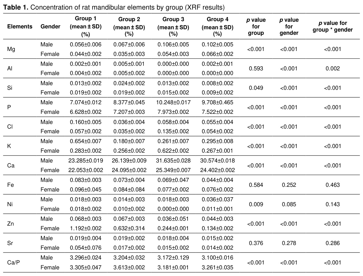
The Ca/P ratio was higher in females than in males at every stage of life. The highest Ca/P ratio was observed in Group 1 in males and in Group 2 in females. The Zn ratio was higher in females than in males in all groups, but it was observed to decrease with age from Group 1 to Group 4 (Table 1).In rat skulls, significant differences were found between genders in Mg, Al, Si, P, Cl, K, Ca, and Zn (p<0.001). Significant differences were also found between groups and genders in Mg, Si, P, Cl, K, Ca, and Zn (p<0.001). Similarly, the Ca/P ratio was statistically significant between groups and genders (p<0.001).
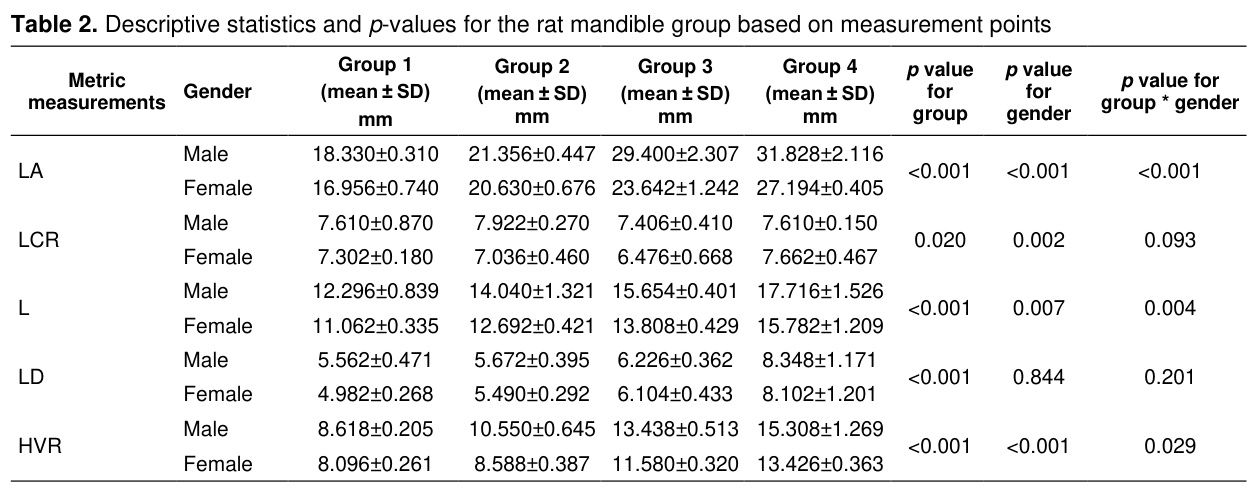
Descriptive statistics and p-values for the rat animal group according to measurement points are given in Table 2. Accordingly, for the LA, L, and HVR measurement points, the group, sex, and group change within sex are statistically significant (p<0.050). That is, developmentally (from group 1 to group 4), the average change is increasing in both females and males. The average measurement value of male rats is higher than that of females. In particular, while no significant difference was observed between females and males in mandibular morphometric measurements until adulthood (group 3), the difference widened in favor of male rats from group 3 onwards. Furthermore, the developmental change in the male animal group differs significantly from that in the female animal group, and this difference stems from the higher mean of males. For the LCR measurement point, both group and sex changes are significant (p<0.050). For the LD variable, only group change is highly significant (p<0.001).
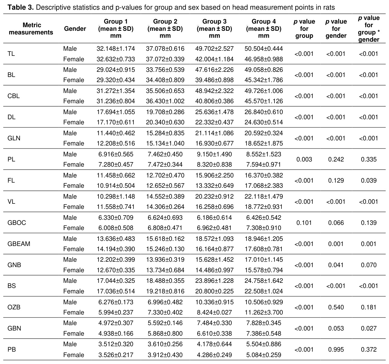
According to Table 3, the measurement points TL, BL, CBL, DL, GLN, VL, and BS are statistically highly significant (p<0.001) in terms of group, gender, and gender-based group variation. Considering all measurement points for these variables, the average developmental change (from group 1 to group 4) is increasing in both females and males.
Furthermore, while the mean developmental change was similar for male and female rats in groups 1 and 2, male rats in groups 3 and 4 showed a higher mean than female rats. The PL variable showed a significant difference only between groups (p= 0.003). In the GBN variable, the findings showed that male animals had a higher average value than females, and this difference was statistically significant (p=0.041).
In some skull measurements (LD, BL, CBL, GBO, BS, OZB, GBN, PB), female rats in group 2 (between adolescence and adulthood) were found to have higher values. Morphometric measurements showed that while there was no significant difference in developmental changes between sexes in rats during the period up to puberty (group 1), Some parameters showed faster development in females during the puberty-adulthood period (group 2), and from adulthood onwards, there was a faster increase in skull bone size in male rats.
Discussion
The results presented in this study provide valuable information regarding the morphometric and XRF characteristics of the skull bones of rats of different age groups and genders.Although numerous morphometric and mineral structure studies have been conducted on the bones of laboratory animals, it is important to examine the effects of pre-puberty, puberty, and adulthood on bone development, and even how these effects differ between genders, by focusing on these specific life stages.
Regarding the effects of age and gender criteria on bone mineral levels; it has been reported that sex is a significant factor in bone element ratios [8,37].
However, it was reported that there was no difference between genders in Fe and Zn ratios, and only Fe and Zn elements were common in the 48 bones examined [6]. Ioannidou [37] reported that in sheep, males had denser bones, while in pigs, sex differences were age-dependent, with females showing higher density when they reached adulthood. Maciejewska et al. [38] also found that Zn and Sr were high in newborn rats and decreased with age, while Fe was slightly higher in 7-day-old rats compared to 14 and 28-day-old rats. Yılmaz et al. [30] reported that the levels of the main bone minerals, Ca and P, were higher in females until adulthood, and higher in males in adulthood; however, the elements Fe, Zn, and Sr were higher in young animalscompared to adults. In addition, a study conducted by Baygeldi et al. [26] on guinea pig skulls reported that Zn and Sr elements were higher in young animals, with a proportional decrease observed as age progressed. Yılmaz et al. [36] found that the Zn ratio in guinea pig femur bone was significantly higher in females than in males throughout all life periods.
In our study, no significant differences in Fe and Sr levels were observed based on sex or age; however, Zn levels were found to be higher in females than in males at every life stage, though they decreased proportionally with advancing age in both sexes. Similarly, while it has been reported that Fe, Zn, and Sr elements are higher in young animals than adults in the long bones of the same animals [30] , in this study this characteristic was only observed for Zn mineral in the flat bones of the same animals.
Buddhachat et al. [7] examined the mineral content of humerus bones in 14 different species (cat, dog, elephant, horse, human, monkey, dolphin, hyena, lion, tapir, pig, sheep, buffalo, and tiger) using XRF analysis; they reported that the average humerus bone Ca percentages ranged from 16.083 (pigs) to 24.727 (horses), and the P percentages ranged from 3.917 (pigs) to 11.448 (cats). Nganvongpanit et al. [6] reported Ca as 21.05% and P as 8.05% in dogs.
Furthermore, Nganvongpanit et al. [8] reported Ca levels as 23.303%, 21.872%, and 21.512% in humans, elephants, and dogs, respectively, and P levels as 10.024%, 8.151%, and 8.885%. Yılmaz et al. [30] found the average percentage of Ca in the femur bone of adult rats to be 25.555 in males and 24.745 in females; they also found the average percentage of P to be 7.672 in males and 7.044 in females.
According to the results of this study, the percentage of calcium (Ca) in skull bones was determined to be 30.574% in adult males and 24.402% in adult females, while the percentage of phosphorus (P) was found to be 9.708% in adult males and 7.522% in adult females. These results show ratios consistent with literature reports. However, unlike the report given for long bones in the same subjects [30], the ratios of Ca and P in male skull bones appear to be higher than in long bones.
Yılmaz et al. [30] reported that calcium levels in the femurs of rats increased rapidly in females during puberty (group 2) and decreased rapidly in young adults (group 3), while the greatest increase in males was observed during young adults (group 3). In this study, it was observed that the Ca ratio reached its highest value in young adults (group 3) for both genders.
Although the Ca/P ratio in bones is not sufficient to definitively determine bone strength, it is an important parameter for osteoporosis [39,40]. Nganvongpanit et al. [6] found that among the 48 dog bones they examined, the highest Ca/P ratio was 4.09 in the patella, the lowest was 2.22 in the left carpal bone, and the average was 2.82. The Ca/P ratio has been reported as 2.21 in humans [41,42], 1.69 in cattle [9], 2.17 in pigs [43], 1.28 in rabbits, and 2.12 in rats [44]. On the other hand, Buddhachat et al. [7] found that the average Ca/P ratio in 14 types of humerus bones was 2.66. In addition, Legros et al. [9] reported a Ca/P ratio of 2.18 in rats. Yılmaz et al. [30] reported values of 3.33 in adult male rats and 3.51 in females. However, in a study conducted by Baygeldi et al. [26] on the skulls of guinea pigs, it was noted that the Ca/P ratio in the guinea pigs ranged from 3.085 to 3.428 throughout the developmental period. In this study, the ratio was determined to be 3.10 in adult male rats and 3.26 in female rats in rat skull bones. These results are similar to those reported by Yılmaz et al. [30] and Baygeldi et al. [26], and higher than other reports. These differences in values may be due to differences in measurement techniques.
Legros et al. [9] also reported that the Ca/P ratio is lower in young rats, increases with age, and has ratios of 1.51 at birth, 1.60 at 30 days of age, and 1.65 at 1 year of age in rats. Our study results differ from those presented in this report.
Some authors [45,46] have reported that, when examining the relationship between morphometric measurements of the bones of male and female rats, male rats exhibited higher measurements than females in both the control and experimental groups. Baygeldi et al. [26] reported that the measurement values of male guinea pigs were higher than those of female guinea pigs, that body development increased with age, and that gender change did not show a statistically significant difference for all measurement points (p>0.05). In the present study as well, morphometric measurements were found to be higher in males. Table 3 shows that the gender factor was significant (p<0.001) for the first five parameters.
In their study, Anbuhl et al. [47] examined the morphological development of the skull from birth to adulthood in 63 guinea pigs (Cavia porcellus) and noted that the skull grew significantly during the first 8 weeks of life. Baygeldi et al. [30] observed that the total skull length gradually increased from the first group to the fourth group. In this study, it was found that skull length (TL) in rats increased gradually with age in both sexes, but that this increase was more rapid in males as they reached adulthood.
Cox and Jeffery [48] reported total skull measurements of adult squirrels, guinea pigs, and rats as 48.22 mm, 57.51 mm, and 43.40 mm, respectively. Baygeldi et al. [26] found the total skull measurement in adult guinea pigs to be 62.564 mm in males and 63.072 mm in females. In this study, rat skull length (TL) was found to be 50.50 mm in elderly males and 46.95 mm in elderly females.
Olude and Olopade [49] reported maximum mandibular height and length in African giant rats as 2.13 cm and 3.62 cm, respectively. Furthermore, they reported that there was a statistically significant relationship (p<0.05) between the two sexes in all four morphometric parameters. In the present study as well, a statistically significant association with gender was observed for the LA and HVR parameters (p<0.001).
In conclusion, morphometric measurements and mineral composition changes in rat skull bones were revealed in both sexes based on adolescence and adulthood.
It was determined that age periods and gender factors influenced the differences in morphometric and mineral composition of the skull bones. We believe this study will be an important resource for future age-related bone structure change studies and drug development research.
References
- Harkness JE, Wagner JE. The Biology and Medicine of Rabbits and Rodents. 4 th. Edition USA: Williams and Wilkins, 1995.
- Dursun N. Veteriner Anatomi 1. 10th Edition, Medisan Yayınevi, 2006.
- Ehrlich PJ, Lanyon LE. Mechanical strain and bone cell function: A review. Osteoporos Int 2002;13(9):688-700.
- Sağlam M, Özer A, Aştı RN. Genel Histoloji. Yorum Matbaacılık Sanayii, 1997.
- Anderson JJB, SCG Garner. Dietary issues of calcium and phosphorus. Calcium and Phosphorus in Health and Disease 1996;123(9):7-25.
- Nganvongpanit K, Buddhachat K, Piboon P, Klinhom S. The distribution of elements in 48 canine compact bone types using handheld X-ray fluorescence. Biol Trace Elem Res 2016;174(1):93-104.
- Buddhachat K, Klinhom S, Siengdee P, et al. Elemental analysis of bone, teeth, horn and antler in different animal species using non-invasive handheld X-ray fluorescence. PLoS One 2016;11(5):e0155458.
- Nganvongpanit K, Buddhachat K, Klinhom S, et al. Determining comparative elemental profile using handheld X-ray fluorescence in humans, elephants, dogs, and dolphins: Preliminary study for species identification. Forensic Sci Int 2016;263:101-106.
- Legros R, Balmain N, Bonel G. Age-related changes in mineral of rat and bovine cortical bone. Calcif Tissue Int 1987; 41(3):137-144.
- Dermience M, Lognay G, Mathieu F, Goyens P. Effects of thirty elements on bone metabolism. J Trace Elem Med Biol 2015;32:86-106.
- Brouwer P. Theory of XRF, PANalytical BV. 3th Edition, Netherlands: Almelo, 2010.
- LL Klepinger. Nutritional Assessment From Bone. Annu Rev Anthropol 1984;13:75-96.
- Ambrose SH, Krigbaum J. Bone chemistry and bioarchaeology. J Anthropol Archaeol 2003;22(3):193-199.
- Reynard LM, Hedges REM. Stable hydrogen isotopes of bone collagen in palaeodietary and palaeoenvironmental reconstruction. J Archaeol Sci 2008;35:1934-1942.
- Gonzalez-Rodriguez J, Fowler GA. Study on the discrimination of human skeletons using X-ray fluorescence and chemometric tools in chemical anthropology. Forensic Sci Int 2013;231(1-3):407.
- Christensen AM, Smith MA, Thomas RM. Validation of X-ray fluorescence spectrometry for determining osseous or dental origin of unknown material. J Forensic Sci 2012;57(1):47-51.
- Kierdorf U, Stoffels D, Kierdorf H. Element concentrations and element ratios in antler and pedicle bone of yearling red deer (Cervus elaphus) stags-a quantitative X-ray fluorescence study. Biol Trace Elem Res 2014;162(1-3):124-133.
- Mukerjee B, Rajan T. Morphometric study of seminal vesicle of rat in normal health and stress conditions. J Anat Soc India 2006;55(1):31-36.
- Van der Merwe M. Discriminating between Thryonomys swinderianus and Thryonomys gregorianus. Afr Zool 2007;42(2):165-171.
- Olayemi A. Taxonomic Evaluation by Morphometric Analysis of Various Muroid Rodents from Parts of Southwestern Nigeria. PhD Thesis, Nigeria: Obafemi Awolowo University, 2007.
- Bezerra AM, de Oliveira JA. Taxonomic implications of cranial morphometric variation in the genus Clyomys Thomas, 1916 (Rodentia: Echimyidae). J Mammal 2010;91(1):260-272.
- Olayemi A, Nicolas V, Hulselmans JAN et al. Taxonomy of the African giant pouched rats (Nesomyidae: Cricetomys): Molecular and craniometric evidence support an unexpected high species diversity. Zool J Linn Soc 2012;165(3):700-719.
- Reisser CM, Marshall BA, Gardner JP. A morphometric approach supporting genetic results in the taxonomy of the New Zealand limpets of the Cellana strigilis complex (Mollusca: Patellogastropoda: Nacellidae). Invertebr Syst 2012;26(2):193-203.
- Endo H, Hayashi Y, Yamazaki K, et al. Geographical variation of mandible size and shape in the wild pig (Sus scrofa) from Taiwan and Japan. Zool Stud 2002;41(4), 452-460.
- Künzel W, Breit S, Oppel M. Morphometric investigations of breed‐specific features in feline skulls and considerations on their functional implications. Anat Histol Embryol 2003;32(4):218-223.
- Baygeldi SB, Yilmaz Y, Güzel BC, et al. Analysis and 3D imaging of morphometric-mineral structure changes in skulls of male and female guinea pigs during and after developmental period. Int J Morphol 2024;42:2.
- Olude MA, Olopade JO, Fatola IO, et al. Some aspects of the neurocraniometry of the African giant rat (Cricetomys gambianus Waterhouse). Folia Morphol 2009;68(4):224-227.
- Ağaç DK, B Onuk, O Gündemir, et al. Comparative cranial geometric morphometrics among wistar albino, sprague dawley, and WAG/Rij rat strains. Animals 2024;14:1274.
- Gürbüz İ, Demiraslan Y. A geometric morphometric study on the rat mandible. Anat Histol Embryol 2025;54(1):e70013.
- Yılmaz S, Baygeldi SB, Kanmaz YA, et al. Physical and chemical characterization of femur during and after body development period in male and female rats. Anat Histol Embryol 2023;52(4):538-545.
- Indrekvam K, Vikøyr I, Lie RT, et al. Age-related differences in chemical composition of rat femur as determinants for strain. Acta Orthop Scand 1991;62(5):455-458.
- Sengupta P. The laboratory rat: Relating its age with human's. Int J Prev Med 2013;4(6):624-630.
- Driesch, Von den A. A guide to the measurements of animal bones from archaeological sites. Bulletin: Peabody Museum, 1976:1-136.
- Beckhoff B, Kanngießer B, Langhoff N, Wedell R, Wolff H. Handbook of Practical X-ray Fluorescence Analysis. Springer Science & Business Media, 2006.
- Özdamar K. Paket programlar ile istatistiksel veri analizi-1: SPSS-MINITAB. İzmir: Kaan Kitabevi, 1999.
- Yılmaz Y, Kanmaz YA, Baygeldi SB, et al. Physical and chemical characterization of the femur during and after the body development period in male and female guinea pigs. Anim Sci J 2023;94(1): e13848.
- Ioannidou E. Taphonomy of animal bones: Species, sex, age and breed variability of sheep, cattle and pig bone density. J Archaeol Sci 2003;30(3):355-365.
- Maciejewska K, Drzazga Z, Kaszuba M. Role of trace elements (Zn, Sr, Fe) in bone development: Energy dispersive X-ray fluorescence study of rat bone and tooth tissue. BioFactors 2014;40(4):425-435.
- Fountos G, Kounadi E, Tzaphlidou M, et al. The effects of inflammation-mediated osteoporosis (IMO) on the skeletal Ca/P ratio and on the structure of rabbit bone and skin collegen. Appl Radiat Isot 1998;49(5-6):657-659.
- Fountos G, Tzaphlidou M, Kounadi E, Glaros D. In vivo measurement of radius calcium/phosphorus ratio by X-ray absorptiometry. Appl Radiat Isot 1999;51(3):273-278.
- Tzaphlidou M, Zaichick V Neutron activation analysis of calcium/phosphorus ratio in rib bone of healthy humans. Appl Radiat Isot 2002;57(6):779-783.
- Tzaphlidou M, Zaichick V. Calcium, phosphorus, calcium-phosphorus ratio in rib bone of healthy humans. Biol Trace Elem Res 2003;93(1):63-74.
- Dickerson JWT. The effect of development on the composition of a long bone of the pig, rat and fowl. Biochem J 1962;82(1):47.
- Tzaphlidou M, Speller R, Royle G, et al. High resolution Ca/P maps of bone architecture in 3D synchrotron radiation microtomographic images. Appl Radiat Isot 2005;62(4):569-575.
- Kara ME. Ratlarda Gelişme Döneminde Sigara İnhalasyonunun İskelet Sistemi Üzerine Etkisinin Morfometrik Yöntemlerle Belirlenmesi. Doktora Tezi, Istanbul Üniversitesi Sağlık Bilimleri Enstitüsü Anatomi Anabilim Dalı, 2002
- Miller JP, German RZ. Protein malnutrition affects the growth trajectories of the craniofacial skeleton in rats. The Journal of Nutrition 1999;129(11):2061-2069.
- Anbuhl KL, Benichoux V, Greene NT, et al. Development of the head, pinnae, and acoustical cues to sound location in a precocial species, the guinea pig (Cavia porcellus). Hear Res 2017;356:35-50.
- Cox PG, Jeffery N. Reviewing the morphology of the jaw-closing musculature in squirrels, rats, and guinea pigs with contrast-enhanced microCT. Anat Rec 2011;294(6):915.
- Olude MA, Olopade JO. Morphometric Studies of the Axial Skeleton of the African Giant Rat (Cricetomys gambianus,Waterhouse) Part (1): Skull Typology. J Vet Anat 2010;3(2):1-12.
- Localization and Surgical Treatment of Parathyroid Lesions
- PARATİROİD LEZYONLARININ LOKALİZASYONU VE CERRAHİ TEDAVİSİ
- Development of Skin During Prenatal and Postnatal Stages In Rats
- Effect Of Nıfedıpıne On Experımental Epılepsy Induced By Penıcıllın In Rat
- Use of Carnitine to Prevent Effectis of Ischemiae in Torsion-Induced Ovarian Ischaemiae