
Mandibula Ramus Bölgesinde Dev Kemik Kitlesi: Osteoma, Olgu Sunumu
Volume 40, Issue 3
November 2026 · Pages 303-307
Mandibula Ramus Bölgesinde Dev Kemik Kitlesi: Osteoma, Olgu Sunumu
Giant Bony Mass in the Mandibular Ramus Region: Osteoma, Case Report
Yakup ŞEN1, Sümeyye COŞGUN BAYBARS1
Çenelerde görülen osteomalar, genellikle belirti vermeksizin büyüdüğünden, erken tanı ve düzenli takip büyük önem taşımaktadır. Bu vakada, 41 yaşında erkek hasta, sol alt çenede birkaç aydır mevcut olan ağrısız şişlik şikayetiyle kliniğimize başvurmuştur. Panoramik radyografta, sol mandibulada sınırları belirgin, oval şekilli radyoopak bir lezyon izlenmiştir. Konik ışınlı bilgisayarlı tomografi (KIBT) incelemesinde, osteoma ile uyumlu, homojen yapıda ve radyoopak mantara benzer bir kitle gözlemlenmiştir. Lezyon, genel anestezi altında ekstraoral yaklaşım ile tamamen eksize edilmiştir ve histopatolojik inceleme sonucunda osteoma tanısı doğrulanmıştır. İyileşme süreci komplikasyonsuz ilerlemiş ve üç yıllık takip süresince nüks gözlenmemiştir.
Osteomas of the jawbones can grow without symptoms, making early diagnosis and regular monitoring crucial. In this case, a 41-year-old male patient presented to our clinic with a painless swelling in the left lower jaw that had been present for several months. Panoramic radiography revealed a well-defined, oval, radiopaque lesion in the left mandible. Cone beam computed tomography (CBCT) examination showed a uniform, pedunculated radiopaque mass consistent with an osteoma. The lesion was completely excised through an extraoral approach under general anesthesia, and the diagnosis of osteoma was confirmed by histopathological examination. The healing process was uneventful, and no recurrence was observed during the 3-year follow-up period
Introduction
Osteomas are slow-growing, benign osteogenic tumors that commonly affect the craniofacial region and, less frequently, the jawbones [1]. They are typically asymptomatic and can be classified as central or peripheral types [2,3]. Osteomas can occur at any age, but they are more frequently observed in patients aged 40 and above. Structurally, they may consist of compact, spongy, or compact-spongy bone combinations [2,4].Clinically, osteomas may present with symptoms such as joint pain, swelling, restricted mouth opening, difficulty swallowing, trismus, and hearing problems. Osteomas are more likely to develop in the mandible than in the maxilla due to the greater susceptibility of the mandible to tension and trauma, as it is the site of attachment for muscles and tendons. They are more commonly seen in the paranasal sinuses, particularly in the frontal and ethmoid sinuses, with sizes typically ranging from 10 to 40 mm. The treatment approach includes observation if the lesion does not cause problems, and surgical excision if it does [2].
This study presents a case of an osteoma located in the left mandibular ramus of a 41-year-old male patient, along with its clinical and radiological findings. Although such cases are relatively rare in the literature, existing reports emphasize the importance of early diagnosis and a multidisciplinary approach in management. The present case is particularly noteworthy due to the unusual localization of the lesion within the mandibular ramus, its considerable size, and its impact on facial symmetry. Furthermore, the preference for an extraoral surgical approach and the absence of recurrence during the follow-up period further highlight the clinical significance and distinctiveness of this case.
Case Presentation
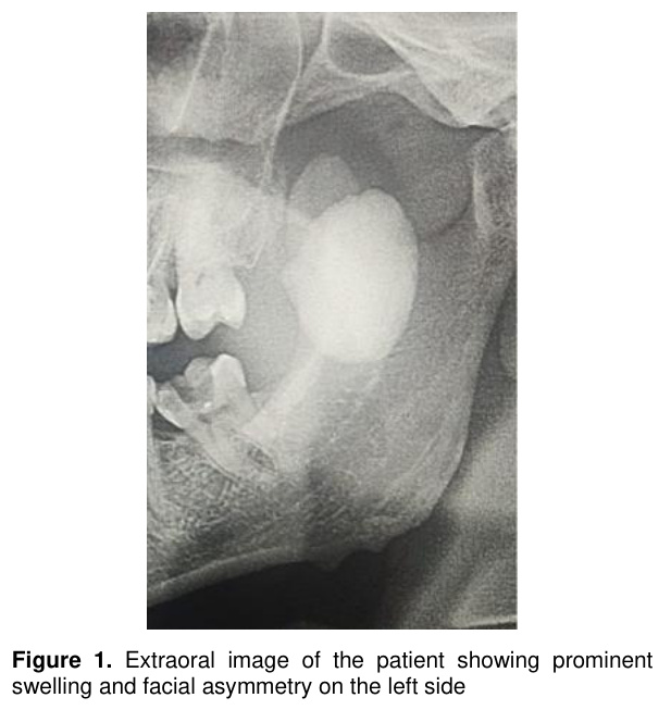
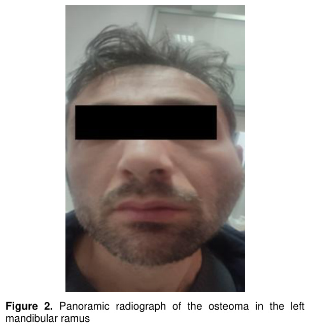
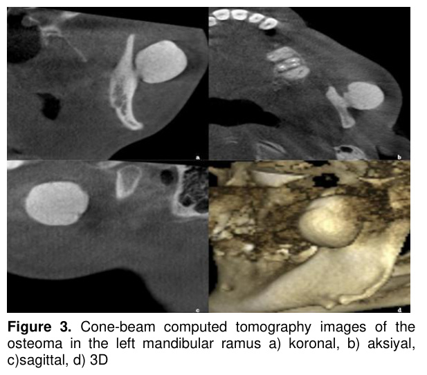
A 41-year-old male patient presented to our clinic in January 2022 with a complaint of facial asymmetry on the left side. The patient reported that the swelling had been present for several months without causing any pain. His medical history was unremarkable, with no systemic conditions and no current medication use. There was no history of infection, trauma, or tooth extraction. No obvious swelling or trismus was observed during the intraoral examination, while the extraoral examination revealed bone hardness and a prominent swelling in the posterior region of the left mandibula(Figure 1). A panoramic radiograph revealed a well-defined, oval radiopacity in the coronoid process region of the left ascending ramus (Figure 2). CBCT imaging was performed using a Planmeca ProMax 3D Mid device (Planmeca Oy, Helsinki, Finland). The scanning parameters were 90 kVp and 10 mA, with a voxel size of 0.2 mm and a field of view (FOV) of 100 × 100 mm. Images were evaluated in axial, sagittal, and coronal planes. Further tomographic evaluation demonstrated a well-defined pedunculated mass, measuring 25×24×22 mm, in the middle portion of the left mandibular ramus, extending laterally and superiorly. The mass was continuous with the bone cortex, as observed on coronal, sagittal, and axial (CxSxA) slices (Figure 3a–d).
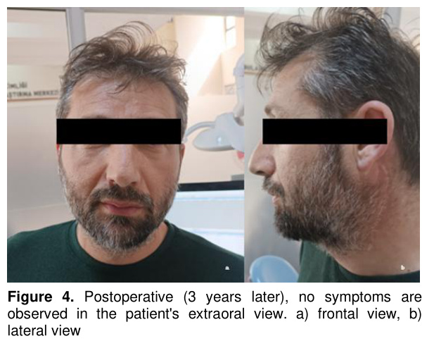
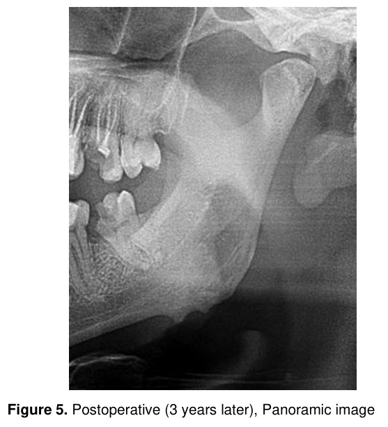
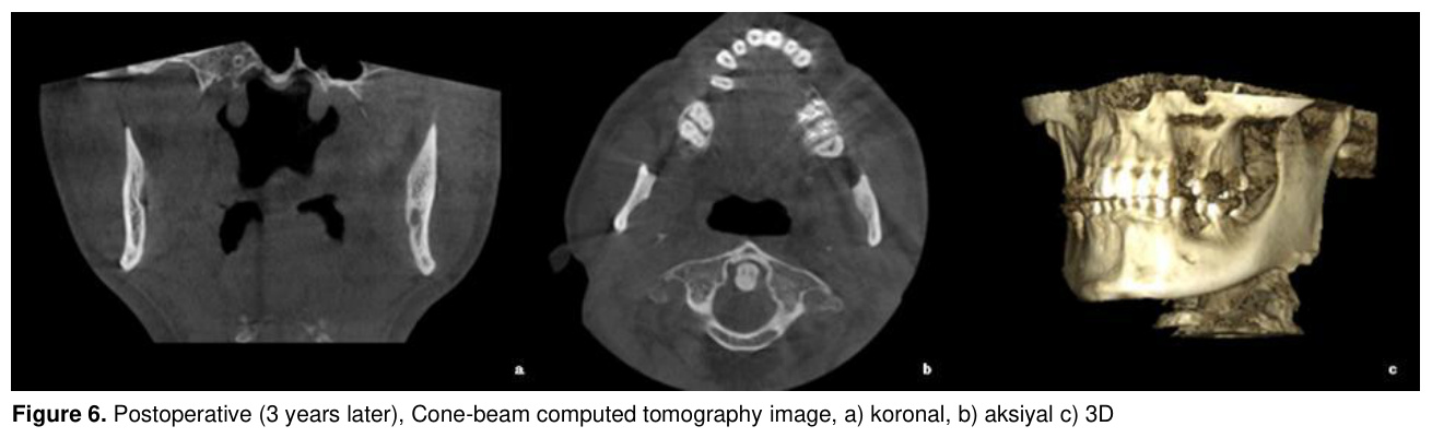
At the Fırat University Hospital ENT clinic, the lesion was completely removed via an extraoral approach under general anesthesia. An extraoral submandibular approach was performed to ensure adequate surgical access. The lesion was exposed, carefully dissected from surrounding tissues, and completely excised at its base. The surgical site was irrigated and closed in layers. The diagnosis of osteoma was confirmed by histopathological examination. The recovery period was uneventful, and no recurrence was observed during the 3-year follow-up. Clinical examination at follow-up demonstrated resolution of the facial asymmetry and no evidence of swelling (Figure 4a, b). Follow-up panoramic radiography showed no signs of recurrence (Figure 5), and CBCT images confirmed complete lesion removal without recurrent bone formation (Figure 6a–c). Informed consent was obtained from the patient.
Discussion
Osteoma is a benign osteogenic tumor composed of well-organized mature osteocytes. Although developmental, infectious, and traumatic causes have been suggested in its etiology, its pathophysiology has not been fully elucidated. The most likely mechanism involves stimulation of osteoblasts or remnants of Malassez cells, leading to the differentiation of osteoprogenitor cells and subsequent tumor formation. In the present case, no evidence of infection or trauma was identified, supporting a non-reactive origin [5].Osteomas are most commonly observed in the craniofacial region, particularly in the paranasal sinuses, orbit, external auditory canal, and temporal bone [6]. Johann et al. [7], in a study of 69 osteoma cases, reported that 6 originated from the maxilla and 63 from the mandible. Among mandibular cases, lesions were most frequently located in the corpus, condyle, and angle, whereas involvement of the mandibular ramus was relatively uncommon. This distribution highlights the rarity of ramus-localized osteomas and underscores the unusual localization observed in the present case. Furthermore, the mean age of occurrence (36.5 years) is consistent with our patient profile [7].
Consistent with previous studies, osteomas are more frequently found in the mandible than in the maxilla [8]. However, atypical localizations have also been reported, including the hard palate and paranasal sinuses [9,10]. These findings indicate that, although osteomas have predilection sites, they may also present in less common anatomical regions, as demonstrated in the current case.
Previous reports of mandibular ramus osteomas have described clinical findings such as facial asymmetry and extraoral swelling, similar to those observed in our patient [11]. However, in these cases, intraoral surgical approaches were generally sufficient for lesion removal. In contrast, the present case required an extraoral approach due to the lesion’s considerable size and anatomical localization, which limited intraoral access. Similarly, Bulut et al. [12] reported a large peripheral osteoma in the mandibular corpus with radiographic characteristics comparable to our case; however, the difference in localization influenced the surgical management strategy. In Nojima et al.'s [13] case involving the mandibular condyle, functional disturbances such as deviation and occlusal imbalance were observed, which resolved following surgical treatment. Kaplan et al. [14], in a 30-year literature review, identified a limited number of osteoma cases, further emphasizing the relative rarity of these lesions.
Aydoğdu et al. [15] reported three osteoma cases involving the condyle and temporal bone, with unclear etiology in most cases. Similarly, Gedük et al. [16] described a peripheral osteoma extending from the ramus to the condyle in a 60-year-old patient and emphasized that such lesions may remain asymptomatic for many years. In the present case, the lesion remained asymptomatic for several months and became clinically evident only after the development of facial asymmetry.
In cases of multiple osteomas, Gardner's syndrome should be considered, as it is associated with intestinal polyposis, desmoid tumors, fibromas, and epidermoid cysts. Given the malignant potential of intestinal polyposis, patients with multiple osteomas should be carefully evaluated. In the present case, only a solitary lesion was identified clinically and radiologically, and no findings suggestive of Gardner’s syndrome were observed [2,17].
Radiographic evaluation plays a critical role in the diagnosis of osteomas. Panoramic radiographs, CT, and CBCT are commonly used imaging modalities, with tomographic methods providing detailed information regarding lesion size, extent, and localization. Typically, osteomas appear as well-defined, oval or round radiopaque masses with unilateral involvement, as observed in the present case [18].
Surgical excision is accepted as the standard treatment for osteomas, and recurrence is rare; however, isolated cases of recurrence have been reported in the literature [19]. While conservative follow-up may be sufficient for asymptomatic lesions [4], surgical intervention is indicated in cases presenting with aesthetic or functional concerns. In the present case, surgical excision was performed due to facial asymmetry, the large size of the lesion, and its relationship with surrounding anatomical structures. Unlike smaller and more accessible lesions that can be managed intraorally, the size and localization of the lesion in the mandibular ramus limited intraoral access, necessitating an extraoral surgical approach. This approach provided improved surgical visibility and control, enabling complete excision while minimizing intraoperative complications.
In the differential diagnosis of osteoma, conditions such as exostosis, complex odontoma, osteoid osteoma, osteoblastoma, peripheral ossifying fibroma, and osteochondroma should be considered [1]. These lesions can be distinguished based on their clinical behavior, growth patterns, and radiographic characteristics. For example, exostoses typically cease growth after puberty, whereas osteoid osteomas and osteoblastomas are usually painful and exhibit more rapid growth. Complex odontomas appear more radiopaque and are surrounded by a thin radiolucent capsule [20], while peripheral ossifying fibromas originate from the periodontal ligament and osteochondromas are cartilage-capped exophytic lesions affecting the condyle or coronoid process [21,22].
Overall, this case contributes to the existing literature by emphasizing the rarity of osteomas located in the mandibular ramus, highlighting the impact of lesion size and localization on clinical presentation and surgical decision-making, and underscoring the importance of individualized treatment planning.
Osteomas may reach considerable sizes while remaining asymptomatic, particularly in uncommon locations such as the mandibular ramus. The present case is of clinical significance due to its atypical localization and notable size, highlighting the importance of careful radiographic evaluation and accurate differential diagnosis in panoramic imaging. Proper identification of such lesions is essential for appropriate surgical planning and to prevent unnecessary interventions. Additionally, patients diagnosed with osteoma should be evaluated for multiple lesions and possible association with Gardner’s syndrome.
References
- Molina T, Rodriguez LM, Virella BR, Gonzalez DM, Moncada UC. A case report of a giant mandibular osteoma. Oral Surg Oral Med Oral Pathol Oral Radiol 2024;138(2):e64.
- Geron ABG, Carvalho VA, Dos Santos JL, et al. Surgical management of traumatic peripheral osteoma of the mandible. J Cranio Surg 2017;28:405-408.
- White SC, Pharoah MJ. Benign tumors. In: White SC, Pharoah MJ, editors. Oral Radiology: Principles and Interpretation. 7th ed. New York, USA: Elsevier Mosby; 2014: 359-401.
- Starch-Jensen T. Peripheral solitary osteoma of the zygomatic arch: a case report and literature review. Open Dent J 2017;11:120-125.
- Güray G, Sümer P. Mandibular osteoma: bilgisayarlı tomografi bulguları ile olgu sunumu. Gaziosmanpaşa Univ J Med 2021;13(1):52-60.
- Garça MF, Miçooğulları ED, Bozan N, Çankaya H. Mandibula yerleşimli periferal osteom: Olgu sunumu. Haseki Med Bull 2012;50(1):30.
- Johann ACBR, De Freitas JB, De Aguiar MCF, De Araújo NS, Mesquita RA. Peripheral osteoma of the mandible: Case report and review of the literature. J Cranio-Maxillofac Surg 2005;33(4):276-281.
- Sayan NB, Üçok C, Karasu HA, Günhan Ö. Peripheral osteoma of the oral and maxillofacial region: A study of 35 new cases. J Oral Maxillofac Surg 2002;60(11):1299-1301.
- Cordeiro TD, Bruzinga FFB, Souza MS, et al. Atypical osteoma in the hard palate. Oral Surg Oral Med Oral Pathol Oral Radiol 2025;139(5):e50-e51.
- Johansyah LC, Irwansyah AC, Pieter NAL, et al Osteoma raksasa sinus maksilaris dan etmoidalis: kasus jarang. Jurnal Riset Multidisiplin 2024;2(3):111-116.
- Torres LHS, Costa Filho JRT, Uchôa CP, et al. Intrabuccal surgical management of a peripheral mandibular osteoma. J Craniofac Surg 2021;32(5):1952-1953.
- Bulut E, Acikgoz A, Ozan B, Gunhan O. Large peripheral osteoma of the mandible: A case report. Int J Dent 2010;2010:123456.
- Nojima K, Niizuma-Kosaka F, Nishii Y, et al. Multidisciplinary treatment of peripheral osteoma arising from mandibular condyle in patient presenting with facial asymmetry. Bull Tokyo Dent Coll 2014;55(1):39-47.
- Kaplan I, Calderon S, Buchner A. A peripheral osteoma of the mandible: a study of 10 new cases and analysis of the literature. J Oral Maxillofac Surg 1994;52(4):467-470.
- Aydoğdu M, Misirlioğlu M, Adişen MZ, Meşhur Z, Ertaş G. Temporomandibular eklem bölgesinde görülen üç osteoma vakası. Turkiye Klin J Dent Sci 2021;27(3):504-508.
- Gedük G, Zengin AZ, Sümer P. Kondil ve ramusta periferal osteoma: vaka sunumu. Atatürk Univ J Dent Sci 2015;26(4):1-3.
- Olivares CM, Francisco PL, Claudio HM, Francisco PH. Multiple mandibular osteomas not associated with Gardner syndrome: Case report and literature review. Res Rep Oral Maxillofac Surg 2020;4(2):38.
- Küçükkurt S, Özle M, Barış E. Peripheral osteoma in an unusual location on the mandible. BMJ Case Rep 2016;2016:bcr2016216554.
- Lowell B. Recurrent peripheral osteoma of mandible: Report of case. J Oral Surg 1971;21(6):446-450.
- .Demirbaş AE, Kaba YN, Akkoyun EF, Alkan A, Amuk M. Mandibula angulusunda soliter periferal osteoma: Nadir bir olgu. EÜ Dişhek Fak Derg 2018;39(2):118-121.
- Çiçek Y, Kilitci A, Hakkı A. Periferal ossifiye fibrom. Turkiye Klin J Int Med Sci 2008;4(1):25.
- Yavuz MS, Büyükkurt MC, Bayramoğlu ZS, Velioğlu Ö, Gürsan N. A conservative surgical approach to osteochondroma of the temporomandibular joint. Atatürk Univ J Dent Sci 2016;27(3):18-22.
- Eksizyonel Biyopsi Sonucu Pilomatrikoma Olan Olguların Retrospektif Değerlendirilmesi
- Severe Soft Tissue Inflamation Caused by Cervical Osteoid Osteoma: A Rare Case Report
- Servikal Osteoid Osteoma Nedeniyle Oluşan Şiddetli Yumuşak Doku İnflamasyonu: Nadir Bir Olgu Sunumu
- Tedavi Gereksinimi Duyulan Antral Retansiyon Kistlerine Multidisipliner Yaklaşım- Vaka Serisi
- Türkiye Nüfusundaki Bireylerin Tüm Diş Gruplarında C-Şekilli Kanal Görülme Sıklığının ve Tiplerinin Konik Işınlı Bilgisayarlı Tomografi Kullanılarak Belirlenmesi