
Alendronatın Rat Tibialarında Titanyum İmplantların Osseointegrasyona Etkisinin Biyomekanik İncelenmesi
Volume 40, Issue 3
November 2026 · Pages 232-238
Alendronatın Rat Tibialarında Titanyum İmplantların Osseointegrasyona Etkisinin Biyomekanik İncelenmesi
Biomechanical Investigation of the Effect of Alendronate on Osseointegration of Titanium Implants in Rat Tibias
Mehmet GÜL1, Delil DOĞAN2, Murat TANRISEVER3, Ümit Koray CAN4, Erhan Cahit ÖZCAN5, Özmen İSTEK6, Merve GÜNEY2, Serkan DÜNDAR2
2Fırat University, Faculty of Dentistry, Department of Periodontology, Elazığ, TÜRKİYE
3Fırat University, Faculty of Veterinary Medicine, Department of Surgical, Elazığ, TÜRKİYE
4Türkiye Jokey Klubü Elazığ Hipodromu, At Hastanesi, Elazığ, TÜRKİYE
5Fırat University, Faculty of Medical School, Department of Plastic, Reconstructive and Aesthetic Surgery, Elazığ, TÜRKİYE
6Muş Alparslan University, Faculty of Health Sciences Department of Nursing, Muş, TÜRKİYE
Amaç: Bu çalışmanın amacı, alendronatın peri-implant kemik oluşumunu destekleme, osteoklastogenezi inhibe etme ve ratlarda ağırlık taşıyan bir implant modelinde osseointegrasyonu artırma potansiyelinin biyomekanik olarak değerlendirilmesidir.
Gereç ve Yöntem: Bu amaç doğrultusunda denekler; kontrol grubu (n=8), alendronat doz 1 grubu (n=8) ve alendronat doz 2 grubu (n=8) olmak üzere üç gruba ayrıldı. Bu deneysel çalışmaya toplam 24 Sprague–Dawley rat dahil edildi. Tüm gruplarda ratların sağ tibia metafiz bölgesindeki kortikokansellöz kemik yapısına 2.5 mm çapında ve 4 mm uzunluğunda tornalanmış yüzeyli titanyum implantlar cerrahi olarak yerleştirildi. Kontrol grubuna herhangi bir ilaç uygulanmazken, deney gruplarına 8 hafta boyunca haftada bir kez intraperitoneal yoldan 1 mg/kg ve 2 mg/kg dozlarında alendronat uygulandı. Sekiz haftalık deneysel sürecin sonunda tüm denekler sakrifiye edilerek implantların mekanik stabiliteleri tersine tork yöntemiyle değerlendirildi. Veriler Kruskal–Wallis ve Mann–Whitney U testi ile analiz edildi.
Bulgular: Elde edilen sonuçlara göre, ortalama tersine tork değerleri kontrol grubunda 6.32 N/cm, alendronat doz 1 grubunda 8.41 N/cm ve alendronat doz 2 grubunda 8.21 N/cm olarak ölçüldü. Alendronat uygulanan her iki grupta da kontrol grubuna göre implant stabilitesinin istatistiksel olarak anlamlı düzeyde arttığı saptandı (p=0.001 ve p=0.003). İki alendronat dozu arasında anlamlı fark bulunmadı (p>0.05).
Sonuç: Bu çalışmanın sınırlı sonuçlarından yola çıkarak sistemik alendronat uygulaması, titanyum implantların osseointegrasyon düzeyini artırarak biyomekanik stabiliteyi güçlendirmiştir. Bu sonuçlar, kemik kalitesi düşük bireylerde alendronatın implant başarısını destekleyici potansiyel bir ajan olabileceğini göstermektedir.
Objective: The aim of this study was to biomechanically evaluate the potential of alendronate to promote peri-implant bone formation, inhibit osteoclastogenesis, and enhance osseointegration in a weight-bearing implant model in rats.
Materials and Methods: For this purpose, subjects were divided into three groups: the control group (n=8), the alendronate dose 1 group (n=8), and the alendronate dose 2 group (n=8). A total of 24 Sprague–Dawley rats were included in this experimental study. In all groups, 2.5 mm diameter and 4 mm long turned surface titanium implants were surgically placed in the corticocancellous bone structure of the right tibial metaphyseal region of the rats. The control group received no medication, while the experimental groups received intraperitoneal alendronate at doses of 1 mg/kg and 2 mg/kg once a week for 8 weeks. At the end of the eight-week experimental period, all subjects were sacrificed, and the mechanical stability of the implants was assessed using the reverse torque method. Data were analyzed using the Kruskal–Wallis and Mann–Whitney U tests.
Results: According to the results, the mean reverse torque values were 6.32 N/cm in the control group, 8.41 N/cm in the alendronate dose 1 group, and 8.21 N/cm in the alendronate dose 2 group. Statistically significant increases in implant stability were found in both alendronate groups compared to the control group (p=0.001 and p=0.003). No significant difference was found between the two alendronate doses (p>0.05).
Conclusion: Based on the limited results of this study, systemic alendronate administration enhanced biomechanical stability by increasing the osseointegration of titanium implants. These results suggest that alendronate may be a potential agent to support implant success in individuals with poor bone quality.
Introduction
Bone tissue is a highly vascularized, dynamic, and mineralized connective tissue [1]. This complex structure consists of three primary cell types: osteoblasts, osteoclasts, and osteocytes. Throughout life, it undergoes a continuous process of renewal dependent on the dynamic balance between bone resorption by osteoclasts and bone formation by osteoblasts. Bone remodeling is a highly regulated biological process that proceeds through the synthesis of new bone matrix by osteoblasts and the resorption of existing tissue by osteoclasts [2,3].Bone healing is a dynamic and progressive regeneration mechanism that begins following injury and involves the coordinated interaction of cellular activities with biological and biomechanical processes [4]. However, various diseases, disorders, and traumas affecting the skeletal system can lead to damage to bone tissue. The resulting damage is associated with increased mortality rates, and this can vary depending on the type of bone affected and its anatomical location. These bone damages may be primary pathologies requiring implants [5,6]. Orthopedic implants are an essential part of medical treatment and are surgically placed in the human body to restore physiological functions. These implants replace or support fractured bones, promote bone union and regeneration, and also increase mechanical stability [7].
Today, titanium and its alloys are among the most widely used materials in dental and orthopedic implantology thanks to their high biocompatibility, adequate mechanical strength, and corrosion resistance. However, new bone formation and integration with the implant surface are significantly slowed, especially in osteoporotic bones. The biologically inert nature of pure titanium can limit the formation of a strong biological bond between the surface and bone tissue. As a result, implant stability decreases in osteoporotic individuals, and osseointegration success may be reduced [8-10].
There is a clear correlation between the aging population and implant surgery. It is well known clinically that geriatric patients are often simultaneously affected by many diseases, such as atherosclerosis, diabetes mellitus, cataracts, cognitive impairment, sarcopenia, osteoarthritis, and osteoporosis. Osteoporosis is the most common metabolic bone disease and can lead to increased bone fragility and a higher risk of associated morbidity [11]. Bisphosphonates have been used clinically since 1995, following regulatory approval of alendronate for the treatment of postmenopausal osteoporosis [12]. Bisphosphonates are widely used for a variety of indications, including Paget's disease, prevention of skeletal-related events in advanced malignancies, malignancy-associated hypercalcemia, and male osteoporosis. Oral or intravenous administration is recommended to reduce the risk of fractures in patients with osteoporosis [13]. The clinical pharmacology of bisphosphonates is based on their ability to inhibit bone resorption by specifically binding to bone mineral surfaces. After adsorbing to the bone surface and being internalized by osteoclasts, these drugs disrupt the function of bone-resorbing cells, leading to osteoclast apoptosis [14,15].
Bisphosphonates inhibit osteoclast-mediated bone resorption by binding to the bone mineral surface and are then integrated into the bone, where they are released only during subsequent resorption cycles. They have also been reported to promote bone formation by promoting osteoblast differentiation [16].
Alendronate is one of the most commonly prescribed bisphosphonates in the treatment of osteoporosis, Paget's disease, and other bone metabolism disorders. It reduces bone resorption by inhibiting osteoclast activity, thereby increasing bone mineral density and reducing the risk of fractures. However, long-term systemic alendronate use can lead to some adverse effects, such as esophageal ulcers, gastrointestinal irritation, rare atypical femur fractures, and osteonecrosis of the jaw. Despite these potential side effects, alendronate remains a fundamental agent in the treatment of osteoporosis and similar diseases thanks to its effects on bone metabolism [17-19]. Osseointegration is a fundamental criterion for the success of dental implants. This concept was first described by Brånemark in 1969 and is characterized by the direct bonding of bone to the dental implant surface and its functional load-bearing capacity [20].
Various experimental and clinical studies have shown that bisphosphonates (BPs) can positively affect the osseointegration process by increasing bone density around dental implants. This effect is thought to occur by suppressing osteoclast activity and reducing bone resorption. In this way, bisphosphonates increase bone mineral density, reduce the rate of bone resorption, and may support the remodeling of bone tissue around implants. This pharmacological effect is particularly noteworthy for enhancing implant stability in individuals with poor bone quality [21,22]. Theoretically, bisphosphonates may promote osseointegration of dental implants by reducing osteoclastic bone resorption and, consequently, slowing bone remodeling. However, data on the possible effects of bisphosphonates on relevant aspects of implant therapy, particularly osseointegration, and implant failure rates are quite limited [23-25].
While the biological effects of bisphosphonates on bone tissue have long been known, the effects of systemic alendronate administration on bone-implant integration at dental and orthopedic implant surfaces have not been adequately elucidated. This highlights the need for controlled experimental studies to elucidate the potential effects of alendronate on peri-implant bone formation, osteoclast activity, and mechanical stability. Accordingly, the aim of this study was to evaluate the biomechanical effects of systemic alendronate administration on the osseointegration process in a load-bearing implant model created in the rat tibia. The anticipated findings are expected to fill the gap in the existing literature regarding the roles of bisphosphonates in implant stability and peri-implant bone regeneration and to provide a scientific basis for future experimental and clinical research.
Material and Method
Research and Publication Ethics: This study was approved by the Fırat University Animal Experiments Local Ethics Committee (Protocol No: 11-01, Date: 2025). All procedures were conducted in accordance with the ethical principles of the Declaration of Helsinki and the "Guide for the Use of Experimental Animals," and the experimental procedures were conducted at the Fırat University Experimental Research Center.Experimental Animals and Study Design: A total of 24 Sprague-Dawley rats, 3.5–4 months old and weighing 250–300 grams, were used. A minimum of eight animals per group was determined to be sufficient based on previous similar experimental studies. The subjects were obtained from the Fırat University Experimental Research Center. All animals were housed in specially ventilated rooms throughout the experiment, under a 12-hour light/12-hour dark cycle, at 25±2°C, with free access to water and standard laboratory chow.
This study was designed to investigate the biomechanical effects of systemic alendronate administration on bone-implant union of titanium implants placed in rat tibias, based on previous scientific findings.
Surgical Procedures: All surgical procedures in the study were performed under sterile conditions and in strict accordance with asepsis and antisepsis guidelines. Anesthesia was achieved by intraperitoneal administration of a combination of ketamine hydrochloride (50 mg/kg, Ketalar®, Pfizer, Turkey) and xylazine hydrochloride (10 mg/kg, Rompun®, Bayer, Germany). After shaving, the surgical field was disinfected with povidone-iodine solution and isolated with sterile drapes.
A skin incision approximately 1.5 cm long was made in the metaphyseal region of the right tibia of each rat, encompassing the corticocancellous bone structure. After the soft tissues and periosteum were carefully removed, a 2.5 mm diameter and 4 mm deep implant cavity was created using a rotary instrument under continuous irrigation with sterile physiological saline. Grade 5 (TiAl6V4) titanium implants (2.5 × 4 mm) with turned surfaces were placed in the created cavities to ensure primary stability.
Following completion of the surgical procedure, the soft tissues were closed with 4/0 Prolene® suture material in accordance with their original anatomic positions. Postoperatively, all subjects received penicillin (40 mg/kg, i.m.) and tramadol hydrochloride (0.1 mg/kg, s.c.) for three days to control infection and pain.
Alendronate Administration Protocol: A total of 24 rats were randomly divided into three groups:
Control group (n=8): No pharmacological agents were administered other than implantation.
Alendronate dose 1 group (n=8): 1 mg/kg alendronate was administered intraperitoneally once weekly for an eight-week experimental period.
Alendronate dose 2 group (n=8): 2 mg/kg alendronate was administered intraperitoneally once weekly for an eight-week experimental period.
Drug administrations were performed on the same day and time each week to ensure consistency between doses. All rats were clinically observed throughout the experimental period; no systemic side effects or complications were observed in terms of behavior, feeding, mobility, or body weight. At the end of the eight-week experimental period, all rats were euthanized, and their tibias were removed.
Biomechanical Analysis: To assess the mechanical strength of osseointegration, all implants were subjected to a reverse torque test. After the tibias obtained from sacrificed animals were carefully removed from the soft tissues, they were prepared to preserve the integrity of the bone tissue surrounding the implant and mounted in a fixed holder.
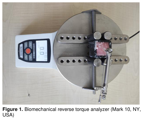
Tests were performed using a digital torque measuring device (Mark-10®, New York, USA) (Figure 1). Torque values for each implant were recorded using a counterclockwise rotation, and the maximum torque resistance (N/cm) was automatically measured by the device at the moment the implant was separated from the bone. Each measurement was repeated three times, and the average of these values was recorded as the final torque value.
The obtained data were evaluated as a biomechanical strength parameter representing implant stability. Differences between groups were compared in statistical analysis to investigate the effect of systemic alendronate administration on implant stability.
Statistical Analysis: Statistical analysis of the obtained data was performed using the SPSS 22.0 (IBM Corp., Armonk, NY, USA) software package. The Shapiro–Wilk test was used to assess the conformity of the data to a normal distribution. Nonparametric tests were used for data that did not show a normal distribution.
The Kruskal–Wallis test was used to compare differences between groups. If significant differences were found between groups, pairwise comparisons were made using the Mann–Whitney U test. In all analyses, a p value of <0.05 was considered statistically significant.
Results
At the end of the eight-week experimental period, all subjects were sacrificed without any complications, and no infection, implant loss, or complications were observed. All tibias were found to be suitable for mechanical testing.

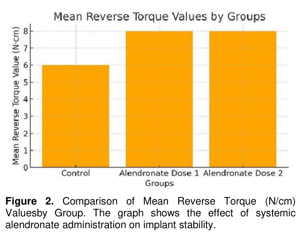
According to the reverse torque test results, the average implant stability values were measured as 6.32 N/cm in the control group, 8.41 N/cm in the alendronate dose 1 group, and 8.21 N/cm in the alendronate dose 2 group. Minimum and maximum values ranged from 5.10 to 7.80 N/cm in the control group, 7.50 to 10.20 N/cm in the alendronate dose 1 group, and 6.70 to 11.90 N/cm in the alendronate dose 2 group, respectively.
Statistical analyses revealed a significant increase in reverse torque values in both alendronate groups compared to the control group (p=0.001 and p=0.003, respectively). However, no statistically significant difference was observed between the two alendronate doses (p>0.05).
These findings suggest that systemic alendronate administration increases biomechanical stability at the implant-bone interface, but increasing the dose does not provide an additional advantage on this effect (Table 1 and figure 2).
Discussion
Bisphosphonates have the potential to increase the stability of bone tissue around implants by reducing bone resorption and thus slowing the rate of bone turnover. For example, recent systematic reviews indicate that anti-resorptive treatments do not significantly affect implant success under normal conditions [26]. However, despite this theoretical benefit, clinical and preclinical data remain variable: some studies have observed positive effects on implant success in patients using bisphosphonates, while others have reported that high doses or long-term use may reduce implant success [27].Recent experimental and clinical studies have extensively investigated the effects of bisphosphonates on bone tissue around dental implants. Some of these studies report that bisphosphonates reduce bone resorption by suppressing osteoclastic activity and promote osseointegration by increasing bone density around the implant.
Yu et al. [24] investigated the effects of bisphosphonates (especially zoledronic acid) on the osseointegration of dental implants using a rabbit model. The findings indicated that the bone-to-implant contact rate was significantly lower in the high-dose zoledronic acid group compared to the control group in the first 4 weeks, but this difference ceased to be significant by 8 weeks. These results suggest that bisphosphonates may suppress peri-implant bone healing in the early period, but this effect diminishes in the long term.
Rebelo et al. [23] conducted a systematic review and examined dental implant failure rates in patients receiving oral or intravenous bisphosphonates. The study reported an average implant failure rate of 49.96% in individuals receiving bisphosphonate therapy. The researchers stated that this may be due to disruption in the bone remodeling cycle, particularly due to high-dose and long-term systemic bisphosphonate administration.
Kniha et al. [28] conducted an animal model study to evaluate the effects of systemic antiresorptive drug administration on osseointegration of dental implants in osteoporotic conditions. The researchers reported that bisphosphonate treatment promoted new bone formation at the bone-implant interface and increased implant stability. These findings suggest that systemic bisphosphonate administration may promote implant integration, particularly in individuals with low bone density.
A comprehensive review by Fiorillo et al. [29] evaluated the effects of bisphosphonate medications on dental implant healing. The researchers emphasized that bisphosphonate use does not necessarily contraindicate implant treatment, but long-term clinical data and the dose-response relationship are still unclear. The study also noted that implant success may vary depending on variables such as the type of bisphosphonate, the duration of application, and the patient's systemic condition.
Bingül et al. [30] conducted an experimental study evaluating the effects of local zoledronic acid application on the osseointegration of dental implants with different surface properties in a rat model. The study reported that local application of zoledronic acid around the implant increased the bone-to-implant contact rate, particularly on implants with rough surfaces, and significantly supported osseointegration. Histological analyses demonstrated that local bisphosphonate application increased bone matrix density and promoted new bone formation. The results of the study suggest that local application of bisphosphonates may produce a more controlled and targeted osseointegrative effect compared to systemic use. This study biomechanically evaluated the effects of systemic alendronate administration on the osseointegration of titanium implants placed in rat tibias. The findings showed a significant increase in reverse torque values in the alendronate-treated groups compared to the control group. This result may be attributed to alendronate's ability to inhibit osteoclastic activity, reduce bone resorption, and create a stronger biomechanical bond at the bone-implant interface. However, no statistically significant difference was found between the groups treated with different alendronate doses.
In a study evaluating the effects of alendronate, Basudan et al. [31] conducted an in vivo study to evaluate the effects of alendronate alone and in combination with simvastatin on implant osseointegration in both healthy and osteoporotic rat models. The researchers reported that systemic alendronate administration significantly increased peri-implant bone volume and bone-to-implant contact ratio, and this effect became even more pronounced when combined with simvastatin. Histomorphometric analyses showed that alendronate promoted new bone formation, densified bone trabecular structure, and accelerated osseointegration, particularly in osteoporotic groups. The study concluded that alendronate alone had a positive effect on osseointegration, but that its use with simvastatin produced a synergistic improvement.
Verzola et al. [32] in their experimental study, bilateral titanium implants were placed in rat tibias following systemic alendronate administration for 120 days, and bone healing and mineralization around these implants were monitored in detail over time. The findings indicated that long-term alendronate use resulted in increased bone volume and high mineralization levels at the bone-implant interface in the early period, while bone remodeling slowed down and bone quality tended to decline with extended follow-up. This study provides important warnings that while alendronate contributes positively to the osseointegration process, the dose-effect ratio may decrease or the bone regeneration rhythm may slow down over time.
Studies in the literature evaluate the effects of alendronate on bone metabolism not only from a pharmacological perspective but also in conjunction with mechanical factors. Chen et al. [33] investigated the effects of osteoporosis and mechanical loading on osseointegration in a rat model in their experimental study. Controlled mechanical loading was applied to rats implanted with titanium implants under osteoporotic conditions at regular intervals, and bone-to-implant contact ratio and bone mineral density were assessed. The findings showed that osteoporosis negatively impacts osseointegration, but regular mechanical loading partially offsets this effect. The researchers emphasized that mechanical stimulation promotes bone remodeling and that this effect, combined with anti-resorptive drugs such as alendronate, may enhance the osseointegration process.
The systemic effects of bisphosphonates can be decisive not only on bone tissue but also on the host response in peri-implant soft tissues. In this context, Kniha et al. [34] evaluated the effects of systemic alendronate and zoledronic acid administration on the peri-implant inflammatory response and clinical parameters in a rat model. The researchers reported that peri-implant bone density significantly increased in the bisphosphonate-administered groups, but also increased inflammatory cell infiltration was observed in the peri-implant tissues. Histological findings revealed that alendronate increased the bone-implant contact rate during the early healing period, while zoledronic acid significantly suppressed bone resorption. However, it was emphasized that high doses and long-term use of both agents may negatively affect peri-implant tissue reactions.
Although numerous studies have demonstrated the positive effects of bisphosphonates on osseointegration, some studies have also reported negative results. Inoue et al. [35] conducted an experimental study evaluating the healing of soft and hard tissues around implants in the rat maxilla under systemic alendronate and dexamethasone combination therapy. The researchers reported that this combination resulted in wound healing disorders characterized by significantly increased necrotic bone area, empty osteocyte lacunae, and dense inflammatory cell infiltration compared to alendronate alone or the control group. The study emphasized that the combined use of alendronate and dexamethasone may negatively impact tissue regeneration around the implant, and this effect is particularly pronounced in the maxilla. Although not routinely used in clinical practice, reverse torque analysis is considered a reliable and objective method for assessing the mechanical strength of osseointegration in experimental and preclinical research. This test provides indirect but robust data on the mechanical integrity of the bone-implant interface by measuring the force required to break the bond between the implant and bone. Various surface modifications are a frequently preferred method for comparing the effects of implant materials or pharmacological agents (e.g., anti-resorptive drugs such as alendronate) on osseointegration [36]. The increase in reverse torque values observed in the alendronate-treated groups in our study suggests that this drug has a biological effect supporting osteogenic activity and mineralization at the bone-implant interface. However, the fact that this increase did not reach statistically significant differences between doses suggests that alendronate supports osseointegration to a certain extent, but that increasing the dose did not significantly enhance this effect.
The findings from this study indicate that systemic alendronate administration may have a supportive effect on implant osseointegration. This result is clinically significant, particularly in terms of increasing implant success in osteoporotic individuals. Alendronate's ability to reduce bone resorption may enhance early-stage mechanical stability. However, considering the potential side effects of systemic use, it is believed that local or controlled-release systems may offer a safer and more targeted treatment option in the future.
This study has several limitations. First, the inclusion of only eight rats in each group limited the sample size. This may have reduced the power of the statistical analyses, making it difficult to statistically demonstrate biologically significant differences. Secondly, the assessments were limited to biomechanical analysis (reverse torque testing). While this method is a reliable assessment tool for implant stability, it falls short in determining parameters such as bone density, degree of mineralization, and microstructural integrity. The absence of micro-CT, histomorphometric examination, or molecular biomarker analyses limited the biological depth of the results. Thirdly, only two different systemic alendronate doses were administered in the study. The lack of testing of different dose intervals or application durations made it difficult to determine the most effective pharmacological level.
Finally, the study included only a short-term evaluation of eight weeks. However, osseointegration is a dynamic process and involves biological responses that change over time. Therefore, the late-term effects of alendronate could not be assessed without longer-term follow-up.
This study evaluated the effect of systemic alendronate administration on osseointegration in titanium implants placed in rat tibias. The findings showed an increase in reverse torque values in the alendronate-treated groups compared to the control group. This suggests that alendronate may increase biomechanical strength at the bone-implant interface. However, no statistically significant difference was found between the two different doses administered. These findings suggest that alendronate may support implant stability within a certain dose range, but increasing the dose does not provide an additional advantage. In conclusion, alendronate has the potential to support osseointegration, and this effect should be investigated over longer periods and with different application protocols.
References
- Blumer MJF. Bone tissue and histological and molecular events during development of the long bones. Ann Anat 2021;235:151704.
- Omi M, Mishina Y. Roles of osteoclasts in alveolar bone remodeling. Genesis 2022;60(8-9):e23490.
- Sun J, Xie W, Wu Y, Li Z, Li Y. Accelerated bone healing via electrical stimulation. Advanced Science 2024;12(24):e2404190.
- Fan S, Sun X, Su C, et al. Macrophages—bone marrow mesenchymal stem cells crosstalk in bone healing. Frontiers in Cell and Developmental Biology 2023;11:1193765.
- Kim T, See CW, Li X, Zhu D. Orthopedic implants and devices for bone fractures and defects: Past, present and perspective. Engineered Regeneration 2020;1:6-18.
- Molitoris KH, Huang M, Baht GS. Osteoimmunology of fracture healing. Current Osteoporosis Reports 2024;22(3):330-339.
- Bohara S, Suthakorn J. Surface coating of orthopedic implant to enhance the osseointegration and reduction of bacterial colonization: A review. Biomaterials Research 2022;26(1):26.
- McClung MR, Pinkerton J, Blake J, et al. Management of osteoporosis in postmenopausal women: The 2021 position statement of The North American Menopause Society. Menopause 2021;28(9):973-997.
- Shen X, Yie KHR, Wu X, et al. Improvement of aqueous stability and anti-osteoporosis properties of Zn-MOF coatings on titanium implants by hydrophobic raloxifene. Chemical Engineering Journal 2022;430:133094.
- Li K, Tao B, Tian H, et al. Titanium implants with antiaging effect to repair senile osteoporosis fracture. Materials & Design 2023;232:112071.
- Föger-Samwald U, Kerschan-Schindl K, Butylina M, Pietschmann P. Age Related Osteoporosis: Targeting Cellular Senescence. International Journal of Molecular Sciences 2022;23(5):2701
- Cummings SR, Santora AC, Black DM, Russell RGG. History of alendronate. Bone 2020;137:115411.
- Billington E, Aghajafari F, Skulsky E, Kline GA. Bisphosphonates. BMJ 2024; 386:e076898.
- Kuźnik A, Październiok-Holewa A, Jewula P, Kuźnik N. Bisphosphonates—much more than only drugs for bone diseases. European Journal of Pharmacology 2020;866:172773.
- Barbosa JS, Almeida Paz FA, Braga SS. Bisphosphonates, old friends of bones and new trends in clinics. Journal of Medicinal Chemistry 2021;64(3):1260-1282.
- Villatoro-Villar M, Kwoh CK. Bisphosphonates, Bone and Joint Pain. Current Osteoporosis Reports 2021;19(4):417-428.
- Mary A, Reddy SG, Kumar BS, Kugabalasooriar S. Novel Approaches to Alendronate Delivery Beyond Oral Administration - A Review. Engineered Science 2024;32:1274.
- Klara J, Lewandowska-Łańcucka J. How efficient are alendronate-nano/biomaterial combinations for anti-osteoporosis therapy? An evidence-based review of the literature. International Journal of Nanomedicine 2022;17:6065.
- Kendler D, Chines A, Clark P, et al. Bone mineral density after transitioning from denosumab to alendronate. The Journal of Clinical Endocrinology & Metabolism 2020;105(3):e255-e64.
- Acikan I, Dundar S. Biomechanical examination of osseointegration of titanium implants placed simultaneously with allogeneic bone transfer. J Craniofac Surg 2022;33(1):350-353.
- Al-Noaman AS. Influence of oral bisphosphonate on dental implant: A review. Medical Journal of Babylon 2022;19(2):180-184.
- Hammouri MH, Salekh KM. Dental implants osseointegration in patients with osteoporosis. RUDN Journal of Medicine 2022;26(4):422-430.
- Rebelo CG, Fernandes JC, Bernardo N, Couto P, Fernandes GV. Bisphosphonates and their influence on the ımplant failure: A systematic review. Applied Sciences [Internet] 2023; 13(6).
- Yu RQ, Wang JY, Rao NJ, Huo L, Zheng LW. Effects of Bisphosphonates on Osseointegration of Dental Implants in Rabbit Model. BioMed Research International 2021;2021(1):6689564.
- Gibreel S, Mohamed HG, Suraj AR, Anil S. Osseointegration of dental implants and osteoporosis. Current Concepts in Dental Implantology-From Science to Clinical Research: IntechOpen; 2021.
- Jung J, Shim GJ, Park JS, Kwon YD, Ryu JI. Effect of anti-resorptive therapy on implant failure: A systematic review and meta-analysis. J Periodontal Implant Sci 2025;55(2):87-103.
- Sulaiman N, Fadhul F, Chrcanovic BR. Bisphosphonates and dental implants: A systematic review and meta-analysis. Materials 2023;16(18):6078
- Kniha K, Hermanns-Sachweh B, Möhlhenrich SC, et al. Effect of systemic antiresorptive medication on the histopathological parameters of implant osseointegration in an in vivo rodent study. BMC Oral Health 2023;23(1):117.
- Fiorillo L, Cicciù M, Tözüm TF, et al. Impact of bisphosphonate drugs on dental implant healing and peri-implant hard and soft tissues: a systematic review. BMC Oral Health 2022;22(1):291.
- Bingul MB, Gul M, Dundar S, Bozoglan A, et al. Effects of the application local zoledronic acid on different dental ımplants in rats on osseointegration. Drug Des Devel Ther 2024;18:2249-2256.
- Basudan AM, Shaheen MY, Niazy AA, et al. Biological effect of single or combined pharmacological therapy using alendronate and simvastatin on implant osseointegration: An in vivo study in healthy and osteoporotic rat models. Applied Sciences 2020;10(12):4298.
- Verzola MHA, Frizzera F, Cerri PS, et al. Effect of long-term treatment with alendronate on bone repair and mineralization around ımplants in rats. Brazilian Dental Journal 2024;35:e23-5751.
- Chen X, Moriyama Y, Takemura Y, Rokuta M, Ayukawa Y. Influence of osteoporosis and mechanical loading on bone around osseointegrated dental implants: A rodent study. Journal of the Mechanical Behavior of Biomedical Materials 2021;123:104771.
- Kniha K, Rink L, Wolf J, et al. Host inflammatory response and clinical parameters around implants in a rat model using systemic alendronate and zoledronate acid drug administrations. Scientific Reports 2022;12(1):4431.
- Inoue M, Matsumoto C, Nakajima K, Kuroshima S, Sawase T. Alendronate/dexamethasone combination therapy worsens soft and hard tissue wound healing around implants in rat maxillae. Bone 2021;148:115942.
- Gunes N, Gul M, Dundar S, et al. Biomechanical evaluation of implant osseointegration after guided bone regeneration with different bone grafts. Journal of Craniofacial Surgery 2021;32(4):1545-8.
- Localization and Surgical Treatment of Parathyroid Lesions
- PARATİROİD LEZYONLARININ LOKALİZASYONU VE CERRAHİ TEDAVİSİ
- Development of Skin During Prenatal and Postnatal Stages In Rats
- Effect Of Nıfedıpıne On Experımental Epılepsy Induced By Penıcıllın In Rat
- Use of Carnitine to Prevent Effectis of Ischemiae in Torsion-Induced Ovarian Ischaemiae